Case Report
We present the case of a patient with a descending aorta aneurysm rupture into the esophagus, which, after aortoplasty with Dacron tube interposition and suture of esophageal laceration, developed a pleural-esophagus fistula on the 3rd postoperative day. She needed re-intervention and intensive care, followed by adequate recovery. Considering this unusual case and the knowledge acquired through its management, we reviewed the literature in order to discuss the best alternative for the correction of this rare and often fatal form of presentation of aortic diseases.
Key Words
Aortic aneurysm; aortic ruptura; esophageal fistula.
Aortic Aneurysm Rupture into the Esophagus
Christiano da Silveira de Barcellos1, Paulo Ceratti de Azambuja1, Marcelo Kunh Momolli1, Clóvis Manfredini Rigoni1, Marcelo Lopes1, Henrique Biavatti1, Wagnes Franceschi1, Claudio Borges Fortes2
Hospital São Vicente de Paulo - Universidade de Passo Fundo, Passo Fundo, RS1; Hospital Divina Providência - Frederico Westphalen, RS2 - Brazil
Mailing Address: Christiano da Silveira de Barcellos •
Rua Teixeira Soares, 885 sala 204 – Centro - 990081-010 - Passo Fundo, RS - Brazil
E-mail: [email protected]
Manuscript received December 03, 2007; revised manuscript received April 08, 2008; accepted April 18, 2008.
Introduction
Aortoesophageal fistulae (AEF) are rare and represent a therapeutic challenge due to the high morbimortality of the surgical treatment. However, the conservative approach inevitably leads to the patient’s death1. Its treatment aims at the control of the hemorrhage, the sepsis management and the food tract reconstruction2. The main causes of aortoesophageal fistulas are the thoracic aorta aneurysms, responsible for 2/3 of the cases, the ingestion of foreign bodies, tumors and esophageal surgeries, as well as surgical interventions in the thoracic aorta1,2.
Case Report
The patient was a 64-year-old Caucasian woman that reported being awakened by intense abdominal pain followed by severe hematemesis. When she was admitted at the hospital, she was pale and hypotensive and was promptly reanimated by an infusion of crystalloids and red blood cell concentrate. The high digestive endoscopy showed concaving of the posterior esophageal wall at 30 cm from the upper dental arch, pulsatile, with a mucous ulcer of a purplish color, measuring around 1 cm of diameter (Figure 1).The patient was then referred to Hospital São Vicente de Paulo de Passo Fundo, state of Rio Grande do Sul, Brazil. Her general status was regular; her mucosae were pale 2+/4+, she was afebrile and presented stage II hypertension (systolic arterial pressure
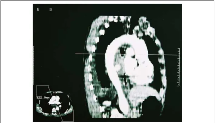
= 160 mmHg). A chest computed tomography (CT) showed a saccular aneurysm of the descending aorta protruding anteriorly at the level of the 6th thoracic vertebra, measuring 4 cm in its largest diameter, very close to the esophagus, but with no signs of rupture into the mediastinum or pleural cavity (Figure 2).
The surgical exploration did not demonstrate rupture of the aneurysm into the mediastinum or pleura. The patient was submitted to descending aortoplasty with femorofemoral circulatory support, interposing a Dacron tube was interposed in the aortic segment related to the saccular aneurysm. The esophagus was debrided, sutured and recovered by the aneurysm wall, in a conservative approach. The patient presented good immediate evolution, with early extubation and minimal hemorrhage through the drains. On the third postoperative day, the patient presented a febrile peak. The chest X-ray showed clearly purulent pleural effusion to the left and multi-septated at the draining. The contrasted study of the esophagus showed a pleural-esophageal fistula. Antibiotic therapy was initiated and the patient was submitted to the reintervention; an esophagectomy with cervical esophagostomy was performed, together with a jejunostomy for nutritional support, pulmonary decortication and a new aortoplasty, as the aorta presented an extensive necrotic segment and signs of imminent rupture close to the anastomoses with the Dacron tube. The new tube was interposed after cavity debridement and washing, without circulatory support. On the 4th day after the reintervention, she developed a thoracic duct fistula and was treated with lipid restriction and prolonged draining; resolution was achieved after 15 days.
There was good response to the performed surgical measures and antibiotic therapy and the patient was discharged 45 days after the first procedure, returning 4 months later for the reconstruction of the digestive tract through gastroplasty and gastric cervical-esophageal anastomosis. The patient has been followed for 3 years and is socially and professionally rehabilitated.
Discussion
The first report describing an AEF due to the ingestion of foreign bodies was published in 1818. Almost a century later, in 1914, Chiari described the classic triad of thoracic pain, sentinel hemorrhage and fatal hemorrhage3.
The causes of AEF described to date are aneurysms and perforating ulcers of the thoracic aorta, esophageal perforation by foreign body, esophageal surgery, mediastinitis due to tuberculosis, radiotherapy and stent implant in the thoracic aorta2,4. The prognosis is poor and the evolution
Case Report
Barcellos et al Aortic aneurysm rupture into the esophagus
Arq Bras Cardiol 2008;91(6):e61-e63
Figure 2 -Chest CT demonstrating saccular aneurysm in the descending aorta, at the level of the 6th thoracic vertebra, very close to the esophagus.
Figure 1 -High Digestive Endoscopy demonstrating mucosal ulcer measuring around 1 cm of diameter.
without surgical treatment is invariably fatal5. The rupture into the esophagus implicates in the contamination of the mediastinum, determining the worst outcome of these patients6. The recommended conducts are not uniform and the experience in the treatment of the AEF is not consistent. For the aortic correction, the most often used technique is the in situ repair, with the extra-anatomic repair being an uncommon alternative2. Although the left atrial-to-femoral or femoro-femoral circulatory support is frequently used associated to the CSF drainage, the urgency of the situation
many times does not allow such measures to be taken and the ischemic clamping is chosen7.
At the second intervention, the septic conditions of the surgical field did not allow the use of circulatory support.
The management of the esophageal laceration must be individualized, by evaluating the lesion extension, time of rupture, the status of the cavity and of the patient7. The conservative approach consists in esophageal suture after laceration debridement and aneurysm wall reinforcement with a diaphragm muscle, omental or pleural flap, associated to cervical esophagostomy and gastrostomy for nutritional support.
However, the primary esophagectomy with cervical esophagostomy and concomitant repair, or at a second surgical time, has been associated with better outcomes2.
Recently, some publications have suggested the endovascular treatment of AEF in patients with contraindications to the conventional surgical treatment. The presented results do not allow the endorsement of this treatment modality, except in situations where the aforementioned surgical treatment modalities are impossible to be carried out1. Large-spectrum antibiotic therapy must be used from the moment of the diagnosis and prolonged for several weeks aiming at preventing graft infection2.
Conclusions
Based on the experience acquired with the reported case, we conclude that, for patients with good general status at the moment of the diagnosis, with no evidence of
Case Report
Barcellos et al
Aortic aneurysm rupture into the esophagus
Arq Bras Cardiol 2008;91(6):e61-e63
References
1. Flores J, Shiiya N, Kunihara T, Yoshimoto K, Yasuda K. Aortoesophageal fistula: alternatives of treatment: case report and literature review. Ann Thorac Cardiovasc Surg. 2004; 10: 242-6.
2. Silva ES, Tozzi FL, Otochi JP, Tolosa EM, Neves CRB, Fortes F. Aortoesophageal fistula caused by aneurysm of the thoracic aorta: successful surgical treatment, case report, and literature review. J Vasc Surg. 1999; 30: 1150-7. 3. Chiari H. Ueber fremdkorpeverletzung des oesophagus mit aortenperforation.
Berlin Klin Wschr. 1914; 51: 7-9.
4. Hance KA, Hsu J, Eskew T, Hermreck AS. Secondary aortoesophageal fistula afterendoluminal exclusion because of thoracic aortic transaction. J Vasc Surg.
2002; 37: 886-8.
5. Sloop RD, Thompson JC. Aorto-esophageal fistula: report of a case and review of the literature. Gastroenterology. 1967; 53: 768-77.
6. Segesser LK, Tkebuchava T, Niederhauser U, Kunzli A, Lachat M, Genoni M, et al. Aortobronchial and aortoesophageal fistulae as risk factors in surgery of descending thoracic aortic aneurysms. Eur J Cardiothorac Surg. 1997; 12: 195-201.
7. Reardon MJ, Brewer RJ, LeMaire SA, Baldwin JC, Safi HJ. Surgical management of primary aortoesophageal fistula secondary to thoracic aneurysm. Ann Thorac Surg. 2000; 69: 967-70.
mediastinal or pleural cavity contamination, the safest option is the esophagectomy associated to cervical esophagostomy with posterior reconstruction of the digestive tract, when sepsis is no longer a risk. We emphasize the need for the surgical management of the esophageal lesion at the moment of the aortoplasty, considering the possible and fatal consequences of the resulting esophageal-pleural fistulas. The surgical treatment is mandatory even in cases with evidence of developing sepsis. The endovascular treatment remains an exception for patients that cannot undergo surgical treatment.
Potential Conflict of Interest
No potential conflict of interest relevant to this article was reported.
Sources of Funding
There were no external funding sources for this study.
Study Association
This study is not associated with any graduation program.