1 . Master’s Degree of the Postgraduation Program of the Cardiology Institute of Rio Grande do Sul/University Foundation of Cardiology (IC/FUC), Porto Alegre, RS, Brazil.
2 . PhD Student of the Postgraduation Program of IC/FUC, Porto Alegre, RS, Brazil.
3 . Cardiac Surgeon of IC/FUC, Porto Alegre, RS, Brazil. 4 . Cardiologist of IC/FUC, Porto Alegre, RS, Brazil.
5 . Biologist and Collaborator Researcher of IC/FUC, Porto Alegre, RS, Brazil.
6 . PhD; Institute of Cardiology of Rio Grande do Sul/University Foundation of Cardiology (IC/FUC) and Federal University of Health Sciences of Porto Alegre (UFSCPA), Porto Alegre, RS, Brazil.
Bruna Eibel
1, Clarissa G. Rodrigues
2, Imarilde I. Giusti
1, Ivo A. Nesralla
3, Paulo R. L. Prates
3, Roberto
T. Sant’Anna
4,
Nance B. Nardi
5,
Renato A. K. Kalil
6Terapia gênica para cardiopatia isquêmica: revisão de ensaios clínicos
Gene therapy for ischemic heart disease: review of
clinical trials
This study was carried out at Cardiology Institute of Rio Grande do Sul/ University Foundation of Cardiology (IC/FUC) and Federal University of Health Sciences of Porto Alegre (UFSCPA), Porto Alegre, RS, Brazil.
Correspondence address: Renato A. K. Kalil
Av. Princesa Isabel, 370 – Santana – Porto Alegre, RS, Brazil – Zip Code: 90620000
-E-mail: [email protected]
Article received on June 14th, 2011
Article accepted on September 5th, 2011
Abstract
Severe ischemic heart disease with refractory angina, occurs in increasing incidence. Alternative forms of treatment, in an attempt to reduce myocardial ischemia and relief of symptoms has been studied. In this context, gene therapy is an option, for the possibility of inducing angiogenesis, establish collateral circulation and reperfuse ischemic myocardium. Several clinical trials have been conducted and, except for specific cases of adverse effects, there is indication of safety, feasibility and potential effectiveness of therapy. The clinical benefit, however, is not yet well established. In this article we review the clinical trials of gene therapy for patients with ischemic heart disease. The approach includes: (1) myocardial ischemia and angiogenesis on the pathophysiological aspects involved, (2) growth factors, dealing with specific aspects and justifying the use in cardiac patients with no option for conventional therapy, (3) controlled clinical trials, where a summary of the main studies involving gene therapy for severe ischemic heart disease is presented, (4) our experience, especially on preliminary results of the first gene therapy clinical trial in Brazil and (5) future prospects.
Keywords: Gene therapy. Myocardial ischemia. Angina pectoris.
Resumo
Cardiopatia isquêmica grave com angina refratária a formas convencionais de tratamento apresenta-se em uma crescente incidência. Para tratar angina refratária, terapias alternativas na tentativa de redução da isquemia miocárdica e alívio de sintomas têm sido estudadas. Neste contexto, a terapia gênica representa uma opção, pela possibilidade de induzir angiogênese, estabelecer circulação colateral e reperfundir miocárdio isquêmico. Diversos ensaios clínicos têm sido conduzidos e, com exceção de casos isolados e específicos de efeitos adversos, há indicação de segurança, viabilidade e potencial eficácia da terapia. O benefício clínico não está bem definido. Neste artigo, revisamos os ensaios clínicos que utilizaram terapia gênica para tratamento de pacientes cardiopatas isquêmicos. A abordagem inclui: (1) isquemia miocárdica e angiogênese, sobre os aspectos fisiopatológicos envolvidos; (2) fatores de crescimento, tratando sobre aspectos específicos e justificando a utilização em pacientes cardiopatas isquêmicos sem opções pela terapêutica convencional; (3) ensaios clínicos controlados, onde é apresentado um resumo dos principais estudos envolvendo terapia gênica para tratamento da cardiopatia isquêmica grave; (4) nossa experiência, especialmente sobre resultados preliminares do primeiro ensaio clínico de terapia gênica do Brasil e (5) perspectivas.
Descritores: Terapia de genes. Isquemia miocárdica. Angina pectoris.
INTRODUCTION
It is estimated that cardiovascular disease (CVD) cause
approximately 17 million deaths worldwide each year, with
higher prevalence in developed countries [1]. CVD should
be leaders in mortality in the developing world within the
next decades, reaching epidemic levels [1]. Coronary artery
disease (CAD) is a problem of increasing prevalence,
especially in large cities and the populations of older age,
its mortality is 80% of deaths from CVD [2,3]. Angina
refractory to traditional forms of treatment in cardiology,
including percutaneous and surgical revascularization and
optimal drug therapy, represents up to 15% of all cases of
angina [2]. According to statistics from the American Heart
Association [3], the prevalence of refractory angina in the
U.S. population is 4.6%, affecting 58% of patients with CAD
and growing rapidly with increasing age. Despite advances
in treatment modalities, it is estimated that the incidence of
patients with refractory angina will increase in coming years
[4,5], pointing to the need for new treatment options.
In this context, gene therapy could be an option, due
to the potential to induce myocardial angiogenesis and
establish collateral circulation [5]. Gene therapy can be
defined as a set of techniques that allow the insertion and
expression of a therapeutic gene in target cells that have
some kind of disorder of genetic origin (not necessarily
hereditary), enabling the correction of inappropriate gene
products that cause diseases, therefore being an
alternative for the treatment of diseases based on the
transfer of genetic material [6-8]. Studies have tested the
effects on ischemic heart disease patients using different
growth factors, several doses, vectors and routes of
administration. It can be emphasized the vascular
endothelial growth factor (VEGF), a regulator of
endothelial cells, which has the property of mediating
angiogenesis during tissue repair [4].
The availability of vectors with tropism for the
myocardium, capable of a long and stable protein expression
[9], and the isolation of progenitor cells with regenerative
and angiogenic potential [10] offers possibilities for
development of therapy based on protection and
regeneration of ischemic myocardium.
Although promising, the clinical effects on the
myocardial vasculature provided by gene therapy remain
to be clarified fully. The aim of this study is to review the
clinical trials of gene therapy for the treatment of ischemic
heart disease patients.
GENE THERAPY APPLIED TO ISCHEMIC
CARDIOMYOPATHY
Myocardial Ischemia and Angiogenesis
In the pathophysiology of ischemic heart disease, two
processes are involved: supply and demand of myocardial
oxygen. Myocardial ischemia occurs when there is
imbalance between supply and demand for oxygen. Two
situations alter the oxygen supply to the myocardium,
ischemia and hypoxia. In some conditions, the impairment
of oxygen is secondary to decreased blood flow and in
other situations, the increase in oxygen demand is the main
responsible for myocardial ischemia [11].
Features of molecular biology and gene therapy have
been developed for application in cardiovascular therapy,
in situations where there are no options, or when these
conventional methods have limitations. The main area of
development of gene therapy in cardiology is the induction
of myocardial angiogenesis with potential benefits in
end-stage ischemic heart disease, after exhaustion of
pharmacological, surgical and interventional resources
using catheter, ie, in those refractory cases to all forms of
treatment, where only the use of cardiac transplantation
would be possible[4].
Angiogenesis, the formation of new vessels from
existing endothelium of blood vessels, has an important
role in embryonic development, tissue repair and progression
of a variety of pathological processes [12,13]. Angiogenesis
induced by administration of growth factors is intended to
promote the formation of new blood vessels, capillaries
and arterioles.
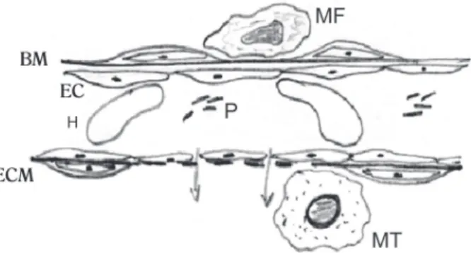
The mechanism of angiogenesis can be initiated by
factors of a mechanical nature, by inflammatory or hypoxic
process (energy imbalance). The process of angiogenesis
occurs in stages (Figure 1) comprising: vessel dilation,
endothelial cell activation, platelet activation, secretion of
plasminogen activators and proteolytic enzymes, mast cell
degranulation, activation of macrophages, disruption of
the basement membrane and increased permeability with
release of fibrin and other proteins. Following, formation of
pseudopodia occurs, degradation of extracellular matrix,
migration of endothelial cells to the extravascular space
with the same proliferation and formation of shoots of
vascular tissue. Finally, they form new basement membrane
and maturation of the new establishment of the vascular
wall to blood flow, formation of tubes and conections,
establishing new vessels [14].
The idea that angiogenic factors may promote
revascularization of ischemic tissues is called therapeutic
angiogenesis [15]. The concept of therapeutic angiogenesis
in humans through clinical trials phase I went ahead with
the idea of testing this strategy in ischemic cardiomyopathy.
Therefore, therapeutic angiogenesis is a strategy designed
to amplify the natural process of angiogenesis and reperfuse
ischemic tissues, which may represent a new process of
revascularization in these high-risk patients [16].
of PR-39macrophage-derived peptide, this inhibits the
degradation of HIF-1
α
(hypoxia-inducible factor 1-
α
) leading
to increased expression of VEGF and its receptors [17].
Inflammation induces the production of cytokines that
promote angiogenesis [18]. In contrast, PR-39 increases the
production of fibroblast growth factors (FGF), which have
angiogenic power. Mechanical factors may act by activating
the same mechanism, resulting in angiogenesis [10].
Vascular Endothelial Growth Factor, Fibroblast Growth
Factor (FGF), hepatocyte growth factor (HGF) Plasmid and
Aenoviral vector.
The formation of new blood vessels responds to the
stimulation of angiogenic factors, which regulate
endothelial migration, proliferation, survival, and proteolytic
activity. Among the factors described in the literature, VEGF
has emerged as a critical regulator of pro-angiogenic
process [19-21]. This molecule promotes the formation of
new vessels and their morphogenesis, through a complex
process of angioregulatory events [22,23].
VEGF, family member of VEGF A, which consists of five
isoforms resulting from alternative divisions of a single
gene, ie, VEGF121, VEGF145, VEGF165, VEGF189 and
VEGF206, is a growth factor specific to the endothelium
[24.25 ]. It acts mainly by activating two Flt-1 tyrosine kinase
receptors (fms-like tyrosine kinase-1, VEGF receptor-1) [26]
and KDR (kinase-insert domain-containing receptor, VEGF
receptor-2) [27] but can also activate other receptors, such
as Neuropilins-1 and 2 [28]. VEGF may represent a new
treatment modality for ischemic heart disease. This is due
to the possibility of developing new blood vessels or
promote the reformation of existing vessels [29]. The
VEGF165 contains 165 amino acids and works by interacting
with specific receptors on endothelial cells, initiating the
cascade of events that culminates in endothelial cell
migration, proliferation and aggregation of microtubules
that will eventually form a network of arterial and venous
systems.
Gene therapy in cardiovascular diseases is not intended
to replace an abnormal gene, but suprarregular the
expression of a useful protein, increasing DNA content. Its
effectiveness depends on the gene vector and method of
administration used [30]. VEGF functions both as an
important marker of endothelial damage, as the mediator of
repair. In cases of injury such as ischemia, inflammation
and infarction have their expression increased. In addition,
it encourages the maintenance, mobilization and recruitment
of endothelial progenitor cells (EPC) from bone marrow [31].
The angiogenic potential of VEGF stimulates endothelial
production of nitric oxide through activation of nitric oxide
synthase (eNOS) (Figure 2).
The endothelium synthesizes important substances,
playing a key role on the vascular control, both in
physiological conditions and in pathological processes
such as acute coronary syndromes. The monolayer of
endothelial cells acts as a nonstick surface for platelets
and leukocytes, producing a variety of important regulatory
factors, such as NO [32]. Thus, influences not only vascular
tone but also its remodeling through the production of
substances promoting and inhibiting their growth [33].
Dysfunction in endothelial cells leads to a loss of
antithrombotic properties of vascular wall and corresponds
to the beginning of the atherosclerotic process [32]. The
reconstruction occurs by endothelial migration and
proliferation of circulating mature endothelial cells.
However, these cells have low proliferative potential and
their ability to repair is limited. Evidence indicates that
peripheral blood contains bone marrow cell subsets, with
properties similar to embryonic angioblastic. EPCs have a
proliferative capacity and differentiate into mature
endothelial cells, and can be induced by various cytokines
or growth factors, acquiring different phenotypes [32].
The FGF family comprises at least nine polypeptides,
including acidic FGF and basic FGF. Unlike VEGF, FGF acts
in the mitogenesis of endothelial cells, fibroblasts and
smooth muscle cells [18]. The increased availability of FGF
provided the use of this gene has been most studied.
Among the experimental studies, we highlight the report
by Kawasuji et al. [34] in a model of acute myocardial
infarction, where the response to FGF demonstrated to
increase the number of capillaries in the border zone and in
the epicardium of the infarcted area, the increased blood
flow in these areas and the improvement in left ventricular
ejection fraction 7 days after infarction.
On the other hand, HGF is a potent mitogen for a wide
variety of cells, and angiogenic, antiapoptotic and possess
antifibrotic properties [35,36]. In a pilot study of gene
therapy in patients with CAD, Yang et al. [37] reported the
growing evidence of the beneficial effects of HGF in
myocardial infarction, heart failure and peripheral arterial
disease. The aim of this study was to assess the effects of
intracoronary administration of an adenovirus vector
encoding the human HGF gene (Ad-HGF) on serum levels
of cytokines and mobilization of CD34 (+) and CD117 (+)
cells in patients with heart disease. Given the findings, it
was concluded that gene therapy with HGF may play an
important role in the regulation of inflammatory cytokines
and induce mobilization of EPCs in patients with CAD.
Genetic vectors are all DNA molecules with potential for
autonomic replication within the host cell in which DNA
sequences can be inserted and expanded. The origin of the
vector plasmid allows to classify in bacteriophage or viral
infections [38]. They are used to transport genes into recipient
cells. They have not only markers for ease of recognition as
well as replicating sequences. Common vectors include
plasmids carriers for transportation of naked DNA and viral
vectors such as adenovirus, retrovirus and lentivirus.
Advantages and disadvantages compared to the vector
used include the size of the inserted gene, the site of
incorporation in the nucleus, the duration of expression,
the transfer efficiency and the degree of body’s immune
response [39]. The plasmid vector is expressed by only a
few days after viral vector administration and shows gene
expression for several weeks [40]. Thus, the clinical studies
that attempt to treat end-stage ischemic disease through
gene therapy may be limited by duration of exposure to
inadequate angiogenic agent [4].
Gene therapy suffered a major setback when the
occurrence of death in a research subject, probably due to
high viral load administered, and there was cancellation of
several clinical projects and return to the laboratory
research. Since 2000, few projects have been developed for
clinical application. Theoretically, before being introduced
into the patient, the viruses used as vectors suffer from
several genetic changes, so that the therapeutic gene is
inserted, while several other genes that confer virulence
are removed or inactivated [7,41,42]. Thus, when binding
and invading the target cell, the viral vectors inject its
genetic material containing the therapeutic gene in the
patient’s DNA, allowing transcription and translation of
the gene to their corresponding functional protein, or using
the molecular machinery of the host cell to express their
genes. However, in specific cases, the virulence became
something uncontrollable, where the therapy became the
cause of death of patients undergoing such intervention.
On the other hand, plasmid vectors have no gene size limit
to be inserted and induces minimal immune response,
resulting in sustained transgenic expression. The
disadvantage is the low rate of transfer of the encoding
gene of the angiogenic factor [43].
The ideal vector would be one that combines low
immunogenicity and a satisfactory safety profile, with high
efficiency of transfection and transgene expression to
specific time periods [4].
Gene transfer to the myocardium has been used as an
alternative strategy to achieve a sustained local expression
of angiogenic proteins [44]. There is a variety of different
methods to replace or repair the genes targeted in gene
therapy. A normal gene can be inserted into a nonspecific
location within the genome to replace a nonfunctional gene,
which is the most common approach, though, an abnormal
gene could be replaced by a normal gene through
homologous recombination, an abnormal gene could be
repaired through selective reverse mutation, which returns
the gene to its normal functions and also the regulation of
a gene can be altered, such regulation corresponding to
the degree to which a gene is active or inactive [6-8].
Controlled Clinical Trials of Gene Therapy
Table 1. Clinical trials involving gene therapy with intramyocardial VEGF by left mini-thoracotomy.
Author and Year
Losordo et al. [45], 1998
Symes et al. [48], 1999
Vale et al. [49], 2000
Reilly et al. [50], 2005
REVASCStewart
et al. [51], 2006
Ruel et al. [52], 2008
Kalil et al. [53], 2010
Vector
VEGF165
VEGF165
VEGF165
VEGF2
Adenoviral VEGF121
Plasmid VEGF165 and supplemental oral L-arginine associated with CABG.
plasmid VEGF165
Patients
5 patients; CAD, refractory angina, underperfused myocardial areas, but viable AC (3/4)
125 ì g/n=10, 250 ì g/ n=10;CAD, refractory angina, reversible ischemia, AC (3/4)
13 patients; CAD, refractory angina, myocardial areas underperfused but viable, AC (3/4)
30 patients; CAD, AC (3/ 4) with no
revascularization options;
Active/placebo group (32/ 35), CAD not eligible for revascularization, refractory angina, AC (2/4)
VEGF/L-arg (5),Placebo/L-ar (6),VEGF/placebo (7),Placebo/placebo (1);CAD with commitment of the ADA
13 patients, ischemic heart disease, LV ejection fraction greater than 25%symptoms of angina and/or IC, myocardial hypoperfusion
Outcomes
Myocardial perfusion and symptoms of disease in 60 days
Safety of the therapy and myocardial perfusion in 180 days
Myocardial perfusion and left ventricular performance
Safety and adverse events at 1 year of therapy
Time on ergometric test and ST segment depression in 1 mm in diameter at 26 weeks
Myocardial perfusion, left ventricular
contractility and AC in 3 months
Myocardial perfusion,
ergometric test time, quality of life, CF according to NYHA in 3 months
Results
Reduction in angina between 10 and 30 days after treatment, reducing the consumption of nitrate in 60 days (P <0.05), no reduction of myocardial ischemia in 60 days
Safety and feasibility of therapy, improvement of myocardial perfusion in 60 days, SSS = 19.4 ± 3.7 versus 15.9 ± 3.4, P = 0.025)
Reduction of ischemia before (15.26 ± 0.98%) versus after (9.94 ± 1.53%, P = 0.004) therapy,
improvement of myocardial function
After 1 year, 3 (11.5%) patients had AC 3 and 23 (88.5%) AC 1 or 2, there were 4 deaths (13.8%), MI 5 (17.2%) 7 CABG (24.1%), 15 hospitalizations and two new cancer diagnoses
Time on ergometric test at 1 mm ST segment depression was significantly higher in VEGF121-ad (P=0,026); AC improved in the group
ad-VEGF121(P<or =0.001)
Group VEGF/L-arg showed improvement in perfusion and contractility of the anterior myocardial wall (P = 0.02)
Improvement of myocardial perfusion SSS (18.38 ± 7.51 versus 15.31 ± 7.29, P = 0.003) and SRS (11.92 ± 7.49 versus 8.53 ± 6.68, P = 0.002); trend towards
improvement in ergometric test time (7.66 ± 4.47 versus 10.29 ± 4.36, P = 0.08), improved quality of life, second IC accordingNYHA class and AC.
Table 2. Clinical trials involving gene therapy with VEGF percutaneously.
Author and year
VEGFVale et al. [54], 2001
Losordo et al. [25], 2002
KATHedman et al. [28], 2003
VIVA TrialHenry et al. [46], 2003
Euroinject One Kastrup et al. [55], 2005
Ripa et al. [56], 2006
NORTHERN TrialStewart et al. [57], 2009
Vector
Intramyocardial of VEGF2 guided by electromechanical mapping (NOGA)
Intramyocardial administration via electromechanical mapping of plasmid VEGF2
Intracoronary administration of adenoviral plasmid VEGF165 and after percutaneous coronary intervention
Intracoronary infusion, followed by intravenous infusion of VEGF
Percutaneous intramyocardial administration via electromechanical mapping of plasmid VEGF165
Percutaneous intramyocardial
administration of VEGF165 using electromechanical mapping and subcutaneous injection of G-CSF
Administration of Plasmid VEGF165 by endocardial catheter through the NOGA electroanatomic catheter
Patients
6 patients, angina refractory to therapy of CAD, reversible ischemia, AC(3/4)
Group active/placebo (12 / 7), CAD not eligible for revascularization, refractory angina, AC (3/4)
VEGF-ad (37), VEGF-P (28), Placebo (38); CAD, AC (2/3), percutaneous coronary intervention
VEGF - high dose (59), VEGF - low dose (56), placebo (63); refractory angina, underperfused myocardium, but viable
Group active/placebo (40/ 40), coronary artery disease not eligible for
revascularization, refractory angina, AC (3/4)
VEGF165 + G-CSF (16), VEGF165 (16), Placebo (16); coronary artery disease not eligible for revascularization, refractory angina, AC (3/4)
A multicenter study: group active/placebo (48/ 45), advanced coronary artery disease, AC (3/4)
Outcomes
Myocardial perfusion, time during ergometric testing, angina episodes in 360 days
Change in AC and exercise tolerance in 12 weeks
% minimal luminal diameter stenosis in coronary angiography at 6 months
Myocardial perfusion time during exercise testing, AC and quality of life in 120 days
Myocardial perfusion, wall motion mapping by NOGA, left
ventriculography and AC in 3 months
Change of perfusion defects, measured by SPECT in 3 months
Myocardial perfusion, time in ergometric time and AC at 6 months
Results
Reduction of episodes of angina, improvement in ergometric test performance from 7 to 127 seconds (72 ± 25 s), improvement in myocardial perfusion at rest and stress
Improvement in AC - VEGF2 group versus placebo (-1.3 versus -0.1, P = 0.04), exercise tolerance (91.8 versus 3.9 seconds)
Clinical restenosis rate: 6%, minimum diameter and percent stenosis were not different between groups, myocardial perfusion showed a significant improvement in the VEGF-ad group
There was no significant improvement in myocardial perfusion, increase in performance under exercise test (high dose versus placebo: 48 versus 23 seconds, P = 0.15), reduction of angina episodes and improved quality of life
Myocardial perfusion was not different between the VEGF165 and placebo group (38 ± 3%, 44 ± 2%), wall motion by NOGA (P = 0.04) and left ventriculography (P = 0.03) improved compared to placebo; improvement in AC with no difference between
There was no improvement in myocardial perfusion in both treated groups and clinical symptoms have not changed
There was no improvement in myocardial perfusion; significant reduction in the ischemic area in both groups, increase in exercise test time and improvement in AC
This study highlights the potential clinical applications
of growth factors in humans, since experimental studies
have shown favorable results and initial clinical studies in
humans do not report adverse events related. Current tests
report that the use of high doses of VEGF, compared with
low doses and placebo, improves myocardial perfusion in
patients with severe angina and provides evidence of a
dose-dependent positive effect [46.47]. Confronted with
evidence, VEGF has been shown to be a potential
angiogenic factor, which benefits in medium and long term
follow-up have been or are being assessed, including the
improvement of quality of life, functional class of heart
failure, angina class, functional capacity and reduction of
myocardial ischemia [25,28,45,46,48-57].
FGF [58-60] and HGF [61] have also been demonstrating
its potential benefits in the induction of myocardial
angiogenesis, and to further develop this promising
therapeutic approach we must critically assess the results
and the experimental protocols, to identify factors that may
have undermined the effectiveness of therapy or
confounding data interpretation [4].
As the route of administration, the intramyocardial route
proved to be more effective and, therefore, have been the
most widely used in studies involving gene therapy in
cardiology [38]. Previous studies suggest that
administration by intramuscular injection offers the
possibility to offer more effective in focal areas of ischemic
muscle [4].
There are questions concerning the safe transfer of
angiogenic factors, and also in relation to time of expression
[4.62], where it is known that plasmids carriers of angiogenic
factors protein, because they have more short expression
and do not incorporate DNA to which the cell will connect
to, have a lower risk of this adverse effect. Since the viral
vectors require care in biosafety, it is unnecessary measure
with non-viral vectors. Studies indicate temporary events
related to use of adenovirus, such as fever or elevated
serum C-reactive protein, liver enzymes and antibody
titration [22]. Hao et al. [63] published in 2007, an
experimental study on myocardial angiogenesis VEGF
165compared with adenoviral plasmid vector. These authors
demonstrated equivalent benefits in terms of ventricular
Table 3. Clinical trials involving gene therapy with FGF.
Author and Year
FGFAGENTGrines et al. [58], 2002
AGENT-2 Grines et al. [59], 2003
AGENT-3 AGENT-4 Henry et al. [60], 2007
Vector
Administration of Ad5-FGF4 by intracoronary via
Administration of Ad5-FGF4 by intracoronary via
Administration of Ad5-FGF4 by intracoronary via
Patients
Group active/placebo (60/19), coronary artery disease, AC (2/3)
Group active/placebo (35/17), coronary artery disease, not eligible for revascularization, refractory angina, AC (2/4)
High dose (175), Low-dose (180), Placebo (177), coronary artery disease and refractory angina, AC (2/4); 3 AGENT: no immediate need for revascularization AGENT 4: not eligible for
revascularization
Outcomes
Safety and time of exercise testing in 12 weeks
Change of perfusion defects, measured by adenosine SPECT in 8 weeks, follow-up of 12 months
Change in time of exercise testing in 12 weeks, follow-up of 12 months
Results
Single administration of Ad5-FGF4 was safe and well tolerated; Ad5-FGF4 group showed improvement in the time of exercise testing in subgroup analysis (1.6 versus 0.6 minutes, P = 0.01, n=50)
Ad5-FGF4 resulted in a significant reduction of ischemia (4.2% absolute, 21%, P <0.001), while placebo did not improve (P = 0.32)
Significant beneficial effect of gender, women showed improvement in exercise test time and improvement of AC
function (P<0.05) for plasmids and adenovirus after 4 weeks,
however, in this study, the TUNEL technique that detects
DNA breaks that occur during the process of apoptosis,
demonstrated an increase in frequency of cardiomyocyte
apoptosis in adenovirus group (P <0.02).
Almost all clinical trials of gene therapy and study
population are patients with end stage ischemic disease,
since the possible increased risk in relation to the benefits
associated with new treatments are acceptable and can be
used as an adjunct to conventional therapy. However, in
very advanced clinical situations, therapy may not lead to
an improvement of great intensity and measurable by
available methods, even when treatment shows some
clinical benefit [4.64].
Local Experience
In the Cardiology Institute of RS/FUC and the Discipline
of Cardiology of UFCSPA, in collaboration with the
Laboratory of Immunogenetics, UFRGS, we previously
developed experimental studies [65-67] and recently
performed the first gene therapy clinical trial in Brazil, using
VEGF165 for refractory angina [53].
In experimental studies, we used a canine model of
myocardial infarction in acute and chronic phases in an
attempt to assess the processes of gene therapy. Recently,
we developed a controlled clinical trial, phase I/II
(ClinicalTrial NCT00744315) [53] in order to clinically assess
the effects of gene therapy with VEGF165 in patients with
advanced coronary artery disease (CAD), not eligible for
revascularization or percutaneous surgical. The thirteen
patients received optimal drug therapy for at least six months
and underwent administration of intramyocardial injections
of 2000 µg of plasmid VEGF
165. Patients were assessed by
myocardial scintigraphy, exercise testing, quality of life
questionnaire (Minnesota) and determination of classes of
heart failure (NYHA) and angina (CCS). In partial results of
3 months of evolution, it was concluded that the therapy
proved to be safe and feasible, tending to improvement in
severity of angina and reducing the intensity of myocardial
ischemia.
CONCLUSIONS
Over 1,000 patients were enrolled in controlled clinical
trials of gene therapy, covering more than a decade and so
far, except for specific cases, no adverse safety signal was
detected, indicating that the therapy is safe, feasible and
potentially effective although they have not produced
conclusive evidence of its benefits definitely. Reports of
retinopathy, cancer or other diseases that could be driven
by vascular growth were perceived as equally distributed
in treated and placebo groups in randomized clinical trials.
More definite conclusions about risks and complications
will require more follow-up time and number of patients
undergoing therapy [4]. Thus, gene therapy has emerged
as a potentially beneficial alternative to ischemic heart
disease patients, when conventional therapies are
exhausted. The definition of angiogenic success, for better
assessment of the results needs to be rethought and defined
by methods of higher sensitivity and specificity.
Traditionally, therapy for the treatment of cardiovascular
disease should demonstrate improvements in morbidity and
mortality. However, for this patient population, the fact of
improving quality of life, and decrease or elimination of
episodes of angina and reduction of events of
hospitalization may be considered the biggest gains on
this new therapy. Knowing that the manifestations of
cardiovascular disease is progressive, the main aim is to
offer patients significant decrease of symptoms and delay
this progression.
The future directions of gene therapy indicate probable
combinations of angiogenic factors or individual factors
(HIF 1-
α
) that activate different pathways of
neovascularization. Combinations of cell therapy and
angiogenic factors, as well as the use of biomaterials to
improve the microenvironment are other promising
strategies for ischemic tissue repair [4].
ACKNOWLEDGEMENTS
The authors acknowledge the participation in group
projects of Gene Therapy of the Institute of Cardiology of
Rio Grande do Sul/University Foundation of Cardiology,
toe following colleagues: Leonardo Karam Teixeira, Felipe
Borsu de Salles, Ana Paula Furlani, Eduardo Mastalir, Paulo
Lavanière Moreno, Sang Won Han, Eduardo Ludwig,
Gabriel Grossman, João Ricardo Michielin Sant’Anna,
Guaracy Fernandes Teixeira Filho, Melissa Medeiros
Markoski, Andrés Delgado Cañedo, Melissa Camassola,
Iran Castro, Maria Cláudia Irigoyen, Luiza Macedo Braga
and Rogério Sarmento Leite.
REFERENCES
of vascular endothelial growth factor augments revascularization in a rabbit ischemic hind limb model. J Clin Invest. 1994;93(2):662-70.
16. Kones R. Recent advances in the management of chronic stable angina I: approach to the patient, diagnosis, pathophysiology, risk stratification, and gender disparities. Vasc Health Risk Manag. 2010;6:635-56.
17. Armstrong EJ, Bischoff J. Heart valve development: endothelial cell signaling and differentiation. Circ Res. 2004;95(5):459-70.
18. Habeck M. Wielding more power over angiogenesis. Mol Med Today. 2000;6(4):138-9.
19. Lewis BS, Flugelman MY, Weisz A, Keren-Tal I, Schaper W. Angiogenesis by gene therapy: a new horizon for myocardial revascularization? Cardiovasc Res. 1997;35(3):490-7.
20. Ripa RS, Wang Y, Jørgensen E, Johnsen HE, Hesse B, Kastrup J. Intramyocardial injection of vascular endothelial growth factor-A165 plasmid followed by granulocyte-colony stimulating factor to induce angiogenesis in patients with severe chronic ischaemic heart disease. Eur Heart J. 2006;27(15):1785-92.
21. Kastrup J, Jørgensen E, Rück A, Tägil K, Glogar D, Ruzyllo W, et al; Euroinject One Group. Direct intramyocardial plasmid vascular endothelial growth factor-A165 gene therapy in patients with stable severe angina pectoris A randomized double-blind placebo-controlled study: the Euroinject One trial. J Am Coll Cardiol. 2005;45(7):982-8.
22. Hedman M, Hartikainen J, Syvänne M, Stjernvall J, Hedman A, Kivelä A, et al. Safety and feasibility of catheter-based local intracoronary vascular endothelial growth factor gene transfer in the prevention of postangioplasty and in-stent restenosis and in the treatment of chronic myocardial ischemia: phase II results of the Kuopio Angiogenesis Trial (KAT). Circulation. 2003;107(21):2677-83.
23. Eppler SM, Combs DL, Henry TD, Lopez JJ, Ellis SG, Yi JH, et al. A target-mediated model to describe the pharmacokinetics and hemodynamic effects of recombinant human vascular endothelial growth factor in humans. Clin Pharmacol Ther. 2002;72(1):20-32.
24. Simons M, Annex BH, Laham RJ, Kleiman N, Henry T, Dauerman H, et al. Pharmacological treatment of coronary artery disease with recombinant fibroblast growth factor-2: double-blind, randomized, controlled clinical trial. Circulation. 2002;105(7):788-93.
25. Losordo DW, Vale PR, Hendel RC, Milliken CE, Fortuin FD, Cummings N, et al. Phase 1/2 placebo-controlled, double-blind, dose-escalating trial of myocardial vascular endothelial growth factor 2 gene transfer by catheter delivery in patients with chronic myocardial ischemia. Circulation. 2002;105(17):2012-8.
26. Sylvén C, Sarkar N, Rück A, Drvota V, Hassan SY, Lind B, et 2. Mannheimer C, Camici P, Chester MR, Collins A, DeJongste
M, Eliasson T, et al. The problem of chronic refractory angina: report from the ESC Joint Study Group on the Treatment of Refractory Angina. Eur Heart J. 2002;23(5):355-70.
3. Lloyd-Jones DM, Hong Y, Labarthe D, Mozaffarian D, Appel LJ, Van Horn L, et al. American Heart Association Strategic Planning Task Force and Statistics Committee. Defining and setting national goals for cardiovascular health promotion and disease reduction: the American Heart Association’s strategic Impact Goal through 2020 and beyond. Circulation. 2010;121(4):586-613.
4. Gupta R, Tongers J, Losordo W. Human studies of angiogenic gene therapy. Circ Res. 2009;105(8):724-36.
5. Kim MC, Kini A, Sharma SK. Refractory angina pectoris: mechanism and therapeutic options. J Am Coll Cardiol. 2002;39(6):923-34.
6. Huard J, Li Y, Peng H, Fu FH. Gene therapy and tissue engineering for sports medicine. J Gen Med. 2003;5(2): 93-108.
7. Karthikeyan BV, Pradeep AR. Gene therapy in periodontics: a review and future implications. J Contemp Dent Pract. 2006;7(3):83-91.
8. Li SD, Huang L. Gene therapy progress and prospects: non-viral gene therapy by systemic delivery. Gene Ther. 2006;13(18):1313-9.
9. Svensson EC, Marshall DJ, Woodard K, Lin H, Jiang F, Chu L. Efficient and stable transduction of cardiomyocytes after intramyocardial injection or intracoronary perfusion with recombinant adeno-associated virus vectors. Circulation. 1999;99(2):201-5.
10. Rafii S, Lyden D. Therapeutic stem and progenitor cell transplantation for organ vascularization and regeneration. Nat Med. 2003;9(6):702-12.
11. Lotufo P. Mortalidade precoce por doenças do coração no Brasil. Comparação com outros países. Arq Bras Cardiol. 1998;70(5):321-5.
12. Kastrup J. Therapeutic angiogenesis in ischemic heart disease: gene or recombinant vascular growth factor protein therapy? Curr Gene Ther. 2003;3(3):197-206.
13. Isner JM. Myocardial gene therapy. Nature. 2002;415(6868):234-9.
14. Rakusan K. Coronary angiogenesis. From morphometry to molecular biology and back. Ann N Y Acad Sci. 1995;752:257-66.
al. Myocardial Doppler tissue velocity improves following myocardial gene therapy with VEGF-A165 plasmid in patients with inoperable angina pectoris. Coron Artery Dis. 2001;12(3):239-43.
27. Rosengart TK, Lee LY, Patel SR, Sanborn TA, Parikh M, Bergman GW, et al. Angiogenesis gene therapy: phase I assessment of direct intramyocardial administration of an adenovirus vector expressing VEGF121 cDNA to individuals with clinically significant severe coronary artery disease. Circulation. 1999;100(5):468-74.
28. Hedman M, Hartikainen J, Syvänne M, Stjernvall J, Hedman A, Kivelä A, et al. Safety and feasibility of catheter-based local intracoronary vascular endothelial growth factor gene transfer in the prevention of postangioplasty and in-stent restenosis and in the treatment of chronic myocardial ischemia: phase II results of the Kuopio Angiogenesis Trial (KAT). Circulation. 2003;107(21):2677-83.
29. Kalil R. Terapia gênica aplicada à cirurgia cardiovascular. Rev da SOCERGS. 2001;3:61-6.
30. Kalil R, Sant’Anna R. Terapia gênica aplicada às doenças cardiovasculares. Rev SOCERGS. 2004;3:213-9.
31. Heiss C, Amabile N, Lee AC, Real WM, Schick SF, Lao D, et al. Brief secondhand smoke exposure depresses endothelial progenitor cells activity and endothelial function: sustained vascular injury and blunted nitric oxide production. J Am Coll Cardiol. 2008;51(18):1760-71.
32. Hristov M, Erl W, Weber PC. Endothelial progenitor cells: mobilization, differentiation, and homing. Arterioscler Thromb Vasc Biol. 2003;23(7):1185-9.
33. Hambrecht R, Wolf A, Gielen S, Linke A, Hofer J, Erbs S, et al. Effect of exercise on coronary endothelial function in patients with coronary artery disease. N Engl J Med. 2000;342(7):454-60.
34. Kawasuji M, Nagamine H, Ikeda M, Sakakibara N, Takemura H, Fujii S, et al. Therapeutic angiogenesis with intramyocardial administration of basic fibroblast growth factor. Ann Thorac Surg. 2000;69(4):1155-61.
35. Azuma J, Taniyama Y, Takeya Y, Iekushi K, Aoki M, Dosaka N, et al. Angiogenic and antifibrotic actions of hepatocyte growth factor improve cardiac dysfunction in porcine ischemic cardiomyopathy. Gene Ther. 2006;13(16):1206-13.
36. Morishita R, Aoki M, Hashiya N, Yamasaki K, Kurinami H, Shimizu S, et al. Therapeutic angiogenesis using hepatocyte growth factor (HGF). Curr Gene Ther. 2004;4(2):199-206.
37. Yang ZJ, Xu SL, Chen B, Zhang SL, Zhang YL, Wei W, et al. Hepatocyte growth factor plays a critical role in the regulation of cytokine production and induction of endothelial progenitor cell mobilization: a pilot gene therapy study in patients with
coronary heart disease. Clin Exp Pharmacol Physiol. 2009;36(8):790-6.
38. Bae J, Cho M. Gene therapy for heart failure. Korean Circ J. 2005;35(5):345-52.
39. Giordano FJ, Ping P, McKirnan MD, Nozaki S, DeMaria AN, Dillmann WH, et al. Intracoronary gene transfer of fibroblast growth factor–5 increases blood flow and contractile function in an ischemic region of the heart. Nat Med. 1996;2(5):534-9.
40. Wright MJ, Wightman LM, Lilley C, de Alwis M, Hart SL, Miller A, et al. In vivo myocardial gene transfer: optimization, evaluation and direct comparison of gene transfer vectors. Basic Res Cardiol. 2001;96(3):227-36.
41. Wilson DR. Viral-mediated gene transfer for cancer treatment. Curr Pharm Biotechnol. 2002;3(2):151-64.
42. Rubanyi GM. The future of human gene therapy. Molecular Aspects Med. 2001;22(3):113-42.
43. Dulak J, Zagorska A, Wegiel B, Loboda A, Jozkowicz A. New strategies for cardiovascular gene therapy: regulatable pre-emptive expression of pro-angiogenic and antioxidant genes. Cell Biochem Biophys. 2006;44(1):31-42.
44. Mack CA, Patel SR, Schwarz EA, Zanzonico P, Hahn RT, Ilercil A, et al. Biologic bypass with the use of adenovirus-mediated gene transfer of the complementary deoxyribonucleic acid for vascular endothelial growth factor 121 improves myocardial perfusion and function in the ischemic porcine heart. J Thorac Cardiovasc Surg. 1998;115(1):168-76.
45. Losordo DW, Vale PR, Symes JF, Dunnington CH, Esakof DD, Maysky M, et al. Gene therapy for myocardial angiogenesis: initial clinical results with direct myocardial injection of phVEGF165 as sole therapy for myocardial ischemia. Circulation. 1998;98(25):2800-4.
46. Henry TD, Annex BH, McKendall GR, Azrin MA, Lopez JJ, Giordano FJ, et al; VIVA Investigators. The VIVA trial: Vascular endothelial growth factor in Ischemia for Vascular Angiogenesis. Circulation. 2003;107(10):1359-65.
47. Hendel RC, Henry TD, Rocha-Singh K, Isner JM, Kereiakes DJ, Giordano FJ, et al. Effect of intracoronary recombinant human vascular endothelial growth factor on myocardial perfusion: evidence for a dose-dependent effect. Circulation. 2000;101(2):118-21.
48. Symes JF, Losordo DW, Vale PR, Lathi KG, Esakof DD, Mayskiy M, et al. Gene therapy with vascular endothelial growth factor for inoperable coronary artery disease. Ann Thorac Surg. 1999;68(3):830-6.
angiogenesis in chronic myocardial ischemia. Circulation. 2000;102(9):965-74.
50. Reilly JP, Grise MA, Fortuin FD, Vale PR, Schaer GL, Lopez J, et al. Long-term (2-year) clinical events following transthoracic intramyocardial gene transfer of VEGF-2 in no-option patients. J Interv Cardiol. 2005;18(1):27-31.
51. Stewart DJ, Hilton JD, Arnold JM, Gregoire J, Rivard A, Archer SL, et al. Angiogenic gene therapy in patients with nonrevascularizable ischemic heart disease: a phase 2 randomized, controlled trial of AdVEGF(121) (AdVEGF121) versus maximum medical treatment. Gene Ther. 2006;13(21):1503-11.
52. Ruel M, Beanlands RS, Lortie M, Chan V, Camack N, deKemp RA, et al. Concomitant treatment with oral L-arginine improves the efficacy of surgical angiogenesis in patients with severe diffuse coronary artery disease: the Endothelial Modulation in Angiogenic Therapy randomized controlled trial. J Thorac Cardiovasc Surg. 2008;135(4):762-70.
53. Kalil RAK, Salles FB, Giusti II, Rodrigues CG, Han SW, Sant'anna RT, et al. Terapia gênica com VEGF para angiogênese na angina refratária: ensaio clínico fase I/II. Rev Bras Cir Cardiovasc. 2010;25(3):311-21.
54. Vale PR, Losordo DW, Milliken CE, McDonald MC, Gravelin LM, Curry CM, et al. Randomized, single-blind, placebo-controlled pilot study of catheter-based myocardial gene transfer for therapeutic angiogenesis using left ventricular electromechanical mapping in patients with chronic myocardial ischemia. Circulation. 2001;103(17):2138-43.
55. Kastrup J, Jørgensen E, Rück A, Tägil K, Glogar D, Ruzyllo W, et al; Euroinject One Group. Direct intramyocardial plasmid vascular endothelial growth factor-A165 gene therapy in patients with stable severe angina pectoris A randomized double-blind placebo-controlled study: the Euroinject One trial. J Am Coll Cardiol. 2005;45(7):982-8.
56. Ripa RS, Wang Y, Jørgensen E, Johnsen HE, Hesse B, Kastrup J. Intramyocardial injection of vascular endothelial growth factor-A165 plasmid followed by granulocyte-colony stimulating factor to induce angiogenesis in patients with severe chronic ischaemic heart disease. Eur Heart J. 2006;27(15):1785-92.
57. Stewart DJ, Kutryk MJ, Fitchett D, Freeman M, Camack N, Su Y, et al. VEGF gene therapy fails to improve perfusion of ischemic myocardium in patients with advanced coronary disease: results of the NORTHERN trial. Mol Ther. 2009;17(6):1109-15.
58. Grines CL, Watkins MW, Helmer G, Penny W, Brinker J, Marmur JD, et al. Angiogenic Gene Therapy (AGENT) trial
in patients with stable angina pectoris. Circulation. 2002;105(11):1291-7.
59. Grines CL, Watkins MW, Mahmarian JJ, Iskandrian AE, Rade JJ, Marrott P, et al; Angiogene GENe Therapy (AGENT-2) Study Group. A randomized, double-blind, placebo-controlled trial of Ad5FGF-4 gene therapy and its effect on myocardial perfusion in patients with stable angina. J Am Coll Cardiol. 2003;42(8):1339-47.
60. Henry TD, Grines CL, Watkins MW, Dib N, Barbeau G, Moreadith R, et al. Effects of Ad5FGF-4 in patients with angina: an analysis of pooled data from the AGENT-3 and AGENT-4 trials. J Am Coll Cardiol. 2007;50(11):1038-46.
61. Powell RJ, Simons M, Mendelsohn FO, Daniel G, Henry TD, Koga M, et al. Results of a double-blind, placebo-controlled study to assess the safety of intramuscular injection of hepatocyte growth factor plasmid to improve limb perfusion in patients with critical limb ischemia. Circulation. 2008;118(1):58-65.
62. Hamawy AH, Lee LY, Crystal RG, Rosengart TK. Cardiac angiogenesis and gene therapy: a strategy for myocardial revascularization. Curr Opin Cardiol. 1999;14(6):515-22.
63. Hao X, Månsson-Broberg A, Grinnemo KH, Siddiqui AJ, Dellgren G, Brodin LA, et al. Myocardial angiogenesis after plasmid or adenoviral VEGF-A(165) gene transfer in rat myocardial infarction model. Cardiovasc Res. 2007;73(3):481-7.
64. Rana JS, Mannam A, Donnell-Fink L, Gervino EV, Sellke FW, Laham RJ. Longevity of the placebo effect in the therapeutic angiogenesis and laser myocardial revascularization trials in patients with coronary heart disease. Am J Cardiol. 2005;95(12):1456-9
65. Kalil RA, Teixeira LA, Mastalir ET, Moreno P, Fricke CH, Nardi NB. Experimental model of gene transfection in healthy canine myocardium: perspectives of gene therapy for ischemic heart disease. Arq Bras Cardiol. 2002;79(3):223-32.
66. Sant’Anna RT, Kalil RAK, Moreno P, Anflor LCJ, Correa DLC, Ludwig R, et al. Gene therapy with VEGF 165 for angiogenesis in experimental acute myocardial infarction. Rev Bras Cir Cardiovasc. 2003;18(2):142-7.
67. Furlani A, Kalil RA, Castro I, Cañedo-Delgado A, Barra M, Prates PR, et al. Effects of therapeutic angiogenesis with plasmid VEGF165 on ventricular function in a canine model of chronic myocardial infarction. Rev Bras Cir Cardiovasc. 2009;24(2):143-9.
![Fig. 1 - Sequence of events in the angiogenesis process [30]](https://thumb-eu.123doks.com/thumbv2/123dok_br/15419985.589802/3.892.94.829.676.1091/fig-sequence-events-angiogenesis-process.webp)