1 . Full Professor. Head of the Pediatric Cardiovascular Surgery Service of São José do Rio Preto - Hospital de Base (HB) - Regional Medical School Foundation (FUNFARME)/ São José do Rio Preto Medical School (FUNFARME).
2 . Pediatrics 3 . Infectologist
4 . Radiologist. Head of the Image Service of Santa Casa de Misericórdia de São José do Rio Preto.
5 . Radiologist. President of Sociedad Latino Americana de Radiología Pediátrica and adjunct professor of São José do Rio Preto Medical School.
6 . Pediactir cardiologist.
7 . Full Professor. Vice-Director of Regional Medical School Foundation (FUNFARME)/ São José do Rio Preto Medical School (FUNFARME).
8 . Pro-rector of Postgraduation at Regional Medical School Foundation (FUNFARME)/ São José do Rio Preto Medical School (FUNFARME). Chief Editor of Brazilian Journal of Cardiovascular Surgery. This study was carried out at Hospital de Base - Regional Medical School Foundation (FUNFARME)/ São José do Rio Preto Medical School (FUNFARME), SP, Brazil.
Ulisses Alexandre Croti
1, Lilian Beani
2, Airton Camacho Moscardini
3, Arthur Soares Souza Júnior
4,
Antônio Soares Souza
5, Sírio Hassem Sobrinho
3, Carlos Henrique De Marchi
3, Moacir Fernandes
de Godoy
6, Domingo Marcolino Braile
7Tomografia computadorizada na avaliação tardia do tratamento cirúrgico da conexão anômala total
de veias pulmonares
Computed tomography in late evaluation of
surgical treatment of pulmonary veins total
anomalous connection
Correspondence address:
Ulisses Alexandre Croti. Av. Brigadeiro Faria Lima, 5544 – Sala 7 – São José do Rio Preto, SP, Brazil – Zip Code: 15090-000
E-mail: uacroti@uol.com.br, uacroti@cardiol.br Potential Conflict of Interest
No relevant potential conflict of interest. Sources of Funding
This study had no external funding sources. The tests of computed tomography were performed for free by Ultra X Clinics Image. Study Association
This article is based on the thesis presented at São José do Rio Preto Medical School (FAMERP) as part of the prerequisites for obtaining the title of Full Professor in Cardiovascular Surgery.
Article received on August 29th, 2011 Article accepted on November 23rd, 2011
Abstract
Objective: To evaluate if the findings of multislice computed tomography (MSCT) are associated with clinical and laboratory tests routinely used in the late follow-up of children undergoing surgical treatment of total anomalous pulmonary venous connection (TAPVC).
Methods: From January 2002 to December 2007, 12 patients operated due to CATVP were evaluated with history, physical examination, chest X-ray, electrocardiogram, echocardiography and MSCT. Specific changes observed in each one of these tests were identified and compared with MSCT qualitative findings.
Results: Eleven patients were in functional class I (NYHA), three had nonspecific murmurs, and three were below the 15th percentile of weight and height. Two had pulmonary field
abnormalities and three had a slight increase of the cardiac area in the X-ray examination. In the electrocardiogram, one patient had right ventricular overload and one had junctional rhythm. All echocardiograms were within the normal range, except for one patient with stenosis between the superior vena cava and right atrium. MSCT was completely normal in four patients, three had compression of the pulmonary veins and four had significant caliber reduction, which correlated with the other findings. Thus, MSCT showed a sensitivity of
INTRODUCTION
The connection of the anomalous pulmonary venous return, commonly called total anomalous connection of pulmonary veins (TAPV) is a rare congenital disease that encompasses a group of changes in which the pulmonary veins (PV) connect directly to the systemic venous circulation, and not the left atrium (LA) [1]. According to the anatomical characteristics, it can be classified into supracardiac, infracardiac or mixed, with the possible use of various surgical techniques for correction of the defect [2,3]. Regardless of the technique used, it is known that the PV can be compromised and do not present proper development, both because the tissue and the constitution of the common pulmonary vein (CPV) or suture lines used during the procedure. This situation is a late complication of most concern because it has important clinical consequences and compromises the prognosis [4,5].
Usually, these patients are followed-up with clinical assessment, chest radiography, electrocardiography (ECG)
and Holter, and echocardiogram that demonstrates or suggests with color Doppler the presence or absence of reduced caliber of PV [4-7].
The gold standard for definitive diagnosis of this complication is still the hemodynamic study. But recently, magnetic resonance imaging and multidetector computed tomography (MDCT) have been employed with greater emphasis on congenital heart disease to replace the invasive procedure [8-10].
This fact attracted our attention to verify if the findings of MDCT are associated with clinical and laboratory tests routinely used in the assessment of children undergoing surgical treatment of TAPV, because until we know how, this is a diagnostic tool that has not been applied for late follow-up to this specific group of patients.
METHODS
Between January 2002 and December 2007, 1492 patients with congenital heart defects underwent surgery, 16 (1.07%)
87.5%, specificity of 0.75%, positive predictive value of 87.5%, negative predictive value of 75% and accuracy of 83.3% to demonstrate anatomic changes compared to changes in the physical examination or other additional tests.
Conclusion: MSTC may provide valuable information and complement the diagnosis of possible anatomical and functional changes in the late follow-up of patients undergoing surgical repair of TAPVC.
Descriptors: Tomography. Heart defects, congenital/ surgery. Treatment outcome.
Resumo
Objetivo: Verificar se os achados da tomografia computadorizada de múltiplos detectores (TCMD) apresentam associação com os parâmetros clínicos e exames complementares rotineiramente empregados na avaliação tardia das crianças submetidas ao tratamento cirúrgico da conexão anômala total de veias pulmonares (CATVP).
Métodos: No período de janeiro 2002 a dezembro de 2007, 12 pacientes operados de CATVP foram avaliados tardiamente com anamnese, exame físico, radiografia de tórax, eletrocardiograma, ecocardiograma e TCMD. Alterações específicas de cada um desses exames foram identificadas e comparadas com os achados qualitativos da TCMD.
Resultados: Onze pacientes estavam em classe funcional I (NYHA), três apresentavam sopros inespecíficos, três estavam abaixo do percentil 15 de desenvolvimento pôndero-estatural. À radiografia de tórax, dois pacientes tinham alteração dos campos pulmonares e três, aumento discreto da área cardíaca. Ao eletrocardiograma, um paciente apresentava sobrecarga ventricular direita e um, ritmo juncional. Todos os ecocardiogramas mostraram-se dentro dos limites de normalidade, exceto em um paciente com estenose entre a veia cava superior e o átrio direito. A TCMD foi totalmente normal em quatro pacientes, em três demonstrou compressão de veias pulmonares e em quatro, redução de calibre considerada significativa, as quais se correlacionaram com os demais achados. Assim, a TCMD para demonstrar alterações anatômicas, quando comparadas a alterações do exame físico ou outros exames complementares testados, apresentou sensibilidade de 87,5%, especificidade de 75%, valor preditivo positivo 87,5%, valor preditivo negativo de 75% e acurácia de 83,3%.
Conclusão: No acompanhamento tardio dos pacientes submetidos à correção cirúrgica de CATVP, a TCMD pode fornecer subsídios valiosos e complementar o diagnóstico de eventuais alterações anatômicas e funcionais.
with TAPV; everyone at the Hospital de Base, São José do Rio Preto, Regional Medical School Foundation (FUNFARME)/ São José do Rio Preto Medical School (FAMERP).
The study protocol was submitted to the Scientific and Ethics Committee for review of the Research Project, São José do Rio Preto Medical School, and was approved by opinion No. 013/2009. The written informed consent was signed by all responsible who participated in the study.
Of the 16 patients, 3 (18.75%) died during the same hospital stay of the operation, being considered a hospital death and could not attend for assessment due to geographical difficulties.
The survival of the total group of 16 patients in 2000 days was 81%, confidence interval (CI) of 95% from 61.2% to 100%.
Thus, 12 (92.31%) patients were assessed and later formed the study group.
The mean follow-up of these 12 patients was 3.73 ± 1.33 years and median of 3.96 (1.34 to 5.43) years. The average age at the time of MDCT was 3.95 ± 1.32 years.
The information found in medical records and intraoperative data of the group are presented in Tables 1 and 2.
The late assessment was based on history, physical examination, chest radiography, ECG, echocardiography and MDCT.
The anamnesis consisted of information on the functional class according to New York Heart Association (NYHA) and the use of drugs. Physical examination was focused on cardiac auscultation and anthropometric measurements of height and weight. At chest radiography, we assessed the transparency of the lung fields, the hemidiaphragm, the size of the area and other changes, if present. ECG provided rhythm and heart rate, PR, QT and QTc intervals, the axes of the P wave and QRS complex, and other changes.
Echocardiography was performed using Phillips brand, model HD11 (Bothell, WA, USA) with pediatric multifrequency electronic transducer of 3 to 8 MHz, o one-dimensional, two-one-dimensional, pulsed Doppler, continuous and color. Echocardiographic patterns were obtained.
Using pulsed Doppler was possible to measure the velocities of peak venous blood flow in the right superior pulmonary vein (RSPV), called S-waves, D and A. The S wave indicated the systolic forward flow, the D wave, the diastolic antegrade flow, and A wave, retrograde flow during atrial contraction. In the other three VP, the measures were not performed due to technical difficulties inherent in the method. Also with the help of the Doppler, it was used to Table 1. Information obtained from medical records in the group
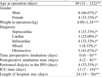
of 12 patients with total anomalous connection of pulmonary veins assessed lately
Age at operation (days) Gender
Male Female
Weight in operation (kg) Diagnosis
Supracardiac Cardiac Infracardiac Mixed Obstructive
Time preoperative intubation (days) Postoperative intubation time (days) Peritoneal dialysis in the IPO (days) ICU time (days)
Length of hospital stay (days)
69 (31 – 122)**
8 (66.67%)* 4 (33.33%)* 4.09±1.24***
4 (33.33%)* 3 (25.00%)* 4 (33.33%)* 1 (8.33%)* 5 (41.67%)*
0 (0 – 0)** 4 (2 – 8)** 4 (33.33%)* 11 (7 – 19)** 24 (19 – 30)**
Kg = kilogram, IPO = immediate postoperative period, ICU = intensive care unit * = data in absolute value (percentage of total), ** = data expressed as median (interquartile range) *** = data expressed in mean ± standard deviation
Table 2. Data from the intraoperative group of 12 patients with total anomalous connection of pulmonary veins assessed lately.
TAPVC
Supracardiac Carddiac Infracardiac Mixed
CPB Temperature (ºC)
22.50±3.32 28.33±3.51 19.50±1.91
26.00*
TAPVC = total anomalous pulmonary veins connection, CPB = cardiopulmonary bypass; C = degrees Celsius; min = minutes; TCA = total circulatory arrest, NO = not observed; * = data in absolute values, because there is only one patient. All other values are expressed as mean ± standard deviation
Perfusion (min) 92.80±15.26 68.67±38.07 90.00±21.28
100.00*
Myocardial ischemia (min)
49.00±27.12 33.80±16.01 49.25±24.09
71.00*
TCA (min) 19.00±12.78
NO 30.75±9.74
calculate right ventricular systolic pressure (RVSP) by physiological tricuspid regurgitation and pulmonary artery mean pressure (MPAP). Both in the RSPV, as in other PV and around the LA, was insistently sought with the help of Doppler locations with the presence of possible reductions in size, which is considered when the flow velocity was greater than 2.0 m / s and pulsed Doppler or color Doppler when there was flow turbulence [4,5,11,12].
Chest MDCT was performed using the GE® brand (General Electric Medical Systems, Milwaukee, WI) model lighspeed, Multicut (multislice) 16-channel images were obtained at intervals between the cuts of 0.625 mm.
With the child in fasting, peripheral vein was punctured with a Jelco 22 or 24 on upper limb and midazolam administered at a dose of 0.1 mg/kg propofol and 0.5% from 2 to 4 mg/kg. Mean arterial pressure and non-invasive pulse oximetry were continuously monitored and the child kept under spontaneous ventilation with oxygen mask to 100% with 2 to 4 l/min.
The standardization of the examination was possible because they are all performed by one radiologist and one radiographer, accompanied by an anesthesiologist, pediatrician, cardiovascular surgeons and nurses.
The contrast used was 350 Optiray® (injectable loversol 74%) containing 350 mg/mL organically bound iodine, or a non-ionic contrast toxicity and low osmolarity (Mallinickrodt Inc. - Raleigh, NC - USA). The dose was based on the child, according to the manufacturer’s instructions and administered in the smallest amount possible to obtain adequate images.
Images were obtained from axial helical mode, which allowed large amounts of data to be reformatted images in sagittal, coronal and oblique plans. With this volume of images acquired by axial cuts, it was possible to obtain reformatting and volumetric three-dimensional reconstructions called MIP (maximum intensity projection) and colorful 3D reconstructions called volume Rendering. The diameter of the mouth of each pulmonary vein was measured essentially based on axial, sagittal, coronal and oblique cuts, in the short and long axes, in order to obtain the area and check for possible reductions in size when compared to qualitative description.
With the diameter of each pulmonary vein, the area was calculated and indexed by the respective patient’s body surface, aiming to standardize the measurements and can compare them. We considered the possibility of size reduction when the value was at least one standard deviation of the mean area of the pulmonary vein studied. The measurements were reconfirmed in three-dimensional reconstructions and associated with descriptive findings, but with illustrative purpose and not scientific.
The LA was measured in length (craniocaudal), width (lateral-lateral) and depth (anteroposterior), and the volume
can be calculated by multiplying the measurements and, in the same way as for PV was indexed to body surface of each patient. We considered the possibility that the LA be undeveloped when the value was at least one standard deviation lower than the average volumes. These data were compared with the descriptive findings.
The lungs were assessed for the presence of parenchymal lesions, vascular markings and signs to indicate abnormalities.
With the images obtained and measurements of PV and LA, a single observer, radiologist, experienced in congenital heart disease, assessed the morphological patterns descriptively (qualitatively) and all data were correlated with the findings of the anamnesis, physical examination, chest radiography , ECG and echocardiography.
In statistical analysis, the continuous quantitative variables with Gaussian distribution were analyzed and represented as mean ± standard deviation. Continuous quantitative variables without Gaussian distribution were represented as median (25-75 percentile). All variables were represented as absolute number (percentage of total).
Survival was expressed by Kaplan-Meyer and significance using the log rank test with Prism 4.0 for Windows®. The alpha error allowed for statistical significance was 5%.
We calculated sensitivity, specificity, positive predictive value, negative predictive value and accuracy of MDCT in clinical evaluations and tests ordered in routine follow-up evaluation. It was considered the gold standard when at least one of these had been changing.
RESULTS
The series of 12 patients are presented descriptively, with the aim of allowing better analysis of the data found. Patients MHDF, EEM, GVR and LLF were suffering from supracardiac type MDCT, LJD, MICP and EBA, cardiac type; JDSA, HVDS, KCRM and JRM, infracardiac type, and VSF, mixed type.
All patients were in functional class I (NYHA), except one (LJD) with cardiac type MDCT, which presented reduced caliber of VP, was reoperated and is under use of acetylsalicylic acid.
Another patient (JDSA) with MDCT of infracardiac type presented with seizures postoperatively and, although with no more symptoms after discharge, still makes use of carbamazepine.
Cardiac auscultation was within normal limits, except in three patients with nonspecific murmurs (MICP, HVDS and KCRM).
(37.50%) below the 15th percentile. All patients were female with a BMI above the 85th percentile.
At chest radiography, most children had normal pleuropulmonary fields with transparency, free diaphragmatic domes and heart size within normal limits. Two patients (GVR and HVDS) had a slight increase in heart size by visual impression, thea little discordant from CI,
and in three children it was noted prominence of the right cavities (GVR, HVDS and JRM).
In one patient (EEM) was observed subsegmental opacity in the right lung base, adjacent to the diaphragm, which may represent subsegmental atelectasis. Another patient (LJD) presented hypertransparency of the left lung, by reduction of vascular markings.
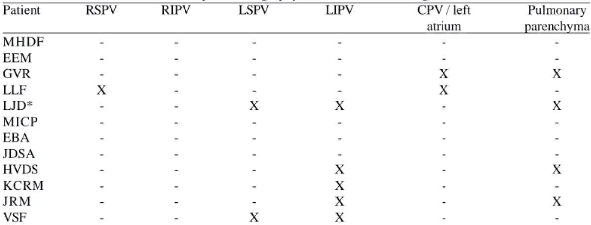
Table 3. The local multi-detector computed tomography found anatomical changes.
Patient MHDF EEM GVR LLF LJD* MICP EBA JDSA HVDS KCRM JRM VSF RSPV -X
-RSPV = right superior pulmonary vein; RIPV = right inferior pulmonary vein, LSPV = left superior pulmonary vein; LIPV = left inferior pulmonary vein, - = within the normal range; X = changed * = patient required reoperation for size reduction of the veins lung and currently with reduced caliber in the superior vena cava
RIPV -LSPV -X -X LIPV -X -X X X X
CPV / left atrium -X X -Pulmonary parenchyma -X -X -X -X
-Table 4. Values obtained by multi-detector computed tomography of the pulmonary vein areas (mm2)/body surface area of each patient (m2).
Patient MHDF EEM GVR LLF LJD MICP EBA JDSA HVDS KCRM JRM VSF M - DP
RSPV (mm2/m2)
133.46 94.05 166.42 20.05 119.47 186.63 207.25 75.70 98.13 39.75 136.96 82.47 56.87
M - SD = mean minus one standard deviation, RSPV = right superior pulmonary vein; RIPV = right inferior pulmonary vein, LSPV = left superior pulmonary vein; LIPV = left inferior pulmonary vein; mm2 = square millimeter; m2 = square meter
RIPV (mm2/m2)
132.13 63.87 124.14 166.20 41.97 88.42 134.76 41.04 45.89 104.03 107.95 134.18 56.46 LSPV (mm2/m2)
74.21 138.32 68.92 84.76 26.32 99.29 57.80 67.65 50.77 39.33 40.36 92.81 39.10 LIPV (mm2/m2)
87.05 77.74 40.65 40.30 15.50 80.52 88.41 57.45 21.60 119.25 35.32 51.18 28.40
Table 5. Values obtained by multi-detector CT of left atrial
volume (cm3) for each patient’s body surface area (m2).
Patient MHDF EEM GVR LLF LJD MICP EBA JDSA HVDS KCRM JRM VSF M - DP
M - SD = mean minus one standard deviation; cm3 = cubic centimetre; m2 = square meter
Most electrocardiograms were within normal limits, with discrete electric conduction disturbances, such as change in ventricular anteroseptal, conduction disturbance in the right branch and right ventricular hypertrophy, apparently without clinical significance.
The echocardiogram showed that the measurement of blood flow velocity in the RSPV was less than 2 m/s in all patients, the largest waves found were D 1 m/s and 1.3 m/s in patients LLF and MICP . There was also no reduction in other PV caliber, as well as the anastomosis between the SVC and LA.
RVSP was considered normal in all patients, the highest found in the patient LJD (37 mmHg). The MPAP can not be measured in two patients, LJD and GVR. In all other patients, the MPAP was also normal.
In one patient (LJD), it was observed narrowing at the SVC mouth with the RA, with venous flow velocity of 1.89 m/s, considered by echocardiography as mild reduction of caliber. Were also observed in this patient significant dilation of the azygos vein and blood flow in the opposite direction than normal.
MDCT analysis of each patient provided so many descriptive details of the anatomical changes in PV, SVC, left atrium and pulmonary parenchyma, among others. Table 3 provides an overview of the sites diagnosed as out of normal standards.
The mean measurements of PV indexed by body surface area and their respective standard deviations were: 113.36 ± 56.49 mm2
/m2
RSPV, 98.72 ± 42.26 VPID mm2
/m2
, LSPV 70.05 ± 30.94 mm2
/m2
, 59.58 ± 31.18 mm2
/m2
LIPV. When we considered at least one standard deviation below the mean, the values that suggested a significant decrease in size and therefore could represent the anatomical impairment VP were, respectively, mm2
/m2
56.87, 56.46 mm2
/m2
, mm2
/m2
mm2
/m2
39.10 and 28.40, as shown in Table 4.
Thus, valuing purely measures, we should consider the significant reduction of caliber patients LLF in RSPV with mm2
/m2
20.05; LJD, in RIPV, LSPV and LIPV with mm2
/m2
41.97, 26.32 mm2
/m2
, 15.50 mm2
/m2
; JDSA in RIPV with mm2
/ m2
41.04; HVDS, in RIPV and LIPV, with 45.89 and 21.60 mm2
/m2
mm2
/m2
, and KCRM in RSPV, mm2
/m2
with 39.75. The descriptive analysis of the findings by a radiologist experienced in congenital heart disease showed a pulmonary vein compression or size reduction in patients LLF, LJD, HVDS, KCRM, JRM and VSF. So, it was the same in four of six patients referred by the measures. Also, with respect to the affected PV there was a discrepancy in patients LJD, JDSA, HVDS and KCRM, suggesting that the measures do not help in correct diagnosis of the presence or absence of injury in PV.
The average LA volume indexed by body surface area and its standard deviation was 6.02 ± 1.70 cm3
/m2
. Considering at least one standard deviation, LA was
considered underdeveloped when the value was less than 4.33 cm3
/m2
. This occurred in two patients, GVR and LLF (Table 5). The descriptive analysis of the LA by the radiologist, with the help of MDCT, suggested appropriate development in all patients, indicating that these measures were not useful.
Four patients had abnormalities suggestive of lesions in the lung parenchyma, all with involvement of the left lung: GVR with hypoattenuation in the posterior basal segments of the upper and lower lobe and the entire upper lobe secondary to atelectasis; LJD with reduced vascular markings; HVDS with opacity in band in the posterior basal segment of lower lobe and middle lobe, suggesting subsegmental atelectasis, and JRM with opacity in band in the lower lobe, also suggesting subsegmental atelectasis (Table 6).
Some examples of these findings are presented below to illustrate the ability of MDCT morphological definition. In MHDF patient, it was found near the big stump together with innominate vein (IV), a remnant of the vertical vein (VV). The four PV were well developed and with no reduction in caliber. There were no signs of increased PT or lesions in the lung parenchyma. These findings were in accordance with the clinical evaluation and other examinations.
In the patient EEM, the MDCT showed no changes, in agreement with the clinical evaluation and laboratory tests, except with the ray, which revealed an image suggestive of atelectasis, but the CT departed this possibility. Thus, for this patient, MDCT was superior and more enlightening.
In the patient GVR, it was noted that the descending aorta seemed to compress the SVC near the mouth of PV to left (Figures 1A to 1C). The left lung opacities had elongated upper and lower lobe, and hypoattenuation in other segments, suggestive of increased air space secondary to atelectasis. The right lung was normal. In clinical evaluation, the patient’s height and weight development below the 15th percentile, the chest radiograph suggested cardiac area at the upper limit of normal and showed no lung lesions. The other tests were normal, suggesting that MDCT for this patient was also higher.
In the patient LJD, who required reoperation for a reduction in caliber of PV, we observed a significant reduction of the caliber of the left SVC and LSPV and LIPV. The right PV, which were amplified with right atrial tissue were normal. The SVC also presented significant reduction in size near its mouth in RA, with the azygos vein opacification by contrast media. There was significant reduction of the vascular markings of the left lung, despite the normal appearance of the tracheobronchial tracheobronchial tree. This was the only patient in functional class II (NYHA), with height and weight development below the 15th percentile in the use of aspirin due to reduced caliber of the SVC at the junction with RA.
Radiography was changed to the left and in ECG there was right ventricular hypertrophy, consistent with the highest RVSP found in the echocardiogram, with D-wave of 0.95 m/s. All these findings were in agreement with those found in MDCT.
In the patient MICP, it was noted that the PV drained directly into the right atrium and the left VP converged on a path of CPV, which showed no reduction in caliber. There was increase in the diameter of the PT, which correlated clinically with systolic murmur at left sternal border, smooth, without irradiation, but no changes were noted on chest radiography, ECG or an increase in RVSP or PMAP. The D
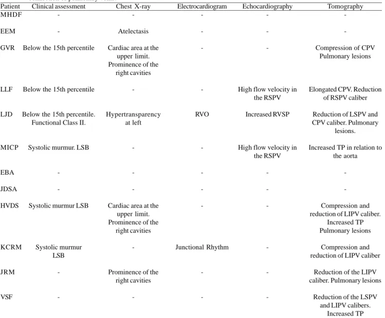
Table 6. Association between multidetector CT and other forms of late assessment for patients undergoing repair of total anomalous connection of pulmonary veins.
Patient MHDF
EEM
GVR
LLF
LJD
MICP
EBA
JDSA
HVDS
KCRM
JRM
VSF
- = Within normal limits; CPV = common pulmonary vein, LA = left atrium, RSPV = right superior pulmonary vein, SVC = superior vena cava; RVO = right ventricular overload; RVSP = right ventricular systolic pressure, PV = pulmonary vein; LUPV = left upper pulmonary vein; LIPV = left inferior pulmonary vein; PT = pulmonary trunk, LVH = left ventricular hypertrophy; LSB = left sternal border
Clinical assessment
-Below the 15th percentile
Below the 15th percentile
Below the 15th percentile. Functional Class II.
Systolic murmur. LSB
-Systolic murmur LSB
Systolic murmur LSB
-Chest X-ray
-Atelectasis
Cardiac area at the upper limit. Prominence of the
right cavities
-Hypertransparency at left
-Cardiac area at the upper limit. Prominence of the
right cavities
-Prominence of the right cavities
-Electrocardiogram
-RVO
-Junctional Rhythm
-Echocardiography
-High flow velocity in the RSPV
Increased RVSP
High flow velocity in the RSPV
-Tomography
-Compression of CPV Pulmonary lesions
Elongated CPV. Reduction of RSPV caliber
Reduction of LSPV and CPV caliber. Pulmonary
lesions.
Increased TP in relation to the aorta
-Compression and reduction of LIPV caliber.
Increased TP Pulmonary lesions
Compression and reduction of LIPV caliber
Reduction of the LIPV caliber. Pulmonary lesions
Reduction of the LSPV and LIPV calibers.
wave was the largest found in all patients, measured at 1.3 m/s and did not correlate with the MDCT images, which clearly demonstrate the excellent mouth of RSPV in LA.
The patient EBA was an example of patient anatomy with normal development of PV. Clinical evaluation as well as all laboratory tests, indicated no change and was consistent in all MDCT findings (Figure 3).
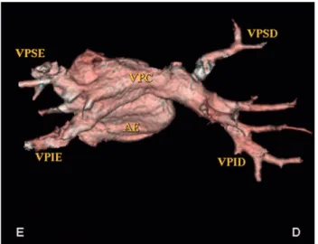
In the patient JDSA the right PV had long extent, but such veins were symmetrical and without reductions in caliber. The pulmonary trunk and pulmonary arteries had normal development and had no signs of pulmonary parenchymal lesions. These data were in agreement with clinical cardiology, chest radiography and ECG, which were normal. In echocardiography, it was found RVSP of 36 mmHg, but with lower MPAP of 7 mmHg. Fig. 1 - Patient GVR. A: Oblique cut with rear view of the LA in MIP. The VP present unique trunks. The descent aorta seems to compress the SVC to the left, B: Oblique reformatting in MIP with CPV on the right shows a normal appearance, C: Oblique reformatting in MIP of the left SVC with detail of the aparent compression by aorta. D: right side of the patient; [E: left side of the patient, LA (AE): left atrium; MIP: maximum intensity projection; PV (VP): pulmonary veins, Ao: aorta, VPC: common pulmonary vein, RSPV (VPSD): right superior pulmonary vein; RIPV (VPID): right inferior pulmonary vein; LSPV (VPSE): left superior pulmonary vein; LIPV (VPIE): left inferior pulmonary vein]
Fig. 2 - Patient LLF. Images of three-dimensional reconstruction. A: Rear view with volume rendering reconstruction, demonstrating reduced in size and dimensions of the RSPV. [D: right side of the patient; E: left side of the patient, LA: left atrium, RSPV (VPSD): right superior pulmonary vein; RIPV (VPID): right inferior pulmonary vein; LSPV(VPSE): left superior pulmonary vein; LIPV (VPIE): left inferior pulmonary vein, CPV (VPC): common pulmonary vein]
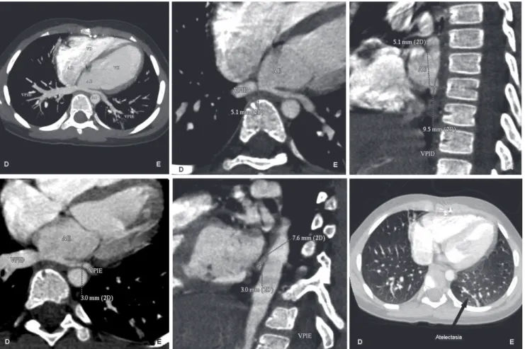
Fig. 4 - Patient HVDS. A: Oblique reformatting in MIP demonstrating inferior VP. Note that the LIPV has a smaller diameter than the RIPV and apparently suffers from compression of the descending aorta; B: Oblique reformatting in MIP measuring the short axis of the LIPV C: Oblique reformatting of short axis measuring the RIPV. D: Oblique reformatting in MIP of long axis measuring the LIPV short-axis, E: Oblique reformatting of short axis of the LIPV; F: Axial opacities in the lung bases with a band-projection of the posterior basal segment of left lower lobe and middle lobe, suggestive of subsegmental atelectasis. [D: right side of the patient; E: left side of the patient; MIP: maximum intensity projection; VP: pulmonary veins, LIPV (VPIE): left inferior pulmonary vein; RIPV (VPID): right inferior pulmonary vein, Ao: aorta, LA (ae): left atrium; RA (AD): right atrium, LV (VE): left ventricle, RV (VD): right ventricle]
The patient HVDS presented the PV less elongated, and the LIPV notably smaller caliber than the RIPV. Apparently, the descending aorta determined the compression of LIPV. The upper PV had normal caliber. The PT and the pulmonary arteries were enlarged caliber in relation to the aorta and opacity was in band in the left basal segment of lower and middle lobe, suggesting subsegmental atelectasis (Figures 4A to 4F). Thus, MDCT findings reflected the clinical assessment with a systolic murmur in left sternal and chest radiography with increased heart size. However, ECG and echocardiogram showed no changes and were not in agreement with all other findings. MDCT also showed lung lesions that were absent in the plain chest radiograph. In KCRM patient, the LIPV was elongated, with reduced size in relation to the contralateral vein, apparently suffering from compression by the descending aorta. There was no reduction in upper PV caliber. This visual interpretation
was different from measurements obtained in PV. In clinical evaluation, there was systolic murmur in left sternal vibration, chest radiography was normal, ECG drew attention by junctional rhythm, the echocardiogram with RVSP of 32 mmHg and wave D of 0.96 m/s showed no change.
In patient JRM, MDCT found reduction in the caliber of LIPV, and opacity in band in the left lower lobe, with the appearance of subsegmental atelectasis. In this patient, clinical evaluation was normal and chest X-ray showed a mild prominence of the right cavities. The ECG and echocardiogram indicated no change. Thus, reduction in the diameter of the LIPV may have some association with these findings.
Clinical evaluation and all other examinations were also normal, indicating that the findings of MDCT should not be interpreted alone.
Based on the findings above, MDCT showed changes in eight patients, seven of these also had alterations in at least one of the other variables evaluated, ie, evaluation of a clinical or laboratory tests routinely used, and in one patient, the other counts were normal.
In the other four patients, MDCT indicated no change, and in three, it was consistent with other data analyzed, and in one patient, there was a change from other examinations.
Thus, we find that the MDCT with respect to clinical and other laboratory tests analyzed had a sensitivity of 87.5% (95% CI 64.6% to 100.0%), specificity of 75.0% (95% 32.6% to 100.0%), positive predictive value 87.5% (95% CI 64.6% to 100.0%), negative predictive value of 75.0% (95% CI 32, 6% to 100.0%) and accuracy 83.3% (95% CI 62.2% to 100.0%), as briefly shown in Table 6.
DISCUSSION
The TAPV is an uncommon disease and case series of 16 patients operated on between 1492 (1.07%) with congenital heart disease in six years, despite being small, as it was expected, in line with other large studies, such as Bohemia Survival Study, which identified 40 children with TAPV among the 815,569 children born between 1980 and 1990, and the Baltimore-Washington Infant Study, which found a prevalence of 0.087 per 1000 invasive births [11,12]. The reduction of the PV caliber, commonly called stenosis, is surely the worst complication that can occur late in a patient operated by TAPVC. It usually manifests itself quickly, leading the patient to reoperation within the first six months after the repair [13]. This need for early approach still remains the cause of high mortality in centers of excellence worldwide [4,5,13-15].
There are several mechanisms that can lead to reduction in size of one or more PV, although it is still incompletely understood [13]. Among these mechanisms are inadequate alignment of the incisions between the CPA and LA, causing shrinkage in the anastomosis, which leads to distortion of PV [16]. Also, some authors attribute the need for reoperation stenosis and intimal hyperplasia with proliferation of thickened inflammatory tissue [17]. In our study, we observed that in some patients, there was a decline of PV caliber, and the LIPV was clearly the most affected. Sometimes, the only PV was compressed by the descending aorta, perhaps because of its anatomical position during formation after the pericardial sac.
Incisions very close to PV can also lead to stenosis of these veins postoperatively. To avoid this complication, good technical option is opening the CPV in the direction
of VV in T or Y, expanding the anastomotic site. This attention should always be in the mind of the surgeon and in surgical patients in the series; such caution was observed, avoiding incisions in PV [17].
Some experts say there is suspicion that many patients operated for TAPVC live normally, however, presented a totally silent lesion in the PV extent [17]. This statement leads us to think that MDCT can be taken with an ability to detect such changes as early as possible, since in our study changes were observed in patients totally asymptomatic. Important to emphasize that this was not the aim of the study, but opens the way for further investigation.
The patient operated of TAPVC evolves, most often, clinically asymptomatic, similar to that found in our study, in which 11 (91.66%) had to be totally asymptomatic. The murmur is usually absent or, when present, is negligible and nonspecific, except in situations that they still have pulmonary hypertension or presence of significant residual defects. The heart murmurs observed in patients in our study were considered non-significant, because all were +/ 6+ or ++/6+ in the left sternal border, smooth and without irradiation. So, it would not be valued as indicative of a change in routine clinical examination, but it was noted that the three patients with murmurs (MICP, KCRM and HVDS) showed abnormalities on MDCT. It is important to remember that there is a problem in one or more PVs, the probability of strong murmur would really be minimal, because it is local low pressure, in addition to the anatomical position being posterior, and therefore hardly audible in the normal cardiac auscultation.
BMI was much lower than expected in three patients (GVR, LJD and LLF), which were in functional class I, not on medication and with normal cardiac auscultation. These patients showed abnormalities on MDCT, whether pulmonary or SVC compression of the descending aorta or decrease in caliber of left PVs, there is a strong association of height and weight development with MDCT findings.
Chest radiography in the late postoperative period should be normal when the patient has not evolved with PV stenosis or stenosis of the anastomotic area, as well as the lung fields should not have changed since the patient did not stay long in need of ventilatory assistance, that has caused injuries such as atelectasis or fibrosis. In our study, all patients who had abnormal chest radiographs were confirmed by MDCT, except one patient who had an image suggestive of atelectasis (MES) and his CT was normal. Therefore, there was a strong association between radiographic images and MDCT.
no significant changes in the conduction system, for example, anteroseptal ventricular repolarization, conduction disorders or right bundle branch block. These findings are consistent with the literature, which shows a high incidence of sinus node dysfunction and low total atrioventricular block, as well as significant atrial and ventricular arrhythmias are uncommon [18].
In all children evaluated by echocardiography, RVSP was within normal limits, and the patient LJD presented the highest value, because we knew the patient had lesions on the left PV. The PMAP was also within normal limits and did not indicate pulmonary hypertension in any child. The venous flow velocity was measured during the RSPV, and although not greater than 1.5 m/s or 2.0 m/s, as recommended by some authors who consider values above these as presence of PV stenosis; in LLF and MICP patients and the speeds were: 1.0 m/s and 1.3 m/s, consistent with changes found in the RSPV under MDCT [4,5].
The correlation of the findings discussed above was correlated with the findings of MDCT.
Chowdhury et al. [19], in 2008, in extensive study on TAPVC of mixed type, presented MDCT image in late evaluation of patients operated for five years, to illustrate that the anastomosis between the left atrial appendage and VV remained patent. This is the first publication we know of using MDCT in the evaluation of a patient with late TAPVC.
MDCT has been considered as an alternative complementary to echocardiography in the preoperative diagnosis of TAPVC, since these patients often find themselves in unfavorable hemodynamic conditions and hemodynamic studies to clarify the anatomy may worsen the clinical picture and provide more risk to the life of child. MDCT has the great advantage of being non-invasive and therefore life-threatening almost negligible, except for a possible anaphylactic shock of the contrast [10].
Kim et al. [9], studying 14 patients with TAPVC demonstrated that the combination of MDCT with three dimensional reconstruction helped in the diagnosis of TAPVC, being a good tool in the preoperative evaluation of neonates and infants [9]. However, it is not sufficiently adequate for visualization of intracardiac structures, due to heart rate and respiratory movements generally high. Indeed, it is virtually impossible to hold the breath in children less than eight months when they are intubated, with this important problem and impediment to implementation of MDCT in neonates. All children in the study were sedated with midazolam and propofol when needed, besides the fact that they are older, with an average age of 3.95 ± 1.32 years.
In this series, we measured the diameters of PV and calculated the LA volume. This had no association with the descriptive findings, and therefore we feel it should be
employed. Already, the measures of PV had some degree of association with the descriptive findings, but it was clear that the interpretation by an experienced physician is important and can complement the other findings of other examinations.
The fact that MDCT is in agreement with other findings in 10 of 12 patients, ie, from the eight MDCT altered, seven also with abnormal tests and four with normal MDCT, with three other tests also normal, makes us believe that the MDCT can be used quite safely in the late follow-up of children operated on TAPVC.
We found discrete and important morphological changes, being higher for the detection of subtle changes, such as simple compression or increased PVs pr PT with respect to the aorta, and it was associated with significant findings for the tests used in everyday practice. Notably, the RSPV was the most affected by compression or the presence of reduced caliber.
Therefore, this analysis suggests that it can be used for follow-up evaluation of patients undergoing total anomalous connection of pulmonary veins in order to anticipate clinical deterioration, allowing more accurate and early assessment, and can anticipate reinterventions when necessary.
Study limitations were the small number of patients, which does not allow statistical comparisons. In the period studied, there was progress in the technical details of experience gained by the surgeon and team. Patients were analyzed not only with ECG and Holter, because most live in other states and could not be in town for a long time. The MDCT findings were compared to non-hemodynamic study because it would not be ethically correct to pergorm invasive test, which theoretically has some life-threatening to the patient. There is also lack of other studies with late MDCT and MRI that have assessed a series of patients undergoing surgical correction of TAPVC.
CONCLUSION
In the late follow-up of patients undergoing surgical correction of TAPVC, the MDCT can provide valuable insights and complement the diagnosis of possible anatomical and functional, with a sensitivity of 87.5%, specificity of 75% and accuracy of 83.3%.
ACKNOWLEDGEMENTS
REFERENCES
1. Hirsch JC, Bove EL. Total anomalous venous connection. MMCTS. 2007;507:2253.
2. Craig JM, Darling RC, Rothney WB. Total pulmonary venous drainage into the right side of the heart; report of 17 autopsied cases not associated with other major cardiovascular anomalies. Lab Invest. 1957;6(1):44-64.
3. Paulista PP, Pedra SRFF. Anomalias de conexão do retorno pulmonar e sistêmico. In: Croti UA, Mattos SS, Pinto Jr. VC, Aiello VD, ed. Cardiologia e cirurgia cardiovascular pediátrica. 1ª ed. São Paulo:Roca;2008. p.203-16.
4. Karamlou T, Gurofsky R, Al Sukhni E, Coles JG, Williams WG, Caldarone CA, et al. Factors associated with mortality and reoperation in 377 children with total anomalous pulmonary venous connection. Circulation. 2007;115(12):1591-8.
5. Lacour-Gayet F, Zoghbi J, Serraf AE, Belli E, Piot D, Rey C, et al. Surgical management of progressive pulmonary venous obstruction after repair of total anomalous pulmonary venous connection. J Thorac Cardiovasc Surg. 1999;117(4):679-87.
6. Chowdhury UK, Airan B, Malhotra A, Bisoi AK, Saxena A, Kothari SS, et al. Mixed total anomalous pulmonary venous connection: anatomic variations, surgical approach, techniques, and results. J Thorac Cardiovasc Surg. 2008;135(1):106-16.
7. Devaney EJ, Chang AC, Ohye RG, Bove EL. Management of congenital and acquired pulmonary vein stenosis. Ann Thorac Surg. 2006;81(3):992-6.
8. Uçar T, Fitoz S, Tutar E, Atalay S, Uysalel A. Diagnostic tools in the preoperative evaluation of children with anomalous pulmonary venous connections. Int J Cardiovasc Imaging. 2008;24(2):229-35.
9. Kim TH, Kim YM, Suh CH, Cho DJ, Park IS, Kim WH, et al. Helical CT angiography and three-dimensional reconstruction of total anomalous pulmonary venous connections in neonates and infants. AJR Am J Roentgenol. 2000;175:1381-6.
10. Sridhar PG, Kalyanpur A, Suresh PV, John C, Sharma R, Maheshwari S. Total anomalous pulmonary venous connection:
helical computed tomography as an alternative to angiography. Indian Heart J. 2003;55(6):624-7.
11. Samanek M, Voriskova M. Congenital heart disease among 815,569 children born between 1980 and 1990 and their 15-year survival: a prospective Bohemia survival study. Pediatr Cardiol. 1999;20(6):411-7.
12. Ferencz C, Rubin JD, McCarter RJ, Brenner JI, Neill CA, Perry LW, et al. Congenital heart disease: prevalence at livebirth. The Baltimore-Washington infant study. Am J Epidemiol. 1985;121(1):31-6.
13. Ricci M, Elliott M, Cohen GA, Catalan G, Stark J, Leval MR, et al. Management of pulmonary venous obstruction after correction of TAPVC: risk factors for adverse outcome. Eur J Cardiothorac Surg. 2003;24(1):28-36.
14. Van Son JA, Danielson GK, Puga FJ, Edwards WD, Driscoll DJ. Repair of congenital and acquired pulmonary vein stenosis. Ann Thorac Surg. 1995;60(1):144-50.
15. Caldarone CA, Najm HK, Kadletz M, Smallhorn JF, Freedom RM, Williams WG, et al. Relentless pulmonary vein stenosis after repair of total anomalous pulmonary venous drainage. Ann Thorac Surg. 1998;66(5):1514-20.
16. Ando M, Takahashi Y, Kikuchi T. Total anomalous pulmonary venous connection with dysmorphic pulmonary vein: a risk for postoperative pulmonary venous obstruction. Interact Cardiovasc Thorac Surg. 2004;3(4):557-61.
17. Lacour-Gayet F, Zoghbi J, Serraf AE, Belli E, Piot D, Rey C, et al. Surgical management of progressive pulmonary venous obstruction after repair of total anomalous pulmonary venous connection. J Thorac Cardiovasc Surg. 1999;117(4):679-87.
18. Tanel RE, Kirshbom PM, Paridon SM, Hartman DM, Burnham NB, McBride MG, et al. Long-term noninvasive arrhythmia assessment after total anomalous pulmonary venous connection repair. Am Heart J. 2007;153(2):267-74.