Arq Bras Cardiol 2001; 77: 59-62.
Ianni et al Chagas' heart disease. ECG and ECHO parameters in indeterminate form
59 59
Heart Institute (InCor) HC-FMUSP, University of São Paulo Medical School Mailing address: Barbara Maria Ianni - InCor - Divisão Clínica, Equipe de Cardiopatias Gerais - Av. Dr. Enéas C. Aguiar, 44 - 05403-900 - São Paulo, Brasil – e-mail: [email protected]
Received to publication in 19/7/00 Accepted in 10/1/01
Objective - To identify and associate potential elec-trocardiographic and echocardiographic changes in pa-tients with the indeterminate form of Chagas’ disease du-ring long-term follow-up.
Methods - One hundred sixty patients underwent standard electrocardiography and two-dimensional gui-ded M-mode echocardiography for left ventricular ejecti-on fractiejecti-on determinatiejecti-on. Patients were followed up for 98.6±30.4 months, undergoing repeat electrocardiogra-phic studies at 6-month intervals and echocardiograelectrocardiogra-phic studies at 12-month intervals.
Results - Based on the electrocardiographic findings, the patients were divided into group I, 125 patients (78.6%) with normal electrocardiograms throughout fol-low-up, and group II, 34 patients (21.3%) who developed electrocardiographic changes. Group II was further divi-ded into group IIA (9 patients, 5.6%) with permanent elec-trocardiographic changes, group IIB (14 patients, 8.8%) with transitory electrocardiographic changes, and group IIC (11 patients, 6.9%) with changes appearing only on the final electrocardiogram. Left ventricular ejection frac-tions remained normal in the entire population studied and did not differ among groups.
Conclusion - The indeterminate form of Chagas’ di-sease clearly represents a benign condition with a favora-ble long-term prognosis. Although some patients develop electrocardiographic changes, left ventricular systolic function is well preserved.
Key words: Chagas’ heart disease, electrocardiographic changes, ejection fraction
Arq Bras Cardiol, volume 77 (nº 1), 59-62, 2001
Barbara Maria Ianni, Edmundo Arteaga, Clovis de Carvalho Frimm, Antonio Carlos Pereira Barretto, Charles Mady
São Paulo, SP - Brazil
Chagas’ Heart Disease: Evolutive Evaluation of
Electrocardiographic and Echocardiographic Parameters in
Patients with the Indeterminate Form
Original Article
Chagas’ disease is an important public health disorder in Latin America. It is an infectious disease caused by the protozoan parasite, Trypanosoma cruzi, which is transmit-ted zoonotically. Infection may occur following skin contact with the excrement of different strains of Triatoma insects, which serve as a vector. The parasite can multiply and trans-form within the Triatoma insect following ingestion via blood meal from an infected host. Chagas’ disease is a chronic disease characterized by cardiovascular and gas-trointestinal involvement. After an often-unrecognized acute phase followed by a generally undetected and leng-thy latent period, cardiac symptoms and dysfunction of the esophagus, the colon, or both may develop.
Data from the World Health Organization state that 25% of the population in Latin American countries, ap-proximately 90 million people, are exposed to this disease. As a consequence, it is estimated that as many as 16 million people are infected 1.
In Brazil, the endemic area is estimated to be around 3.6 million km2, the largest in South America 2,3. According to published data, 5 to 6 million people in this region are in-fected 2,3.Studies in endemic areas show that 25 to 35% of the infected population manifests cardiac symptoms, with severe myocardial damage in 10%. As many as 6000 cardiac deaths are reported each year 4.
60 60
Ianni et al
Chagas' heart disease. ECG and ECHO parameters in indeterminate form
Arq Bras Cardiol 2001; 77: 59-62.
electrocardiographic changes on patient prognosis (i.e. the favorable prognosis) is unclear 7. We hypothesized that these changes are not accompanied by myocardial dys-function.
The objective of this study was to prospectively evaluate patients with the indeterminate form of Chagas’ di-sease and relate the likely appearance of electrocardiogra-phic changes during follow-up to potential changes in left ventricular function.
Methods
We studied 160 patients with the indeterminate form of Chagas’ disease from 1979 until 1994, encompassing a mean follow-up interval of 98.6±30.4 months. The population in-cluded 98 women and 62 men, ranging in age from 17 to 61 years, with a mean age of 36.5±8.8 years. A diagnosis of the indeterminate form was made in asymptomatic patients based on the presence of two positive serologic tests for Chagas’ disease in combination with a normal electrocardio-gram and chest radiograph with no evidence of cardiac en-largement. Barium studies of the esophagus and the colon in such patients are also normal 12,13.
Subjects represented individuals from the general po-pulation in whom serologic tests were performed prior to blood donation. They came from endemic areas and were li-ving in São Paulo. To assure that they were not reinfected after enrollment, none of them returned to their former ho-mes. This study was approved by the Ethics Committee at our institution. All patients were educated about the objec-tives of the study and provided written informed consent.
Clinical follow-up was performed at a 3-month interval, and the same cardiologist always conducted the outpatient visits. Standard electrocardiograms were performed at six-month intervals and were analyzed independently by two cardiologists. Determinations of electrocardiographic changes were based on internationally recognized criteria 14. Annual M-mode two-dimensional guided echocardio-graphy was performed with 2.5 to 3.5 MHz transducers and standard equipment. Tracings were recorded on strip chart paper at 50 m.s-1. Left ventricular internal dimensions at end-systole and end-diastole were obtained with the help of simultaneous electrocardiographic registration, according to the recommendations of the American Society of Echocar-diography 15. End-systolic and end-diastolic volumes, and the corresponding ejection fraction, were calculated based on the cube method for left ventricular volume determination 16.
LVEF3, the ejection fraction corresponding to the echo-cardiogram obtained at the end of the study, was compared with the LVEF1 obtained during enrollment. In addition to the annual studies, a supplemental echocardiogram was ob-tained, and the LVEF2 was determined, whenever an electro-cardiographic change was detected.
Statistical analysis of the data was performed using the analysis of variance for repeated measures. An a value of 0.05 was chosen to establish significant differences. Sta-tistical calculations were made using SAS software 17.
Results
Only one patient was excluded from the study because of having suffered an acute myocardial infarction 104 months following enrollment. A total obstruction of the an-terior descending coronary artery was demonstrated angio-graphically before successful thrombolysis.
Based on electrocardiographic changes, the remai-ning 159 patients were classified into two groups: group I and group II. Group I corresponded to 125 patients (78.6%) without changes on follow-up electrocardiograms. It inclu-ded 78 women, ranging from 20 to 57 years in age, with a mean age of 36.0±8.3 years. Group II consisted of 34 pati-ents (21.3%) with electrocardiographic changes that deve-loped during the follow-up interval. It included 20 women, ranging from 17 to 61 years, with a mean age of 38.2±10.4 years. The total period of follow-up in group I averaged 97.5±28.5 months (range: 48 to 175 months) and in group II averaged 102.8±36.8 months (range: 48 to 177 months).
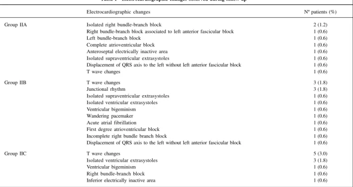
Group II was further divided into three additional sub-groups: group IIA, 9 patients (5.6%), with permanent electro-cardiographic changes; group IIB, 14 patients (8.8%), with transitory electrocardiographic changes; and group IIC, 11 patients (6.9%) in whom the electrocardiographic changes appeared only at the final evaluation period. The types of electrocardiographic alteration are summarized in Table I.
Table II depicts mean ages within subgroups. As can be seen, group IIA and group IIC patients were older than group IIB and group I patients (p=0.0008).
Table III shows the ejection fractions determined via echocardiography for the entire population studied. These ejection fractions did not significantly change during fol-low-up, either in group I or in any of the three group II sub-groups. Initial (LVEF1) and final (LVEF3)ejection fractions were similar between group I and group II patients. Further-more, ejection fractions determined in group II patients clo-se to the appearance of an electrocardiographic change (LVEF2) did not statistically differ from those obtained either at the beginning or end of the study.
Discussion
The results of the present study indicate that only a portion of patients with the indeterminate form of Chagas’ disease develop significant electrocardiographic changes. Furthermore, these changes do not appear to be accompa-nied by corresponding left ventricular dysfunction.
Arq Bras Cardiol 2001; 77: 59-62.
Ianni et al Chagas' heart disease. ECG and ECHO parameters in indeterminate form
61 61
block, right bundle- branch block accompanied by left anteri-or divisional block, left bundle-branch block, eventual high-degree atrioventricular blockade, or all of these. Despite this, all electrocardiographic abnormalities were taken into ac-count in the present analysis, including the nonspecific ones commonly reported in the general population 3,6,8,11,18-28. This likely led to an overestimate of the frequency of electrocardio-graphic changes herein reported.
Echocardiographic data on indeterminate form pati-ents are numerous, but sometimes conflicting, due to a ge-neralized lack of strict criteria used to characterize this latent form of the disease. Many reports include patients with only minor electrocardiographic changes, which likely represent mild, but true, myocardial damage 29,30. This may explain so-me earlier reports of either segso-mental 30 or diffuse impairment of left ventricular contractility 32,33.
However, data are lacking on long-term echocardiogra-phic function in patients with the indeterminate form of the disease. To the best of our knowledge, the sole study publi-shed in this regard documented the development of global left ventricular systolic dysfunction in a small number of pa-tients with previously documented diastolic dysfunction 34. To date, the presence of diastolic dysfunction does not rule out the diagnosis of the indeterminate form of Chagas’ di-sease, which is based solely on electrocardiographic crite-ria, as already stated. Indeed, it is conceivable that diastolic dysfunction could represent the initial stage of myocardial damage, and antedate global left ventricular systolic dys-function. Unfortunately, diastolic properties were not sys-tematically evaluated in our study from the beginning. Ho-wever, the most recent echocardiograms performed in our patients did not show changes in diastolic function (data not shown), indicating that diastole was not initially impai-red. As a consequence, this may explain the lack of evolving systolic dysfunction in our patients in contrast with the findings in the previously reported study.
Interestingly, patients with permanent electrocardio-graphic changes or changes observed only at the final eva-luation, were older than those with or without transitory ab-normalities. Whether or not the changes observed on the final electrocardiographic examination represent permanent
Table I - Electrocardiographic changes observed during follow-up
Electrocardiographic changes Nº patients (%)
Group IIA Isolated right bundle-branch block 2 (1.2)
Right bundle-branch block associated to left anterior fascicular block 1 (0.6)
Left bundle-branch block 1 (0.6)
Complete atrioventricular block 1 (0.6)
Anteroseptal electrically inactive area 1 (0.6)
Isolated supraventricular extrasystoles 1 (0.6)
Displacement of QRS axis to the left without left anterior fascicular block 1 (0.6)
T wave changes 1 (0.6)
Group IIB T wave changes 3 (1.8)
Junctional rhythm 3 (1.8)
Isolated supraventricular extrasystoles 1 (0.6)
Isolated ventricular extrasystoles 1 (0.6)
Ventricular bigeminism 1 (0.6)
Wandering pacemaker 1 (0.6)
Acute atrial fibrillation 1 (0.6)
First degree atrioventricular block 1 (0.6)
Incomplete right bundle branch block 1 (0.6)
Displacement of QRS axis to the left without left anterior fascicular block 1 (0.6)
Group IIC T wave changes 5 (3.0)
Isolated ventricular extrasystoles 3 (1.8)
Ventricular bigeminism 1 (0.6)
Right bundle-branch block 1 (0.6)
Inferior electrically inactive area 1 (0.6)
Table II - Mean ages of patients with indeterminate form of Chagas’ disease by group
Group Nº of patients Age (years)
I 125 36.0±8.3
IIA 9 46.4±4.5*
IIB 14 32.4±8.7
IIC 11 39.1±11.3*
* p = 0.0008
*p = 0.0008; significant increase compared with group I and group IIB.
Table III - Evolutive echocardiographic ejection fraction
Group Nº of patients LVEF1* LVEF2 LVEF3*
I 125 0.73±0.05 - 0.74±0.04
IIA 9 0.73±0.03 0.71±0.08 0.73±0.07 IIB 14 0.72±0.04 0.73±0.06 0.72±0.08 IIC 11 0.73±0.04 0.71±0.03 0.71±0.03 - p=0.6886 p=0.7474 p=0.0601 *p=0.6698
62 62
Ianni et al
Chagas' heart disease. ECG and ECHO parameters in indeterminate form
Arq Bras Cardiol 2001; 77: 59-62.
or only transitory changes awaits further evaluation. It is li-kely, however, that because these changes developed late, and in older subjects, they may be permanent. Neverthe-less, the time-related appearance of electrocardiographic abnormalities suggests that the evolution of the indetermi-nate form of Chagas’ disease is influenced by patient age.
On the other hand, the present results clearly suggest that patients with the indeterminate form of Chagas’ disea-se, in general, have a very good prognosis. The factors rela-ted to this favorable outcome have yet to be determined, but a normal ejection fraction despite electrocardiographic changes may be relevant in this regard.
Unfortunately, it is a common practice in our country to perform serological tests for Chagas’ disease as part of the clinical evaluation for employment admission. Subjects with positive results are often relegated and have difficulty getting hired, even when applying for bureaucratic jobs. We hope that the present results will contribute to a better un-derstanding of this form of Chagas’ disease and reverse this situation. Our view is that these subjects should be advised not to exclude themselves from normal daily activities and that they are fully able to apply for and to perform all kinds of jobs, including those requiring muscle strength.
1. World Health Organization. Control of Chagas’ disease. Report of the WHO Expert Committee. Geneva. WHO Technical Report Series 1991; 811: 1-95. 2. Dias JCP, Dias E. Doença de Chagas’. In: Brasil. Ministério da Saúde - SUCAM.
Doença de Chagas: texto de apoio. Brasília, 1989: 13-20.
3. Maguire JH, Hoff R, Sherlock I, et al. Cardiac morbidity and mortality due to Chagas’ disease: prospective electrocardiographic study of a Brazilian community. Circulation 1987; 45: 1140-5.
4. Brasil. Ministério da Saúde. Estatísticas de Mortalidade. Brasília, 1987. 5. Chagas C. Processos patogênicos da tripanosomíase americana. Mem Inst
Oswaldo Cruz (Rio de Janeiro) 1916; 8: 7-36.
6. Laranja FS, Dias E, Nóbrega E, Miranda A. Chagas’ disease: a clinical, epidemiologic, and pathologic study. Circulation 1956; 14: 1035-60. 7. Dias JCP. História natural. In: Cançado JR, Chuster M - Cardiopatia chagásica.
Belo Horizonte, Fundação Carlos Chagas, 1985: cap.11: 99-113.
8. Coura JR, Abreu LL, Pereira JB, Willcox HP. Morbidade da doença de Chagas: IV - Estudo longitudinal de dez anos em Pains e Iguatama, Minas Gerais, Brasil. Mem Inst Osw Cruz 1985; 80: 73-80.
9. Carrasco HA, Parada H, Guerrero L, Duque M, Durán D, Molina C. Prognostic implications of clinical, electrocardiographic and hemodynamic findings in chronic Chagas’ disease. Intern J Cardiol 1994; 43: 27-38.
10. Espinosa R, Carrasco HA, Belandria F, et al. Life expectancy analysis in patients with Chagas’ disease: prognosis after one decade (1973-1983). Intern J Cardiol 1985; 8: 45-56.
11. Pereira JB, Willcox HP, Coura JR. Morbidade da doença de Chagas. III. Estudo longitudinal de 6 anos em Virgem da Lapa, Minas Gerais, Brasil. Mem Inst Osw Cruz 1985; 80: 63-71.
12. Camargo ME, Oshino-Shimizu S, Macedo V, Peres BA, Castro C. Diagnóstico so-rológico da infecção humana pelo Trypanosoma cruzi. Estudo comparativo de testes de fixação do complemento, imunofluorescência, hemaglutinação e flocula-ção em 3.624 soros. Rev Inst Med Trop São Paulo 1977; 19: 254-60. 13. Primeira Reunião de Pesquisa Aplicada em Doença de Chagas. Validade do
con-ceito da forma indeterminada. Rev Soc Bras Med Trop 1985; 18: 46.
14. Cookey JD, Dunn M, Massie E. Clinical Vectorcardiography and Electrocardio-graphy. 2nd ed. Chicago: Year Book Medical Publishers Inc., 1977.
15. Sahn DJ, DeMaria NA, Kisslo J, Weyman A. The Committee on M-mode Standar-dization of the American Society of Echocardiography. Recommendations regar-ding quantitation in M-mode echocardiography: results of a survey of echocar-diographic measurements. Circulation 1978; 58: 1072-83.
16. Pombo JF, Troy BL, Russell Jr. RO. Left ventricular volumes and ejection fraction by echocardiography. Circulation 1971; 43: 480-90.
17. Timm NH. Multivariate analysis with applications in education and psychology. Monterey, California: Brooks/Cole Publishing Company, 1975: 444-59.
18. Porto CC. O eletrocardiograma no prognóstico e evolução da doença de Chagas. Arq Bras Cardiol 1964; 17: 313-46.
19. Dias JCP, Kloetzel K. The prognostic value of the electrocardiographic features of chronic Chagas’ disease. Rev Inst Med Trop São Paulo 1968; 10: 158-62. 20. Mady C, Cardoso RHA, Pereira-Barretto AC, Luz PL, Bellotti G, Pileggi F.
Survival and predictors of survival in patients with congestive heart failure due to Chagas’’ cardiomyopathy. Circulation 1994; 90: 3098-102.
21. Brasil A. A mutabilidade electrocardiográfica na cardiopatia crônica chagásica. Rev Assoc Med Minas Gerais 1953; 4: 149-52.
22. Hiss RG, Lamb LE. Electrocardiographic findings in 122,043 individuals. Circulation 1962; 25: 947-61.
23. Rotman N, Triebewasser JH. A clinical and follow-up study of right and left bundle branch block. Circulation 1975; 51: 477-84.
24. Averill KH, Lamb LE. Electrocardiographic findings in 67,375 asymptomatic subjects. I. Incidence of abnormalities. Am J Cardiol 1960; 6: 76-83. 25. Grupi CJ, Sosa E, Carvalho JF, Antonelli RH, Bellotti G, Pileggi F. Variabilidade
espontânea da extrassistolia ventricular na cardiopatia chagásica crônica. Arq Bras Cardiol 1991; 56: 445-50.
26. Fosmoe RJ, Averill KH, Lamb LE. Electrocardiographic findings in 67,375 asymptomatic subjects. Am J Cardiol 1960; 6: 84-95.
27. Pereira JB, Coura JR. Morbidade da doença de Chagas. Estudo seccional em uma área endêmica, Virgem da Lapa, MG. Rev Soc Bras Med Trop 1986; 19: 139-48. 28. Brodsky M, Wu D, Denes P, Kanakis C, Rosen KM. Arrhythmias documented by
24-hour continuous electrocardiographic monitoring in 50 male medical students without apparent heart disease. Am J Cardiol 1977; 39: 390-5. 29. Mady C, Pereira-Barretto AC, Ianni BM, Lopes EA, Pileggi F. Right ventricular
endomyocardial biopsy in indeterminate form of Chagas’ disease. Angiology 1984; 35: 755-9.
30. Pereira-Barretto AC, Mady C, Arteaga E, et al. Right ventricular endomyocardial biopsy in chronic Chagas’ disease. Am Heart J 1986; 111: 307-12. 31. Ortiz J, Pereira-Barretto AC, Matsumoto A, et al. Alteração contrátil segmentar na
forma indeterminada da doença de Chagas. Estudo ecocardiográfico. Arq Bras Cardiol 1987; 49: 217-20.
32. Friedmann AA, Armelin E, Nelken JR, Zerbini CAF, Coimbra MA, Serro-Azul LG. Estudo ecocardiográfico do desempenho ventricular em fase pré-clínica da doença de Chagas. Rev Hosp Clin Fac Med S Paulo 1980; 35: 165-70. 33. Friedmann AA, Armelin E, Leme LEG, et al. Desempenho ventricular na doença
de Chagas. Relações ecocardiográficas na miocardiopatia com distúrbio dromótropo e na fase pré-clínica. Arq Bras Cardiol 1981; 36: 23-7.
34. Cunha CLP, Urbanetz LAGT, Souza AM, et al. Evolutive Doppler echocardio-graphic changes in the indeterminate phase of Chagas’ disease. Eur Heart J 1993; 14: 135.