From Division of Cardiac Surgery of Instituto Nacional de Cardiologia Laranjeiras National Institute of Cardiology Laranjeiras - Ministério da Saúde Health Ministery, Rio de Janeiro
Mailing address: Antônio Sérgio Cordeiro da Rocha – Rua Roberto Dias Lopes, 220/201 – 22010-110 – Rio de Janeiro, RJ – E-mail: [email protected]
Objective - To evaluate whether left ventricular end-systolic (ESD) diameters ≤ 51mm in patients (pt) with severe chronic mitral regurgitation (MR) are predictors of a poor prognosis after mitral valve surgery (MVS).
Methods - Eleven pt (aged 36±13 years) were stu-died in the preoperative period (pre), median of 36 days; in the early postoperative period (post1), median of 9 days; and in the late postoperative period (post2), mean of 38.5±37.6 months. Clinical and echocardiographic data were gathered from each pt with MR and systolic diameter
≥51mm (mean = 57±4mm) to evaluate the result of MVS. Ten patients were in NYHA Class III/IV.
Results - All but 2 pt improved in functional class. Two pt died from heart failure and infectious endocarditis 14 and 11 months, respectively, after valve replacement. According to ejection fraction (EF) in post2, we identified 2 groups: group 1 (n=6), whose EF decreased in post1, but increased in post2 (p=0.01) and group 2 (n=5), whose EF decreased progressively from post1 to post2 (p=0.10). All pt with symptoms lasting ≤ 48 months had improvement in EF in post2 (p=0.01).
Conclusion - ESD ≥51mm are not always associated with a poor prognosis after MVS in patients with MR. Symptoms lasting up to 48 months are associated with im-provement in left ventricular function.
Key words: mitral regurgitation, left ventricular dysfunction, surgical correction, mitral valvar correction
Arq Bras Cardiol, volume 80 (nº 1), 13-18, 2003
Antônio Sérgio Cordeiro da Rocha, Nazareth de Novaes da Rocha, Rita de Cássia Villela Soares, Marialda Coimbra, Rosana Grandelle Ramos, Clara Weksler, Fernando Eugênio Cruz Filho,
Celso Garcia da Silveira, Paulo Roberto Dutra da Silva
Rio de Janeiro, RJ - Brazil
Improvement in Left Ventricular Dysfunction After Surgical
Correction of Mitral Regurgitation
Left ventricular function is the main predictive factor of morbidity and mortality after surgical correction of severe chronic mitral regurgitation 1. Several indexes of left
ventri-cular function assessment have been studied to help the prognosis of this disease after mitral valve replacement 1-7.
Even small enhancements in end-systolic left ventricular diameter in the preoperative period imply an increased risk of left ventricular dysfunction in the postoperative period 1,4.
Because of the poor surgical results found in patients with mitral regurgitation and end-systolic diameter ≥ 51mm, an al-ternative treatment has been proposed for these patients 4.
The present study was performed to assess clinical evolve-ment and left ventricular function through echocardiography, after mitral valve replacement in patients with mitral regurgi-tation and systolic diameter ≥ 51mm and to verify which clinical, echocardiographic, and surgical factors influenced the results.
Methods
Between January 1988 and May 1997, 253 patients un-derwent valvar replacement at the Cardiac Surgery Division in of the National Cardiology Laranjeiras Institute, Health Ministry, State of Rio de Janeiro. Fifty-three patients under-went mitral valve replacement for isolated or predominant mitral regurgitation (mitral valve area ≥1.7cm2). All 53
pa-tients had adequate color Doppler or bidimensional Doppler echocardiography. Of the 53 patients, 11 who had systolic diameter ≥51 mm (mean = 57±4mm, ranging from 51 to 65mm) were selected for the study. Mitral regurgitation was consi-dered severe based on the clinical picture and on Doppler echocardiography evaluation 8,9. Exclusion criteria included
in-cluded in the study. Preoperative clinical data and some re-levant surgical data are found in table I. The group was com-posed by 5 women and 6 men with a mean age of 36±13 years. The duration of symptoms was defined as the interval between the beginning of dyspnea Functional Class (FC) II of the New York Heart Association (NYHA) and the date of surgery. One patient was in FC II, 2 were in FC III, and 8 were in FC IV. Nine patients had atrial fibrillation. Four had rheumatic mitral regurgitation, and 7 had degenerative mitral regurgitation (5 patients with chordal rupture) based on echocardiographic, surgical, and anatomicopathological findings. All patients older than 40 years underwent cardiac catheterization to exclude coronary artery disease.
All patients received digoxin, diuretics, and angio-tensin-converting enzyme inhibitors in the postoperative period. Warfarin was used in the first 3 months after surgery in those patients who received biological prostheses and in-definitely in those patients who received mechanical pros-theses, had atrial fibrillation, or received medical prescription. All patients underwent bidimensional transthoracic color or conventional Doppler echocardiography, with commercially available equipment (Apogee CX200 and In-terspect). The semiquantitative assessment of mitral regur-gitation was determined on a scale of 1+ to 4+ degrees 8,9.
Echocardiographic measures performed in the unidimen-sional mode were guided by the bidimenunidimen-sional mode. Left ventricular end-diastolic and end-systolic diameter and ventricular wall thickness were obtained at the papillary muscles level in the parasternal plane. Left atrium diameter was assessed during systole. Cardiac diameters and left ventricular mass were indexed through body surface. Left ventricular ejection fraction was assessed according to the recommendations of the American Association of Echocar-diography 10,11. In patients with atrial fibrillation, a mean of 5
beats was obtained. Mitral valvar area was assessed by the pressure-half-time method. Echocardiography included the last preoperative examination (pre), median of 29 days (ranging from 12 to 120 days); early postoperative examina-tion (post1), median of 9 days (ranging from 6 to 140 days); and the most recent available examination in the postopera-tive follow-up (post2), mean 38.5±37.6 months. In post2, no signs existed of mitral regurgitation. Two patients with rheumatic mitral regurgitation had mild aortic regurgitation in the preoperative period that remained unchanged in post1 and post2.
Surgical procedures were performed with cardiopul-monary derivation and moderate systemic hypothermia (between 28 and 32ºC). Myocardial protection was obtained with a hypothermic blood solution (between 10 and 15º) and enriched with potassium aspartate and potassium glutama-te at a 24mEq/L concentration. A cardioplegic solution was infused intermittently for 3 minutes every 15 minutes; the last infusion was previously heated at 37ºC with a potassium concentration of 10 mEq/L. One patient underwent mitral valve repair with the Carpentier technique 12,13, which
con-sisted of the quadrangular resection of valvar tissue follo-wed by suture and placement of a Carpentier ring. The other
10 patients underwent mitral valve replacement, with chordal preservation in 9 patients (preservation of chodal tendineae to the posterior leaflet in all patients). Nine biolo-gical prostheses of bovine pericardium (Labcor) and one bileaflet mechanical prostheses (Carbomedics) were im-planted. Prostheses size was 33 in 5 patients and 35 in the other 5. Three patients underwent tricuspid annuloplasty with the De Vega technique. Two patients needed left atrium reduction surgery (atrial tissue resection around the atrial ostia) due to a left aneurysmatic atrium. Precocious death did not occur (precocious death was defined as death that occurred in the first 30 days after surgery or during hospita-lization). Reoperation was necessary in one patient because of bleeding in the postoperative period.
Clinical data were prospectively collected by one of the authors, and the follow-up data were collected during the ambulatory visit. Patients were followed-up until death or until the last scheduled ambulatory visit. Mean follow-up was 39±18.7 months (ranging from 9 to 69 months).
Data are expressed as mean ± SD. Statistical computer software EpiInfo 6.0 and SPSS 8.0 for Windows were used for statistical analysis. Paired or unpaired Student t test for continuous data and the analysis of variance (ANOVA) were used to compare continuous variables. Scheffe´s method was used to discriminate significant differences found at ANOVA. Fisher´s exact test was used for compari-son between categoric variables. Pearcompari-son´s correlation was used to correlate the variables. Probability of survival and survival free from severe symptoms (NYHA FC ≥ 3) was es-timated with the Kaplan-Meier method. A p<0.05 was consi-dered significant.
Results
Functional class improved in the postoperative period in all but 2 patients. One remained in Functional Class IV (severe congestive heart failure) and died 14 months after surgery. Another patient was in Functional Class III 44.7 months after surgery (table I). In the last ambulatory visit, 8 patients were in Functional Class I and 1 was in Functional Class II, including one whose Functional Class improved in the postoperative period, but who died due to infective en-docarditis 11 months after surgery. Survival rates at 5-year follow-up was 81.8±11.6% (95% confidence interval raging from 16 to 99%), and the survival rates for patients free from significant symptoms at 5-year follow-up was 67.5±23.7% (95% confidence interval ranging from 13 to 96%).
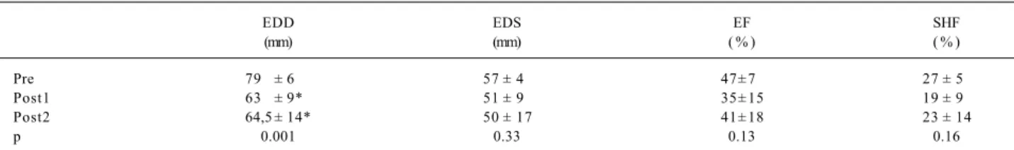
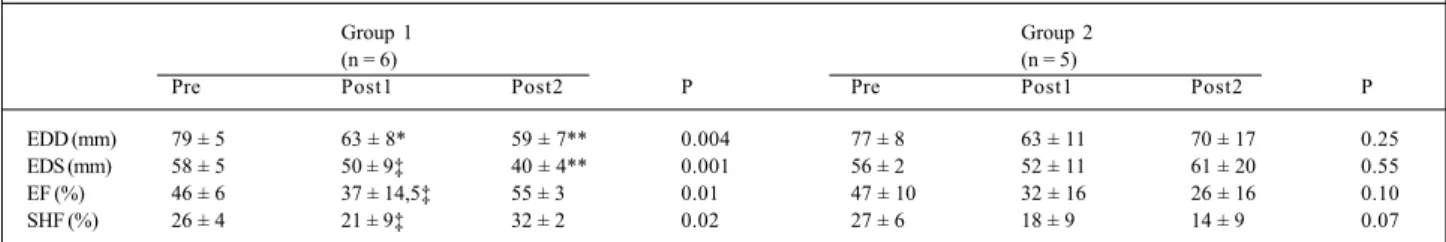
ejection fraction in post2, we can analyze 2 groups of patients (table III). In 6 patients, group 1, a significant and continuous reduction in the diastolic (p=0.0004) and systo-lic diameters occurred (p=0.001) from pre to post2. In this group, ejection fraction did not decrease significantly from pre to post1, but increased significantly from post1 to post2 (p=0.01). In the other 5 patients, group 2, a nonsignificant decrease in diastolic (p=0.25) and systolic diameters (p=0.55) occurred in post1, followed by a nonsignificant in-crease in post2. In this group, ejection fraction dein-creased gradually and not significantly from pre to post1 and from post1 to post2 (p=0.10).
No correlation was found between echocardiographic data in pre and post1 or post2. No difference existed between sex, age, Functional Class, body surface, cause of mitral re-gurgitation, atrial fibrillation, tricuspid valve disease, left atrium dimension, diastolic diameter, systolic diameter, ejection fraction, shortening fraction, septal thickness, and left ventricular posterior wall thickness, left ventricular mass in the pre, aortic clamping time, chordal preservation, tricus-pid annuloplasty, left atrium shortening surgery, and size of the prostheses implanted in the patients of groups 1 and 2. However, persistence of symptoms in patients from group 1, 30±12 months, was significantly lower than that in those patients from group 2, 79±38.5 months (p=0.01). A good in-verse correlation was found between the persistence of
symptoms and the ejection fraction in post2 (r= -0.82, p<0.01), and a direct correlation was found between the du-ration of symptoms and the systolic diameter (r=0.94, p<0.001). All 5 patients with symptoms lasting ≤ 48 months had improved ejection fraction in post2, whereas only 1 of the 6 patients with symptoms lasting > 48 months had ejec-tion fracejec-tion improvement in post2 (p=0.01).
Discussion
Results of this study indicate that some patients with severe symptomatic chronic mitral regurgitation and severe preoperative left ventricular dysfunction may improve in Functional Class and left ventricular function, later, after mitral valve replacement. These results are not in accor-dance with those of previous studies, which demonstrated a poor prognosis in patients with severe mitral regurgitation and severe left ventricular dysfunction after mitral valve re-placement. Starling 14, studying 15 patients with chronic
mitral regurgitation 1 year after mitral valve replacement, de-monstrated the persistence of left ventricular dysfunction in those patients whose end-diastolic and end-systolic vo-lumes were increased and whose ejection fraction was de-creased in the preoperative period. Wisenbaugh et al 4,
reporting the postoperative results of 61 patients with mitral regurgitation, demonstrated that systolic diameter
Table I - Surgical and clinical characteristics
Patients G A Causes Symptoms FC FA SC Type of Chordal Tricuspid TCA CF
(months) Pre (m2) Surgery(*) preservation annuloplasty (min) post2
1 F 50 deg 60 III yes 1.38 MVR - yes 84 II
2 F 26 deg 120 IV yes 1.59 MVRe(33) yes yes 55 IV
3 F 54 deg 120 IV yes 1.43 MVRe(33) yes no 80 III
4 M 53 deg 172 IV no 1.75 MVRe(35) no no 80 II
5 F 24 rheu 72 IV yes 1.75 MVRe(35) yes no 84 I
6 M 31 deg 48 IV yes 1.62 MVRe(35) yes no 13 I
7 F 29 rheu 12 IV yes 1.39 MVRe(33) yes yes 49 I
8 M 25 deg 36 IV yes 1.85 MVRe(35) yes no 50 I
9 M 57 deg 24 III no 1.60 MVRe(33) yes no 65 I
10 M 31 rheu 24 II yes 1.93 MVRe(35) yes no 54 I
11 M 22 rheu 60 IV yes 1.55 MVRe(33) yes no 70 I
Mean 36 68 1.62 62.1
SD 13 49.7 0.1 21.2
A- age in years; F- Female gender; M- male gender; deg-degenerative MR; rheu-rheumatic MR; symptoms-symptoms duration; FC- New York Heart Association
Functional Class; AF- atrial fibrillation; BS- body surface; MVR- mitral valve repair; MVRe- mitral valve replacement; ACT- aortic clamping time; PRE- preoperative period; POST- postoperative period; G- gender; (*)= prostheses
Table II - Dimensions and left ventricular function in the preoperative period (pre), early postoperative period (post1), and late postoperative (post2)
EDD EDS EF SHF
(mm) (mm) ( % ) ( % )
Pre 79 ± 6 57 ± 4 47± 7 27 ± 5
Post1 63 ± 9* 51 ± 9 35± 15 19 ± 9
Post2 64,5 ± 14* 50 ± 17 41± 18 23 ± 14
p 0.001 0.33 0.13 0.16
was the most useful predictor of death, heart failure, and left ventricular dysfunction. Of 8 patients with systolic dia-meter ≥ 51mm, 6 died and 1 remained with severe heart failure. One patient remained with severe left ventricular dysfunction but had an improvement in Functional Class after a mean follow-up of 24 months. Based on these results, they consider surgery in these patients, even with chordal preservation, “heroic”, and they suggest that an alternative surgery should, therefore, be considered for these patients. In our patients, although they had the same level of preoperative left ventricle dysfunction, we did not observe the same results in the postoperative period. The rate of survivors at 5 years was 81.8%. We observed that only 2 patients remained with advanced heart failure (1 died 14 months after surgery), whereas the 9 remaining patients improved their Functional Class. The other death observed resulted from infective endocarditis in 1 patient who had improved Functional Class and left ventricular function. Although they had improved Functional Class, 2 other patients still had severe left ventricular dysfunction in post2. Therefore, the rate of survivors free from important symp-toms, in 5 years, was 67.5%. In contrast with the South African study 4, rheumatic disease was not the only cause
found in our patients that could explain the difference in our results. However, our patients with mitral regurgitation had a decrease in systolic diameter and improvement in Func-tional Class.
Ejection fraction reduction is a common finding after mitral valve replacement, even with chordal preservation and mitral valve repair 4,15-18. Ejection fraction and
shorte-ning fraction decrease in the postoperative period is also observed in those patients with preserved left ventricular function in the preoperative period 4,19,20. It has been
believed that in patients with “noncompensated” mitral re-gurgitation, cardiac chambers enlargement persists after mitral valve replacement because of the increase in final sys-tolic stress, which further decreases syssys-tolic function. Ho-wever, in this study, we have demonstrated that in some pa-tients with noncompensated mitral regurgitation a decrease in left ventricular dimensions occurred, and therefore a conse-quent improvement in left ventricular function was observed. Bonow et al 21, studying patients with severe aortic
re-gurgitation, observed that prolonged left ventricular
dys-function (≥1 year) resulted in a greater chance of left ventri-cular dysfunction after aortic valve replacement. Recently, Krishman et al 22 , studying children operated on for severe
mitral regurgitation, reported an early worsening, followed by a late improvement in left ventricular function. The im-provement was explained by a relatively shorter overall du-ration of mitral insufficiency or a greater potential for reco-very in younger myocardium. These results are reco-very similar to ours, especially in group 1, whose left ventricular dys-function decreased precociously (post1) but improved later (post2) after mitral valve replacement, suggesting a level of contractile reserve in these patients. They had a lower dura-tion of symptoms compared with that in those whose left ventricular function decreased in the postoperative period (30±12 and 79±38.5 months, respectively, p=0.01). We have also observed that patients whose symptoms persisted for
≤ 48 months had an improvement in ejection fraction in post2 compared with that in those with symptoms lasting > 48 months (p=0.01). In addition, we verified a good rela-tionship between symptoms duration and ejection fraction in post2 (r= -0.82, p<0.01). More recently, Tribouilloy et al 23,
studying the influence of Functional Class on the surgical results in patients with mitral regurgitation, demonstrated that patients in Functional Class III/IV had higher operative mortality and lower late survival than those patients in Functional Class I/II, regardless of left ventricular function, age, or other clinical features. Although no systematic studies that correlate the presence and the severity of symptoms with the level grade of left ventricular dys-function are available in severe chronic mitral regurgitation, we should consider the findings of Tribouilloy et al 23 where
patients with mitral regurgitation in Functional Class IV had lower ejection fraction than those patients in Functional Class I/II. In our study, no differences existed in left ventri-cular function between the patients in different Functional Classes, because of the selection criteria used and the small number of enrolled patients. Fuster et al 24 demonstrated
that patients with severe chronic mitral regurgitation and advanced symptoms, and in Functional Class III/IV, had more myocardial interstitial tissue than those patients in Functional Class I/II without heart disease. Unfortunately, neither Tribouilloy et al 23 nor Fuster et al 24 mentioned the
duration of symptoms of the patients involved in their
Table III - Preoperative data (pre), early postoperative (post1), and late postoperative (post2) in patients from group 1 and group 2
Group 1 Group 2
(n = 6) (n = 5)
Pre Post1 Post2 P Pre Post1 Post2 P
EDD (mm) 79 ± 5 63 ± 8* 59 ± 7** 0.004 77 ± 8 63 ± 11 70 ± 17 0.25
EDS (mm) 58 ± 5 50 ± 9‡ 40 ± 4** 0.001 56 ± 2 52 ± 11 61 ± 20 0.55
EF (%) 46 ± 6 37 ± 14,5‡ 55 ± 3 0.01 47 ± 10 32 ± 16 26 ± 16 0.10
SHF (%) 26 ± 4 21 ± 9‡ 32 ± 2 0.02 27 ± 6 18 ± 9 14 ± 9 0.07
studies. Therefore, it is possible to confirm that the amount of myocardial interstitial tissue is less intense in patients with a shorter duration of symptoms. This may be the expla-nation for the improvement observed in our patients with a duration of symptoms ≤ 48 months.
According to our findings, conventional mitral valve surgery, with chordal preservation, or mitral valve repair must be recommended to patients with mitral regurgitation and systolic diameter ≥ 51mm, even though some of them may remain with severe left ventricular dysfunction, but with a great chance of improving in Functional Class. Addi-tionally, we may evaluate the late postoperative result and decide on an alternative treatment, such as cardiac trans-plantation. Duration of symptoms ≤ 48 months identifies the patients with greater chances of left ventricular function improvement after mitral valve replacement in patients with mitral regurgitation and systolic diameter ≥ 51mm.
In this study, we did not assess systolic stress indexes 25,
left ventricle cavity elastance 14, ejection fraction variance,
or systolic diameter during exercise 7; therefore, we do not
know whether they could influence the postoperative results. However, we do not know any studies that have de-monstrated that these indexes have greater predictive value than the diastolic diameter. We do not know either how long left ventricular dysfunction persisted in the patients included in the study, because a few patients had echocar-diographic examinations at the onset of their diseases. Ad-ditionally, our results do not apply to asymptomatic or to minimally symptomatic patients with chronic mitral
regurgi-tation, because most of our patients had severe symptoms (Functional Class III/IV) before surgery. We could not assess the influence on the results of the surgical technique performed, once the number of patients is small to enable such a comparison. We do not know either whether the use of angiotensin-converting enzyme inhibitors played any role in the improvement of left ventricular function or in Functional Class determination. However, all patients from both groups used these medications during the postopera-tive follow-up. Further studies involving a greater number of patients are necessary to confirm our results. We want to point out that the population studied was composed of a special group of patients with severe mitral regurgitation in an advanced stage of the disease who ought medical treatment. In our service, we recommend mitral valve sur-gery for patients with mitral regurgitation that become sym-ptomatic and for those with severe mitral regurgitation asymptomatic or minimally symptomatic, when a real chance exists of mitral valve repair regardless of left ventri-cular dimensions.
In summary, systolic diameter ≥ 51mm is not always associated with a poor prognosis in the postoperative period. In most patients with severe chronic mitral regurgita-tion and systolic diameter ≥ 51mm, Functional Class impro-ves regardless of the response of left ventricular function after surgery. Duration of symptoms ≤ 48 months is asso-ciated with a greater chance of improvement in left ventri-cular function.
References
1. Stewart WJ. Choosing the “Golden Moment” for operation in the era of valve repair for mitral regurgitation. ACC Highlights 1995; 10: 2-7.
2. Zile MR, Gaasch WH, Carrol JD, Levine HJ. Chronic mitral regurgitation: predic-tive value of preoperapredic-tive echocardiographic indexes of left ventricular function and wall stress. J Am Coll Cardiol 1984; 3: 235-42.
3. Borow KM, Green LH, Mann T, et al. End-systolic volume as a predictor of posto-perative left ventricular performance in volume overload from valvular regurgita-tion. Am J Med 1980; 68: 655-63.
4. Wisenbaugh T, Skundicky D, Sareli P. Prediction of outcome after valve replace-ment for rheumatic mitral regurgitation in the era of chordal preservation. Circu-lation 1994; 89: 191-7.
5. Nakano S, Sakai K, Tamiguchi K, et al. Relation of impaired left ventricular function in mitral regurgitation to left ventricular contractile state after mitral valve repla-cement. Am J Cardiol 1994; 73: 70-4.
6. Pai RG, Bausal RC, Shah PM. Doppler-derived rate of left ventricular pressure rise: its correlation with the postoperative left ventricular function in mitral re-gurgitation. Circulation 1990: 82: 514-20.
7. Leung DY, Grifin BP, Stewart WJ, Cosgrove DM, Thomas JD, Marwick TH. Left ventricular function after valve repair for chronic mitral regurgitation: predictive value of preoperative assessment of contractile reserve by exercise echocardio-graphy. J Am Coll Cardiol 1996; 28: 1198-205.
8. Abbasi AS, Allen MW, Decristofaro D, Ungar I. Detection and estimation of the degree of mitral regurgitation by range-gated pulsed Doppler echocardiography. Circulation 1980; 6i: 143-7.
9. Helmcke F, Nanda NC, Hsiung MC, et al. Color Doppler assessment of mitral re-gurgitation with orthogonal plans. Circulation 1987; 75: 175-83. 10. Schiller NB. Two-dimensional echocardiographic determination of left
ventri-cular volume, systolic function and mass: summary and discussion of the 1989
re-commendations of the American Society of Echocardiography. Circulation 1991; 84(suppl): 1280-7.
11. Teichholz LE, Kreulen T, Herman MV, Gorlin R. Problems in echocardiographic volume determinations: echocardiographic-angiographic correlations in the presence or absence of asynergy. Am J Cardiol 1976; 37: 7-11.
12. Carpentier A, Deloche A, Dauptain J, et al. A new reconstructive operation for correction of mitral and tricuspid insufficiency. J Thorac Cardiovasc Surg 1971; 61: 1-13.
13. Carpentier A. Cardiac valve surgery: the “French correction”. J Thorac Cardio-vasc Surg 1983; 86: 323-37.
14. Starling MR. Effects of valve surgery on left ventricular contractile function in pa-tients with long-term mitral regurgitation. Circulation 1995; 92: 81-818. 15. Schuler G, Peterson KL, Johnson A, et al. Temporal response of left ventricular
performance to mitral valve surgery. Circulation 1979; 59: 1218-31. 16. Crawford MH, Souchek J, Oprian CA, et al, and participants in the Department of
Veterans Affairs Cooperative Study on Valvular Heart Disease. Determinants of survival and left ventricular performance after mitral valve replacement. Circu-lation 1990; 81: 1173-81.
17. Enriquez-Sarano M, Tajik AS, Schaff HV, et al. Echocardiographic prediction of left ventricular function after correction of mitral regurgitation: results and cli-nical implications. J Am Coll Cardiol 1994; 24: 1536-43.
18. Bonow RO, Nikas D, Elefteriades JA. Valve replacement for regurgitant lesions of the aortic and mitral valve in advanced left ventricular dysfunction. Cardiol Clin 1995; 13: 73-83.
19. Enriquez-Sarano M, Schaff HV, Orszulak TA, Tajik AJ, Bailey KR, Frye RL. Valve repair improves the outcome of surgery for mitral regurgitation. A multivariate analysis. Circulation 1995; 91: 1022-8.
contractile function in patients with long-term mitral regurgitation and normal ejection fraction. J Am Coll Cardiol 1993; 22: 239-50.
21. Bonow RO, Rosing DR, Maron BJ, et al. Reversal of left ventricular dysfunction after aortic valve replacement for chronic aortic regurgitation. influence of dura-tion of preoperative left ventricular dysfuncdura-tion. Circuladura-tion 1984; 70: 570-9. 22. Krishman U S, Gersony WM, Berman-Rosenzweig E, Apfel HD. Late left
ventri-cular function after surgery for children with chronic symptomatic mitral regurgi-tation. Circulation 1997; 96: 4280-5.
23. Tribouilloy CM, Enriquez-Sarano M, Schaff HV, et al. Impact of preoperative
sym-ptoms on survival after surgical correction of organic mitral regurgitation: ratio-nale for optimizing surgical indications. Circulation 1999: 99: 400-5. 24. Fuster V, Danielson MA, Robb RA, Broadkent JC, Brown AL, Elveback LR.
Quantitation of left ventricular myocardial fibre hypertrophy and increased vo-lume and intersticial tissue in humans hearts with chronically increased vovo-lume and pressure overload. Circulation 1977: 55: 504-8.