Universidade Federal do Rio de Janeiro - Cintilab
Mailing address: Márcia Maria Sales dos Santos – Av. Epitácio Pessoa, 4986/501 – 22471-001 – Rio de Janeiro, RJ, Brazil – E-mail: [email protected] English version by Stela Maris C. e Gandour
Arq Bras Cardiol, volume 80 (nº 1), 25-30, 2003
Márcia Maria Sales dos Santos, Eduardo Cwajg, Mauricio da Rocha Pantoja
Rio de Janeiro, RJ - Brazil
Prognostic Value of Technetium-99m-Labeled Single-Photon
Emission Computerized Tomography in the Follow-Up
of Patients After their First Myocardial
Revascularization Surgery
The surgical treatment of obstructive atherosclerotic coronary disease has been definitively established as an important therapeutical tool to fight ischemic heart disease. However, progression of the atherosclerotic disease in native and grafted vessels limits the duration of the be-nefits obtained with surgery.
Isolated clinical assessment does not provide a clear idea of the anatomical conditions of the vessels (degree of obstruction) and of the risk of future complications due to the influence of several factors.
Most studies on myocardial perfusion scintigraphy and cardiac surgery available in the medical literature were performed with planar Thallium-201 scintigraphy aiming at diagnosing graft disease.
Planar scintigraphy comprises 2-dimensional images of the heart in 3 views: anterior and left anterior oblique at 40 and 70 degrees. With technological advancement, rota-tional gamma cameras (SPECT) were developed, enabling the acquisition of multiple views of myocardial radionuclide activity and providing an increase in the diagnostic sensiti-vity of the method.
During approximately 15 years, Thallium-201 was the major radiopharmaceutical tracer used in myocardial per-fusion studies. The discovery of new Technetium-99m-labeled myocardial perfusion tracers (sestamibi, teboro-xime, tetrofosmin, furifosmin) with better radiolabeled sym-metric characteristics favored the generation of excellent-re-solution images.
Despite the relevance of the subject and the increase in the amount of myocardial revascularization surgeries per-formed, little has been published about the use of more ad-vanced scintigraphic techniques, such as myocardial tomo-graphy and the new radiopharmaceuticals labeled with Technetium-99m.
This study aimed at assessing the prognostic value of
Objective - To assess the prognostic value of Techne-tium-99m-labeled single-photon emission computerized tomography (SPECT) in the follow-up of patients who had undergone their first myocardial revascularization.
Methods - We carried out a retrospective study of 280 re-vascularized patients undergoing myocardial scintigraphy under stress (exercise or pharmacological stress with dipy-ridamole) and at rest according to a 2-day protocol. A set of clinical, stress electrocardiographic and scintigraphic va-riables was assessed. Cardiac events were classified as “ma-jor” (death, infarction, unstable angina) and “any” (major event or coronary angioplasty or new myocardial revascu-larization surgery).
Results - Thirty-six major events occurred as follows: 3 deaths, 11 infarctions, and 22 unstable anginas. In regard to any event, 22 angioplasties and 7 new surgeries occurred in addition to major events, resulting a total of 65 events. The sensitivity of scintigraphy in prognosticating a major event or any event was, respectively, 55% and 58%, showing a negative predictive value of 90% and 83%, respectively. Dia-betes mellitus, inconclusive stress electrocardiography, and a scintigraphic visualization of left ventricular enlargement were significant variables for the occurrence of a major event. On multivariate analysis, abnormal myocardial scintigraphy was a predictor of any event.
Conclusion - Myocardial perfusion tomography with Technetium-99m may be used to identify high-risk patients after their first myocardial revascularization surgery.
Technetium-99m-labeled single-photon emission compute-rized tomography (SPECT) in the follow-up of patients who had previously undergone their first myocardial revascula-rization surgery.
Methods
We carried out a retrospective study designed from a database of a private nuclear cardiology laboratory, from which we selected 1,243 surgically revascularized individuals who underwent myocardial perfusion scintigraphy with the tomographic technique from April 1992 to April 1998.
Technetium-99m-labeled sestamibi or tetrofosmin were the radiopharmaceuticals used. The examinations comprised 2 stages performed in 2 different days: stress (exercise or pharmacological stress with dipyridamole) scin-tigraphy and scinscin-tigraphy at rest.
A single-detector gamma camera with a low-energy high-resolution collimator was used; 32 images with a rota-tion arch of 180º were obtained. The images were processed and analyzed in 3 cuts: short axis and vertical and horizontal long axes. The interpretation of the scintigraphies was sub-jective and semiquantitative (percentage myocardial distri-bution relative to the pixel of greater intensity) and perfor-med by an independent experienced observer, who ignored the patients’ clinical and laboratory data. Doubtful cases required the second opinion of an experienced observer.
Patients with the following characteristics were exclu-ded from the study: third-degree left bundle-branch block; a definitive pacemaker; a history of acute myocardial infarc-tion, percutaneous transluminal coronary angioplasty, or more than 1 myocardial revascularization surgery prior to myocardial scintigraphy; systemic disease that could inter-fere with the prognosis; patients who had undergone dobu-tamine stress scintigraphy, and those with a submaximum physical exertion level on exercise testing.
During examination, demographic and clinical data were collected, as were those regarding the risk factors for coronary atherosclerotic disease.
The electrocardiographic findings of the exercise test were classified as negative, positive, or inconclusive for the diagnosis of myocardial ischemia. The test was negative when no change was observed in the ST segment as compa-red with that on the baseline electrocardiogram. When de-pression of the ST segment m 1 mm (ascending, descending, or rectified) was observed 80 ms from the J point, the tracing was considered positive for ischemia. The interpretation of the tracing was considered inconclusive when the baseline electrocardiogram was altered (for example, left ventricular hypertrophy and use of medication).
Myocardial perfusion scintigraphy was considered normal or abnormal (ischemia, fibrosis, or fibrosis associa-ted with ischemia). The examinations with a homogeneous distribution of the radiotracer in the entire left ventricular myocardium on stress and resting images were considered
negative for ischemia. Fixed perfusion defects present in both images were interpreted as fibrosis; transient perfu-sion defects present on stress images and absent on resting images were interpreted as ischemia. When the recovery of these defects was only partial, the simultaneous existence of fibrosis and ischemia was considered.
Left ventricular size was subjectively assessed, based on the cardiothoracic ratio present in the original scintigra-phic image of acquisition.
Based on the topography of the myocardial perfusion deficit, a correlation with coronary anatomy was establis-hed, and the segments were classified as belonging or not belonging to the region irrigated by the anterior descen-ding artery.
Follow-up was performed through contact with the patient or the assistant physician. The following terminal cardiac events were individually considered: sudden or nonsudden cardiac death; acute myocardial infarction; uns-table angina requiring hospitalization; percutaneous trans-luminal coronary angioplasty; and new myocardial revas-cularization surgery. These events were categorized into 2 subgroups as follows: “major event” (cardiac death, acute myocardial infarction, and unstable angina) and “any event” (a major event or 1 myocardial revascularization pro-cedure). For the purpose of analysis, only the first event of each patient was considered.
The time interval between revascularization surgery and end of the follow-up (TCABG_END) and that between revascularization surgery and myocardial scintigraphy (TCABG_MS) were analyzed with the Mann-Whitney test. Data underwent univariate and multivariate analysis. The variables with a p value < 0.05 and a trend value of 0.05≤p≤0.1 were considered statistically significant. The sensitivity, specificity, and positive and negative predictive values of scintigraphy for cardiac events were calculated. Actuarial curves of survival determined with the Kaplan-Meier method were compiled.
Results
Of the 1,243 revascularized patients undergoing myo-cardial perfusion scintigraphy, 879 patients were excluded. Of the 364 remaining patients, 280 (77%) were followed up. Lack of evolution information on 84 (23%) patients of the initial sample did not create any type of bias, because the distribution of the variables analyzed was homoge-neous between the 2 groups.
Analysis of temporal data showed that 74% of the patients had a TCABG_END above 5 years. No correlation was observed between TCABG_MS or TCABG_END and the type of scintigraphic result or the occurrence of cardiac events.
Thirty-six major events occurred as follows: 3 cardiac deaths, 11 myocardial infarctions, and 22 unstable anginas. Of 65 cardiac events classified as “any event,” 29 were new myocardial revascularization procedures as follows: 22 angioplasties and 7 reoperations.
No correlation was observed between the type of stress (physical or pharmacological) used, the result of the exercise electrocardiography (positive, negative, or incon-clusive), and the result of the myocardial perfusion scinti-graphy (normal or abnormal).
The 3 variables that correlated with an abnormal scin-tigraphic result are shown in figure 1. Left ventricular enlar-gement on scintigraphy (p<0.0001) and unstable angina (p=0.01) and coronary angioplasty (p=0.04) were associated with the presence of an abnormal scintigraphic finding.
The scintigraphic finding of ischemia (p=0.04) predo-minated among the patients who reported the presence of angina pectoris during follow-up. A correlation between the finding of fibrosis (p<0.001) and that of left ventricular enlar-gement on myocardial scintigraphy was observed.
The analysis of the occurrence of cardiac events and a specific type of abnormal scintigraphic finding (ischemia, fibrosis, and fibrosis with ischemia) identified no association. On assessing the occurrence of major events, patients with major events were observed to have a trend towards a greater proportion of left ventricular enlargement as compared with that observed in patients with no major event (p=0.08).
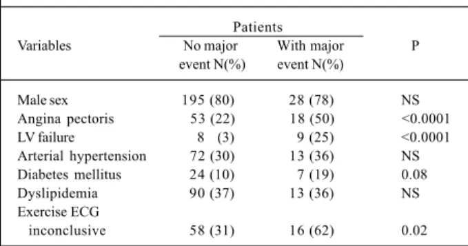
The remaining important variables related to the oc-currence of a major event are shown in table I. The sub-group of diabetic patients showed a trend towards a higher proportion (p=0.08). An association between the presence of angina pectoris (p<0.001) and left ventricular failure (p<0.001), reported during follow-up, and the incidence of a major event was also observed.
Among the patients with any cardiac event, no
predo-minance of the demographic variables studied was obser-ved. The presence of angina pectoris (p<0.001) and left ven-tricular failure (p<0.001) during follow-up related to the inci-dence of “any event.”
An altered scintigraphic result showed a good correla-tion with the occurrence of “any event” (tab. II).
On multivariate analysis, the presence of diabetes mellitus and left ventricular enlargement showed a trend to-wards stressing the probability of the occurrence of major events. For the “any cardiac event” model, the altered scin-tigraphic result had a significant statistical value (tab. III).
To analyze the prognostic value of myocardial perfu-sion scintigraphy, sensitivity, specificity, and the negative and positive predictive values were calculated (tab. IV).
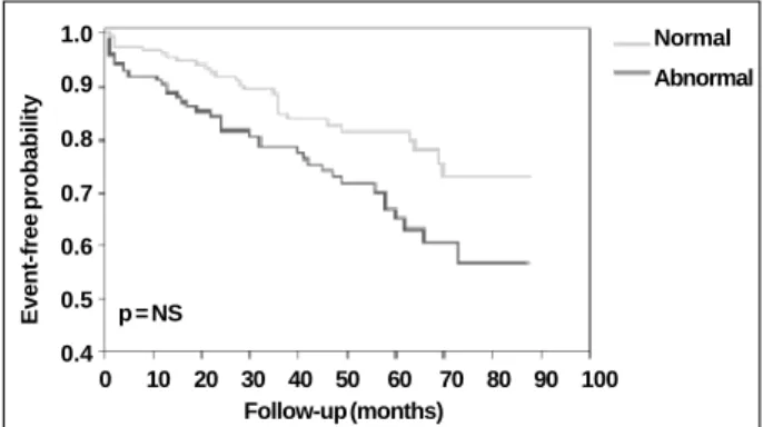
The actuarial survival curves free from a “major cardiac event” or “any cardiac event” are shown in figures 2 and 3.
Discussion
The major applications of nuclear cardiology in the follow-up of revascularized patients comprise the diagno-sis of intraoperative myocardial injury and atherosclerotic disease of the native or graft bed, the documentation of re-duction of the ischemic area, the analysis of ventricular function in the postoperative period, and the prediction of cardiac events.
During the follow-up of the revascularized patients, those at risk for developing future cardiac events need to be identified. On detecting patients at higher risk for cardiac
Table I – Comparison of clinical covariables and major cardiac events
Patients
Variables No major With major P
event N(%) event N(%)
Male sex 195 (80) 28 (78) NS
Angina pectoris 53 (22) 18 (50) <0.0001
LV failure 8 (3) 9 (25) <0.0001
Arterial hypertension 72 (30) 13 (36) NS Diabetes mellitus 24 (10) 7 (19) 0.08
Dyslipidemia 90 (37) 13 (36) NS
Exercise ECG
inconclusive 58 (31) 16 (62) 0.02
LV- left ventricle; ECG- electrocardiogram.
Table II - Comparison of the result and impaired area on myocardial scintigraphy and any cardiac event
Myocardial Patients
scintigraphy result Without any event With any event P
N(%) N(%)
Normal 129 (83) 27 (17) NS
Abnormal 86 (69) 38 (31) 0.009
Non AD irrigated area 43 (50) 19 (50) NS AD irrigated area 43 (50) 19 (50) NS
AD – anterior descending artery Fig. 1- Comparison of clinical and scintigraphic covariables with the result of
myocardial scintigraphy. LV- left ventricle.
% of patients
Angioplasty
Normal scintigraphy Abnormal scintigraphy
Unstable angina
events, the subgroup that benefits most from a more invasi-ve treatment is identified, due to an improinvasi-vement in quality of life and a reduction in mortality.
Basically, the results were published of 4 studies 1-4 that
analyzed the prognostic power of exercise myocardial scin-tigraphy in the follow-up of patients undergoing myocardial revascularization surgery.
The first study was performed by Palmas et al 1, who
assessed 294 patients undergoing exercise testing and Thallium-201 SPECT. On multivariate analysis, the scinti-graphic variables of statistical significance for the occur-rence of events were the reversibility score and increased pulmonary uptake. Another important finding was that scintigraphic information had a greater prognostic value than the association of clinical history and exercise testing. Nallamothu et al. 2 carried out the second study in
which the prognostic value of Thallium-201 myocardial scintigraphy was assessed. The extension of the perfusion defect, the perfusional changes in multiple areas, and the in-crease in pulmonary uptake were independent predictors of cardiac events (a 9.3% risk per year).
In a study by Miller et al 3 using exercise tomographic
scintigraphy with Thallium-201, the extension of the
perfu-sion defect was the only independent variable able to predict events. The patients with normal myocardial perfu-sion had an annual event ratio of 1.6%.
In another study carried out at the Cleveland Clinic 4
with tomography and Thallium-201 in asymptomatic revascularized patients, the group with abnormal scintigra-phic findings had a greater incidence of cardiac events (11%) than that with normal imaging (4%).
In our study, strict inclusion and exclusion criteria were defined aiming at minimizing the possible clinical situations that could interfere with the interpretation of and determina-tion of the value of myocardial perfusion scintigraphy. This is the case of patients with a previous history of acute myo-cardial infarction in whom a myomyo-cardial perfusion deficit in imaging compatible with the infarcted area may occur. The same occurs in third-degree left bundle-branch block or pace-maker rhythm, in which a decreased uptake pattern, respec-tively, at the level of the ventricular septum and inferior wall may be visualized even in the absence of obstructive co-ronary artery disease. However, in most published studies, this preoccupation was not observed.
During follow-up, the total rate of major events (cardiac death, myocardial infarction, or unstable angina) was 12.8%, and the total rate of any cardiac event (major events, angioplasty, or revascularization surgery) was 23.2%. It is worth noting that revascularized patients com-prise a subgroup, in which the treatment of coronary artery disease has already been instituted and a lower rate of cardiac events is expected.
Of the clinical and demographic variables evaluated by the univariate analysis, no association with the type of scintigraphic result was observed. This finding is in accor-dance with those of other published studies.
Univariate analysis identified a trend of the diabetes mellitus risk factor towards being associated with the occur-rence of major events (p=0.08). This probable importance of diabetes mellitus in predicting major events has also been re-confirmed on multivariate analysis (p=0.07). The importance of the diabetes mellitus risk factor in the severity of atheros-clerotic disease of the native bed 5-8 has been well
demons-trated. A more diffuse and extensive atherosclerotic impair-ment of coronary arteries is believed to be the motive.
Fig. 2 - Actuarial curve of survival free from major cardiac event with normal and abnormal scintigraphy.
0 10 20 30 40 50 60 70 80 90 100
Event-free probability
1.0
0.9
0.8
0.7
0.6
0.5
0.4
Normal
Abnormal
p = NS
Follow-up (months)
Table III - Independent predicting variables of any cardiac event
Predicting variables P value Odds ratio 95% confidence
of any event interval
Abnormal myocardial
scintigraphy 0.008 2.11 1.20-3.72
Table IV - Prognostic value of myocardial scintigraphy
Myocardial scintigraphy Major event Any event
Sensitivity (%) 56 58
Specificity (%) 57 60
Predictive value (%)
positive 16 31
negative 90 83
Accuracy (%) 57 60
Fig. 3 - Actuarial curve of survival free from any cardiac event with normal and abnormal scintigraphy.
0 10 20 30 40 50 60 70 80 90 100
Event-free probability
1.0
0.9
0.8
0.7
0.6
0.5
0.4
Normal
Abnormal
p = NS
On univariate analysis, the cardiac symptoms present during follow-up, such as angina pectoris (p=0.001) and the clinical manifestations of left ventricular failure (p=0.001), appeared as important variables in the subgroup of patients with major events.
The determination of the value of precordialgia after myocardial revascularization surgery is extremely complex. The pain may be caused by osteoarticular, neurological, and muscular factors proper to the surgical procedure. Ho-wever, the presence of typical angina has been reported as a strong indicator of ischemia and of the presence of signifi-cant coronary lesions on the hemodynamic study 9.
On assessing the modality of stress used (physical or pharmacological with dipyridamole), no difference was observed in the scintigraphic result and in event predic-tion. However, when studying separately the electrocardio-graphic pattern of the exercise test (normal, ischemic, or inconclusive), the subgroup of patients with major events showed a greater proportion of inconclusive exercise elec-trocardiogram results (p=0.02). The patients with an incon-clusive exercise test may have been those with more signi-ficant disease and, consequently, with persistent changes on the resting electrocardiogram.
On univariate analysis, the patients undergoing per-cutaneous angioplasty had a greater association with abnormal images (p=0.04). Interference of the result of the test with the decision-making process of the assistant phy-sician could not be evaluated.
When the scintigraphic variables were studied on uni-variate and multiuni-variate analysis, the patients with the scin-tigraphic pattern of left ventricular enlargement were obser-ved to have a greater proportion of altered myocardial per-fusion imaging and major events.
Left ventricular dysfunction is an independent marker of poor prognosis. It is a fact that left ventricular size and the dysfunctional degree of the ischemic cardiac muscle are directly correlated with the dimension and intensity of de-creased myocardial uptake on scintigraphy and with the ex-tension and severity of the coronary artery disease on co-ronary angiography 6,7.
Postexercise Thallium-201 pulmonary uptake is a signal of ventricular dysfunction and extensive atheroscle-rotic disease. Transient left ventricular dilation on exercise imaging is caused by ischemic ventricular dysfunction and indicates the presence of severe atherosclerotic disease 6,7.
The scintigraphic visualization of left ventricular enlar-gement, similarly to other indirect signs of left ventricular dysfunction like Thallium-201 pulmonary uptake, transient left ventricular ischemic dilation, extension of decreased myocardial uptake greater than 15% of the left ventricular area, and defects of myocardial uptake in multiple areas, are inde-pendent markers of poor prognosis and cardiac mortality 10-12.
The association of the presence of an abnormal scinti-graphic result and the occurrence of any cardiac event had statistical significance (p=0.009). No specific type of scinti-graphic result (ischemia, fibrosis, or fibrosis with ischemia)
predominated in predicting cardiac events. These findings did not partially agree with those reported in other studies in which ischemia was an important scintigraphic finding.
The event-free actuarial curves clearly show the influ-ence of the appearance of myocardial scintigraphy in the occurrence of events throughout time. They illustrate that the presence of a normal scintigraphy is a positive indication of event-free survival, which is in accordance with the im-portance of the negative predictive value of the examination. In our study, sensitivity, specificity, positive predictive value, and mainly negative predictive value cited in the li-terature could not be specifically reproduced. Discordance with previous publications may be due to differences in sample selection and limitations inherent in the study design. The major limitation of this study might have been the lack of application of quantitative methods of analysis and interpretation on scintigraphy. The interpretation of myo-cardial perfusion images was subjective, identifying im-paired left ventricular myocardial areas. However, according to the literature, this type of analysis has a performance simi-lar to that of quantitative analysis 5-7. We should not also
neglect the prognostic clinical importance of assessing the extension and intensity of the myocardial uptake deficit and of the reversibility of the decreased perfusion. This quanti-tative analysis may have strengthened the prognostic accu-racy of myocardial scintigraphy in this study.
The use of ventricular functional assessment software could have increased the prognostic value of myocardial scintigraphy found in this study. Currently, with computer programs, left ventricular endocardial and epicardial borders may be defined on the acquisition of tomographic myocardial perfusion images (Thallium-201 or Tc-99m-labe-led agents) synchronized with the cardiac cycle by the elec-trocardiographic QRS complex (gated SPECT).
The software of the gated SPECT allows the evalua-tion of systolic thickening, segmentary and global contrac-tion, and the calculation of left ventricular volumes and ejection fraction. This facilitates the differentiation between artifactual and actual defects, with a consequent reduction in the number of false-positive examinations and an in-crease in the specificity of the method.
Unfortunately, anatomic information about the coro-nary circulation and the aortocorocoro-nary grafts, which would complement the functional evaluation, could not be ob-tained. However, it is worth noting that the objective of this study was not to evaluate graft patency on scintigraphy, but the prognosis.
1. Palmas W, Bingham S, Diamond GA, et al. Incremental prognostic value of exercise Thallium-201 myocardial single-photon emission computed tomography late after coronary artery bypass surgery. J Am Coll Cardiol 1995; 25: 403-9.
2. Nallamothu N, Johnson JH, Bagheri B, et al. Utility of stress single-photon emis-sion computed tomography (SPECT) perfuemis-sion imaging in predicting outcome after coronary artery bypass grafting. Am J Cardiol 1997; 80: 1517-21. 3. Miller TD, Christian TF, Hodge DO, et al. Prognostic value of exercise
Thallium-201 imaging performed within 2 years of coronary artery bypass graft surgery. J Am Coll Cardiol 1998; 31: 848-54.
4. Lauer MS, Lytle B, Pashkow F, et al. Prediction of death and myocardial infarction by screening with exercise Thallium testing after coronary-artery-bypass gra-fting. Lancet 1998; 351: 615-22.
5. Braunwauld E. Heart disease: A Textbook of Cardiovascular Medicine. 5th ed. Philadelphia: WB Saunders, 1997: 1996p.
6. Zaret BL, Beller GA. Nuclear Cardiology: State of the Art and Future Directions. 2nd ed. Philadelphia: Mosby, 1999: 640p.
7. Iskandrian AS, Verani MS. Nuclear Cardiac Imaging: Principles and Applica-tions. 2nded. Philadelphia: F.A. Davis Co., 1996: 451p.
8. Topol E, Callif RM. Textbook of Cardiovascular Medicine. Philadelphia: Lippin-cott, Willlians & Wilkins, 1997: 2760p.
9. Pimentel CX, Paranandi SN, Goodhart DM, et al. Clinicals markers, management, and long-term follow-up of early ischemia after coronary artery bypass grafting. Am J Cardiol 1995; 76: 967-72.
10. Gibson RS, Watson DD. Value of planar Tl-201 imaging in risk stratification of patients recovering from acute myocardial infarction. Circulation 1991; 84(suppl I): I-148-62.
11. Brown KA. Prognostic value of Thallium-201 myocardial perfusion imaging. Circulation 1991; 83: 363-81.