2
https://doi.org/10.1590/0004-282X20170179 ARTICLE
Non-inflammatory cerebrospinal fluid delays
the diagnosis and start of immunotherapy in
anti-NMDAR encephalitis
La ausencia de hallazgos de inflamación en líquido cefalorraquídeo retrasa el inicio de
inmunoterapia en la encefalitis anti-NMDAR
Mariana Espinola-Nadurille1,2, Paola Bautista-Gomez1, Jose Flores3, Veronica Rivas-Alonso3,
Rodrigo Perez-Esparza4, Rodolfo Solís-Vivanco5,6, Steven Vargas-Cañas3
1Instituto Nacional de Neurología y Neurocirugía, Unidad de Neuropsiquiatría, Ciudad de México, México
2Universidad Nacional Autónoma de México, Facultad de Medicina, División de Posgrado, Ciudad de México, México 3Instituto Nacional de Neurología y Neurocirugía, Subdirección de Neurología, Ciudad de México, México
4Instituto Nacional de Neurología y Neuricrugía, Laboratorio de Investigación en Adicciones, Ciudad de México, México 5Instituto Nacional de Neurología y Neurocirugía, Departamento de Neuropsicología, Ciudad de México, México 6Universidad Nacional Autónoma de México, Facultad de Psicología, Ciudad de México, México.
Correspondence: Steven Vargas-Cañas; Subdirección de Neurología. Instituto Nacional de Neurología y Neurocirugía. Insurgentes Sur 3877. Tlalpan. ZC 14269 Mexico City; E-mail: [email protected]
Conflict of interest: There is no conlict of interest to declare.
Received 03 August 2017; Received in inal form 26 September 2017; Accepted 29 September 2017.
ABSTRACT
Anti-N-methyl-D-aspartate receptor (anti-NMDAR) encephalitis is a form of autoimmune encephalopathy that presents with a wide variety of symptoms, including neuropsychiatric manifestations. The authors’ aim for this study was to analyze the results of paraclinical studies of patients with a diagnosis of anti-NMDAR encephalitis and the association between symptom onset and diagnosis, and start of immunotherapy. Retrospective data of 29 patients with anti-NMDAR encephalitis were gathered and analyzed. Abnormal EEG was found in 27 patients (93.1%), whereas MRI was abnormal in 19 patients (65.5%). In contrast, an inlammatory pattern on CSF analysis was found in only 13 patients (44.8%). The absence of pleocytosis or increased proteins in the CSF was associated with a longer time from symptom onset to diagnosis and treatment (p = 0.003). The authors conclude that noninlammatory CSF may delay the correct diagnosis and start of immunotherapy in anti-NMDAR encephalitis. In the presence of suggestive clinical features, extensive studies including EEG are recommended.
Keywords: anti-n-methyl-d-aspartate receptor encephalitis; cerebrospinal luid; diagnosis.
RESUMEN
La encefalitis por receptor anti-N-metil-D-aspartato (anti-NMDAR) es una encefalopatía autoinmune con una amplia variedad de síntomas, incluyendo manifestaciones neuropsiquiátricas. Nuestro objetivo en este estudio fue analizar los resultados paraclínicos de pacientes diagnosticados con encefalitis anti-NMDAR y la asociación entre inicio de sintomatología, el diagnóstico y el inicio de inmunoterapia. Encontramos un EEG anormal en 27 pacientes (93.1%), así como IRM anormal en 19 de ellos (65.5%). En contraste, el análisis de LCR mostró un patrón inlamatorio en tan solo 13 pacientes (44.8%). La ausencia de pleocitosis o proteínas incrementadas en el LCR se asoció con un mayor tiempo desde el inicio de la sintomatología hasta el inicio del tratamiento (p=0.003). Concluimos que el LCR no inlamatorio puede retrasar el diagnóstico correcto y el inicio de tratamiento en encefalitis anti-NMDAR, por lo que se recomienda la realización de estudios exhaustivos, incluyendo EEG, ante la presencia de indicadores clínicos sugerentes del padecimiento.
Palabras clave: encefalitis antirreceptor n-metil-d-aspartato; líquido cefalorraquídeo; diagnóstico.
Encephalitis is an inlammation of brain parenchyma
associated with neurologic dysfunction that causes sub
-stantial morbidity and mortality worldwide. Speciic etiolo
-gies are identiied in less than 50% of cases1
. In 2013, in an attempt to better deine cases, the International Encephalitis Consortium proposed the Diagnostic Criteria for Encephalitis
3 Espinola-Nadurille M et al. Normal CSF delays diagnosis in anti-NMDA encephalitis
of documented fever ≥ 38°C (100.4°F), generalized or partial
seizures, or new onset of focal neurologic indings; and para
-clinical features of cerebrospinal luid (CSF) white blood cell
count ≥ 5/mm3, abnormality of brain parenchyma on neuro
-imaging, or electroencephalogram (EEG) abnormality1
. Anti-N-methyl-D-aspartate receptor encephalitis (anti-NMDAR encephalitis) is an autoimmune encephalopathy
characterized by the presence of serum and CSF IgG anti
-bodies against the GluN1 subunit of NMDAR, a neuronal sur
-face synaptic receptor, afecting children, adolescents and
adults2. In adults, it presents with psychiatric, behavioral, and
cognitive symptoms at onset and, less frequently, seizures3,4,5.
Over the irst four weeks, most patients develop movement disorders, loss of consciousness, autonomic dysfunction,
and central hypoventilation. Early diagnosis and immuno
-therapy have consistently been reported to improve the out
-come4,6,7. Since antibody testing for a deinite diagnosis of
anti-NMDAR encephalitis is not readily accessible in many institutions, recent guidelines have recommended starting immunotherapy in the face of suggestive clinical symptoms, in addition to supportive indings in diagnostic tests such as CSF analysis, EEG and magnetic resonance imaging (MRI),
and reasonable exclusion of other disorders8.
he National Institute of Neurology and Neurosurgery (NINN) of Mexico is a neurological referral center for adults
that continuously receives patients with new onset psychi
-atric or behavioral symptoms at the emergency department, transferred from psychiatric and general hospitals in Mexico City and nearby provinces, in order to rule out neurological etiologies, particularly encephalitis. he general assessment
includes a clinical history, vital signs, a neurological exami
-nation, and paraclinical investigations such as general labo
-ratory studies, brain computed tomography scans, and cyto
-chemical CSF analysis. Electroencephalography and MRI are not readily available for the assessment of all patients in the emergency department. In the absence of indings suggestive
of neurological disease, patients are referred back to the hos
-pitals of origin.
We describe the paraclinical results of patients with anti-NMDAR encephalitis diagnosed at the NINN, and their
association with the time of onset of symptoms and pre
-liminary diagnosis of anti-NMDAR, as well as the start of immunotherapy.
METHODS
From January 2014 to December 2016, 29 patients diag
-nosed with deinite anti-NMDAR encephalitis according to previously-suggested criteria9 were included in this analy
-sis. As part of their assessment, CSF cytochemical analysis, EEG and MRI were performed in all patients. he CSF was obtained upon patient arrival, but due to availability, the time to EEG and MRI depended on clinical and CSF indings.
Serum and CSF antibodies against the NR1 subunit of the NMDA glutamate receptor were processed for all patients at Labco Nous Diagnostics, Barcelona, Spain with rat brain immunohistochemistry and cell-based assays with NMDA
expressing cells. Other indings such as the presence of sys
-temic tumors and CSF oligoclonal bands were assessed. he study protocol was revised and approved by the Ethics Committee of the NINN, and it conforms to the provisions of the Declaration of Helsinki in 1995 (revised in Edinburgh,
2000). Statistical analysis included descriptive data (cen
-tral tendency and dispersion measures, and frequencies as needed). Also, we compared the time from symptom onset
to start of treatment between positive and negative paraclini
-cal studies using nonparametric tests (Mann-Whitney U). Statistical signiicance was established in p < 0.05. All statis
-tical analyses were done using SPSS 20.0.
RESULTS
Our sample included 16 women (55.2%). he mean age of all participants was 26.1 ± 9 years and mean education was
10.7 ± 3 years. Complete description of the clinical presenta
-tion of this sample will be part of a future report. Cytochemical CSF analysis acquired 12.6 ± 8 days from symptom onset, revealed an inlammatory pattern (with pleocytosis, and/or
increased proteins) in only 13 (44.8%) patients. he CSF oli
-goclonal band screens were performed in 19/29 patients and were found in 11. he EEGs acquired 22.7 ± 13 days from symptom onset were abnormal (with focal or difuse slow activity, epileptic activity, or extreme delta brush) in 27 (93.1%) patients, and MRIs acquired 26.3 ± 12 days from symptom onset were abnormal (unilateral or bilateral medial
temporal lobes, focal cortical and/or insulae hyperintensi
-ties on T2-FLAIR sequence) in 19 (65.5%) patients. Table 1 shows the relationship of paraclinical results and days from onset of symptoms to preliminary diagnosis of anti-NMDAR encephalitis. Normal CSF results delayed not only the start of immunotherapy, but also, as expected, the MRI and EEG examinations (MRI with normal CSF = 33.9 ± 10 vs. MRI with abnormal CSF = 16.3 ± 6, U = 14, p < 0.001; EEG with normal CSF = 33.1 ± 7 vs. EEG with abnormal CSF = 10.8 ± 6, U = 2, p < 0.001). Serum and CSF samples for antibodies against the NR1 subunit of NMDA glutamate receptor were acquired
24.2 ± 15 days from symptom onset, but since antibod
-ies against NMDAR are processed abroad, the results were received 6–12 weeks after the samples were taken. Serum anti-NMDAR antibodies were positive in 22 (75.8%) patients, and CSF antibodies were positive in 29 (100%). All patients were screened for systemic tumors and four female patients underwent resection of ovarian teratoma.
he two patients with normal EEG also showed nonin
-lammatory CSF and a normal MRI. In one patient, an abnor
4 Arq Neuropsiquiatr 2018;76(1):2-5
hypometabolism more prominent in the hippocampi, amygda
-lae, cingulum and occipital lobes, along with frontal asymmet
-ric hypermetabolism in the left dorsolateral and orbitofrontal cortex, prompted the decision to start treatment. In the other patient, speciic treatment was initiated two months after symptom onset when antibody testing results were received.
DISCUSSION
Anti-NMDAR encephalitis is a neuroimmunological dis
-order that usually presents with psychiatric and/or behav
-ioral symptoms. Since antibody testing takes several weeks
to obtain at the NINN, immunotherapy is initiated with a pre
-liminary diagnosis based on clinical symptoms and support
-ive indings in CSF, EEG and MRI as recommended9.
One of the irst case series of anti-NMDAR encephalitis
reported by Dalmau et al.9, stated that many patients with
anti-NMDAR encephalitis were initially assessed by psychi
-atrists or admitted to psychiatric centers, but subsequently
developed complex neurological symptoms requiring multi
-disciplinary care that later facilitated the correct diagnosis
in conjunction with CSF pleocytosis. More recently, it has
been recognized that autoimmune encephalitis can pres
-ent with core symptoms resembling an infectious etiology,
but typically does not present with fever or CSF pleocyto
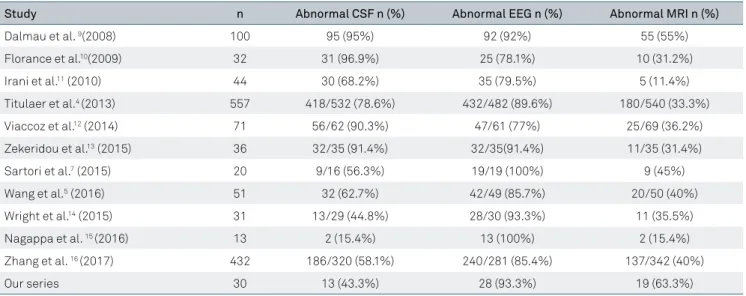
-sis2,8. In other reported case series, as reviewed in Table 2,
initial noninlammatory CSF varied widely, in 15-96% of the patients. In our case series, more than half the patients did not show inlammatory CSF at the irst evaluation, which
is a concern as they may have been misdiagnosed as hav
-ing a primary psychiatric disorder. his is supported by the
inding that in patients who presented with an initial non
-inlammatory CSF, it took twice as long to perform the EEG and MRI compared with the patients with abnormal CSF.
Also, in the former cases, it also took twice as long to initi
-ate immunotherapy (Table 1). Misdiagnosing anti-NMDAR
as a primary psychiatric disorder can have deleterious con
-sequences. For instance, Lejuste et al.3 reported that more
than 40% of their patients were initially hospitalized in psy
-chiatric departments with a primary psy-chiatric diagnosis,
even though most had presented with a neurologic symp
-tom at their irst emergency department evaluation. Besides delaying the correct diagnosis and speciic treatment, many
Table 1. Comparison of paraclinical results with days from onset of symptoms to preliminary diagnosis of anti-NMDAR encephalitis.
Variable n (%) Mean days from onset to
diagnosis (SD) Range (95% CI) p value*
CSF cytochemical
Inlammatory 13 (44.8) 22.9 ± 13 10–50
Noninlammatory 16 (55.2) 40.2 ± 15 13–85 0.003
EEG
Abnormal 27 (93.1) 30.5 ± 14 10–59
Normal 2 (6.9) 58.5 ± 37 32–85 0.18
MRI
Abnormal 19 (65.5) 30.1 ± 13 11–56
Normal 10 (34.5) 36.9 ± 22 10–85 0.60
CSF: Cerebrospinal luid; EEG: Electroencephalogram; MRI: Magnetic resonance imaging; *Mann-Whitney U test.
Table 2. Results of abnormal CSF, EEG and MRI indings in different case series.
Study n Abnormal CSF n (%) Abnormal EEG n (%) Abnormal MRI n (%)
Dalmau et al. 9(2008) 100 95 (95%) 92 (92%) 55 (55%)
Florance et al.10(2009) 32 31 (96.9%) 25 (78.1%) 10 (31.2%)
Irani et al.11 (2010) 44 30 (68.2%) 35 (79.5%) 5 (11.4%)
Titulaer et al.4 (2013) 557 418/532 (78.6%) 432/482 (89.6%) 180/540 (33.3%)
Viaccoz et al.12 (2014) 71 56/62 (90.3%) 47/61 (77%) 25/69 (36.2%)
Zekeridou et al.13 (2015) 36 32/35 (91.4%) 32/35(91.4%) 11/35 (31.4%)
Sartori et al.7 (2015) 20 9/16 (56.3%) 19/19 (100%) 9 (45%)
Wang et al.5 (2016) 51 32 (62.7%) 42/49 (85.7%) 20/50 (40%)
Wright et al.14 (2015) 31 13/29 (44.8%) 28/30 (93.3%) 11 (35.5%)
Nagappa et al. 15 (2016) 13 2 (15.4%) 13 (100%) 2 (15.4%)
Zhang et al. 16 (2017) 432 186/320 (58.1%) 240/281 (85.4%) 137/342 (40%)
Our series 30 13 (43.3%) 28 (93.3%) 19 (63.3%)
5 Espinola-Nadurille M et al. Normal CSF delays diagnosis in anti-NMDA encephalitis
patients receive antipsychotic medications for the treat
-ment of behavioral symptoms, which may lead to the devel
-opment of neuroleptic malignant syndrome with complica
-tions reported in 47%3.
hus, together with assessing for discreet neurological
symptoms relevant to encephalitis, we recommend extend
-ing paraclinical investigations in patients with an acute epi
-sode of psychiatric symptomatology or behavioral changes if (a) the presentation is of less than 10 days in a
previously-healthy individual, and/or (b) speciic symptoms such as dis
-orientation, attention disturbances, decreased awareness of the environment, catatonic symptoms, and history or signs of speech diiculties are present.
he EEG could be a diagnostic aid, given that in most case series, as in ours, it was abnormal in more than 90% of
the cases (Table 2). However, a normal EEG does not neces
-sarily exclude anti-NMDAR encephalitis, and neither does
psychiatric, cognitive or behavioral symptomatology starting
months or even years before assessment3.
In conclusion, we report the presence of noninlammatory
CSF in almost 50% of patients with anti-NMDAR encephali
-tis. We propose cautious neurological decision making based on CSF results when excluding the presence of encephalitis, as the absence of CSF pleocytosis and/or increased proteins
may delay not only diagnosis, but the ordering of complemen
-tary paraclinical investigations and the length of time to ini
-tiate immunotherapy in patients with delayed conirmation.
ACKNOWLEDGEMENTS
he authors thank Prof. Angela Vincent FRS F Med Sci Nuield Department of Clinical Neurosciences, Oxford UK for her helpful suggestions to the manuscript.
References
1. Venkatesan A, Tunkel AR, Bloch KC, Lauring AS, Sejvar J, Bitnun A et al. Case deinition, diagnosis algorithms, and priorities in encephalitis: consensus statement of the international encephalitis consortium. Clin Infect Dis. 2013;57(8):1114-28. https://doi.org/10.1093/cid/cit458
2. Leypoldt F, Armangue T, Dalmau J. Autoimmune encephalopathies. Ann N Y Acad Sci. 2015;1338(1):94-114. https://doi.org/10.1111/nyas.12553 3. Lejuste F, Thomas L, Picard G, Desestret V, Ducray F,
Rogemond V et al. Neuroleptic intolerance in patients with anti-NMDAR encephalitis. Neurol Neuroimmunol Neuroinlamm. 2016;3(5):e280. https://doi.org/10.1212/NXI.0000000000000280 4. Titulaer MJ, McCracken L, Gabilondo I, Armangue T, Glaser C,
Iisuka T et al. Treatment and prognostic factors for long-term outcome in patients with anti-NMDA receptor encephalitis: an observational cohort study. Lancet Neurol. 2013;12(2):157-65. https://doi.org/10.1016/S1474-4422(12)70310-1
5. Wang W, Li JM, Hu FY, Wang R, Hong Z, He L et al. Anti-NMDA receptor encephalitis: clinical characteristics, predictors of outcome and the knowledge gap in southwest China. Eur J Neurol. 2016;23(3):621-9. https://doi.org/10.1111/ene.12911
6. Byrne S, Walsh C, Hacohen Y, Muscal E, Jankovic J, Stocco A et al. Earlier treatment of NMDAR antibody encephalitis in children results in a better outcome. Neurol Neuroimmunol Neuroinlamm. 2015;2(4):e130. https://doi.org/10.1212/NXI.0000000000000130 7. Sartori S, Nosadini M, Cesaroni E, Falsaperla R, Capovilla G, Beccaria
F et al. Paediatric anti-N-methyl-D-aspartate receptor encephalitis: the irst Italian multicenter case series. Eur J Paediatr Neurol. 2015;19(4):453-63. https://doi.org/10.1016/j.ejpn.2015.02.006 8. Graus F, Titulaer MJ, Balu R, Benseler S, Bien CG,
Cellucci T et al. A clinical approach to diagnosis of
autoimmune encephalitis. Lancet Neurol. 2016;15(4):391-404. https://doi.org/10.1016/S1474-4422(15)00401-9
9. Dalmau J, Gleichman AJ, Hughes EG, Rossi JE, Peng X, Lai M et al. Anti-NMDA-receptor encephalitis: case series and analysis
of the effects of antibodies. Lancet Neurol. 2008;7(12):1091-8. https://doi.org/10.1016/S1474-4422(08)70224-2
10. Florance NR, Davis RL, Lam C, Szperka C, Zhou L, Ahmad S et al. Anti-N-methyl-D-aspartate receptor (NMDAR) encephalitis in children and adolescents. Ann Neurol. 2009;66(1):11-8. https://doi.org/10.1002/ana.21756
11. Irani SR, Bera K, Waters P, Zuliani L, Maxwell S, Zandi MS et al. N-methyl-D-aspartate antibody encephalitis: temporal progression of clinical and paraclinical observations in a predominantly non-paraneoplastic disorder of both sexes. Brain.
2010;133(Pt 6):1655-67. https://doi.org/10.1093/brain/awq113 12. Viaccoz A, Desestret V, Ducray F, Picard G, Cavillon G, Rogemond
V et al. Clinical speciicities of adult male patients with NMDA receptor antibodies encephalitis. Neurology. 2014;82(7):556-63. https://doi.org/10.1212/WNL.0000000000000126
13. Zekeridou A, Karantoni E, Viaccoz A, Ducray F, Gitiaux C, Villega F et al. Treatment and outcome of children and adolescents with N-methyl-D-aspartate receptor encephalitis. J Neurol. 2015;262(8):1859-66. https://doi.org/10.1007/s00415-015-7781-9 14. Wright S, Hacohen Y, Jacobson L, Agrawal S, Gupta R, Philip
S et al. N-methyl-D-aspartate receptor antibody-mediated neurological disease: results of a UK-based surveillance study in children Arch Dis Child. 2015;100(6):521-6. https://doi.org/10.1136/archdischild-2014-306795
15. Nagappa M, Bindu PS, Mahadevan A, Sinha S, Mathuranath PS, Taly AB. Clinical features, therapeutic response, and follow-up in pediatric anti-n-methyl-D-aspartate receptor encephalitis: experience from a tertiary care university hospital in India. Neuropediatrics. 2016;47(1):24-32. https://doi.org/10.1055/s-0035-1569464 16. Zhang L, Wu MQ, Hao ZL, Chiang SM, Shuang K, Lin MT et al.