Homocysteine and Methylenetetrahydrofolate Reductase in Subjects
Undergoing Coronary Angiography
Luciana Moreira Lima, Maria das Graças Carvalho, Ana Paulo Fernandes, Adriano de Paula Sabino, Andréia Assis
Loures-Vale, Cirilo Pereira da Fonseca Neto, José Carlos Faria Garcia, Jamil Abdala Saad, Marinez Oliveira Sousa
Universidade Federal de Minas Gerais, Hospital Socor, Belo Horizonte, Minas Gerais - Belo Horizonte, MG - Brazil
Mailing Address: Marinez Oliveira Sousa •
Av. Antônio Carlos, 6627 – UFMG – Faculdade de Farmácia - 31270-901 Belo Horizonte, MG - Brazil
E-mail: [email protected]
Manuscript received January 23, 2006; revised manuscript received March 8, 2006; accepted May 11, 2006.
Summary
Objective: To determine plasma homocysteine levels and the incidence of methylenetetrahydrofolate reductase (MTHFR) gene C677T polymorphism in a group of subjects who underwent coronary angiography, in an attempt to establish a correlation between these parameters and the severity of coronary artery disease (CAD), as well as investigate the correlation between hyperhomocysteinemia and the presence of polymorphism.
Methods: Twenty subjects with no coronary atheromatosis (controls), fourteen subjects with mild/moderate atheromatosis, and twenty-nine subjects with severe atheromatosis were evaluated.
Results: Significant differences were observed in mean homocysteine levels between the control and the severe atheromatosis groups (p < 0.001). No significant differences were observed among the other groups. The severe atheromatosis group showed rates of 62.0% and 6.9% for the C677T MTHFR gene polymorphism, in heterozygous and homozygous subjects, respectively. However, there was no correlation between the presence of mutation and hyperhomocysteinemia. A positive correlation of 41.91% (p < 0.001) was found between hyperhomocysteinemia and CAD.
Conclusion: The most important finding of this study was the association between hyperhomocysteinemia and coronary stenosis > 70%; yet, whether elevated plasma homocysteine worsens atherosclerosis or is a consequence remains to be established.
Key words:Homocysteine, methylenetetrahydrofolate reductase (MTHFR), coronary angiography.
Introduction
Homocysteine (Hcy), an amino acid derived from methionine metabolism, is considered an independent risk factor for atherosclerosis. Studies demonstrate that increased homocysteine levels have toxic effects on vascular endothelium1. Homocysteine elevation has
multifactorial causes, including high methionine intake, decreased metabolism, genetic changes, and enzyme (cystathionine E-synthase or methylenetetrahydrofolate reductase - MTHFR) or vitamin deficiency (B12, B6 and folic acid), all of them required for its metabolism. These factors may lead to homocysteine accumulation and, as a result, vascular endothelial damage2, thereby triggering
atherosclerotic lesions. MTHFR is an enzyme involved in the transmethylation pathway by which homocysteine is converted to methionine. The C677T mutation in the MTHFR gene produces a thermolabile variant that may interfere in enzyme activity of homocysteine metabolic pathways, predisposing to hyperhomocysteinemia (HHcy)1,3.
Subjects homozygous for MTHFR C677T mutation may show homocysteine plasma levels twice as high as those of heterozygous subjects4.
Some studies5-7 have demonstrated an association between
hyperhomocysteinemia and peripheral vascular disease, cerebrovascular disease and CAD, showing that this amino acid concentration was significantly higher than that found in healthy subjects. Hyperhomocysteinemia, therefore, is considered a risk factor for the aforementioned clinical conditions. Investigations on different factors, such as smoking8,
lipid profile9, advanced age10, arterial hypertension10, diabetes
mellitus11, and obesity12, in addition to homocysteine, have
shown that their plasma concentration may be regarded as independent risk factors for atherosclerotic disease. It should be noted that homocysteine values seem to be paramount also as predictors of mortality in CAD patients, besides showing a relationship with the degree of atherosclerosis13-15.
Methods
Sixty-three subjects of both genders, ages ranging from 46 to 68, were evaluated. These subjects were selected during three months at the Hemodynamics Department of the Hospital Socor in Belo Horizonte, Minas Gerais, after undergoing coronary angiography. Patient selection emphasized homogeneity in terms of gender, age, socioeconomic status, and body mass index (BMI). Subjects were divided into three groups based on coronary angiography results: control (n = 20), mild/moderate atheromatosis (n = 14), and severe atheromatosis (n = 29). The study protocol was approved by the Research Ethics Committees of Hospital Socor and the Federal University of Minas Gerais.
All selected subjects were informed about the study’s objectives, and those who agreed to participate signed an informed consent. Cardiologists at the Hemodynamics Department filled out a case report form on each subject containing identification, demographic data, family history, and coronary angiography results.
Patients with intercurrent diseases, such as coagulation disorders, renal, liver and autoimmune diseases, and cancer, were excluded from the study.
The presence of smoking, sedentariness, and family history of CAD was established according to the recommendations of the III Brazilian Guidelines for Dyslipidemias and Atherosclerosis Prevention16. Subjects previously diagnosed
ZLWK GLDEHWHV PHOOLWXV DQG IDVWLQJ JOXFRVH PJG/ considered diabetic17. Subjects with systolic blood pressure
PP+J RU GLDVWROLF EORRG SUHVVXUH PP+J or were taking antihypertensive drugs were classified as hypertensive18.
Venous blood samples were collected after a twelve-hour fast. These patients were instructed to refrain from vigorous physical activity and avoid drinking alcohol 24 and 72 hours, respectively, preceding blood collection, in an attempt to obtain biological samples of patients in metabolic steady state. Ten mL venous blood samples (5 mL without anticoagulant and 5 mL in EDTA) were collected into Vacutainer® tubes (Becton Dickinson). Blood samples without anticoagulant were immediately centrifuged at 2500 rpm for 10 minutes; the separated serum was aliquoted and stored at – 70 °C until homocysteine measurement. The DNA was extracted from the blood samples containing EDTA, which remained stored at – 20 °C until genetic analysis was performed.
Serum homocysteine was measured using the Axsym® Homocysteine assay (Abbott® Laboratories – Germany) based on polarized immunofluorescence technology, according to the manufacturer’s instructions. The assay was performed in the Axsym® (Abbott® Laboratories – Germany) analyzer using three commercial control sera in order to check its performance. Presence of C677T mutation in the MTHFR enzyme was investigated by PCR-RFLP using oligonucleotides and restriction endonucleases described previously3. PCR
reactions were performed in a PT100 PCR thermal cycler (MJ Research, Waltham, USA) using 1 pMol from each primer (Invitrogen®, São Paulo, S.P), 0.2 mM dNTPs (GIBCO BRL®, São Paulo, SP), and 1 unit of Taq polymerase (Phoneutria®- Belo Horizonte, Minas Gerais, Brazil). PCR reactions were
run for 40 cycles, each consisting of one minute at 94 °C for denaturation, one minute at 64 °C (C677T) for primer annealing, and two minutes at 72 °C for primer extension. The PCR products were digested with restriction endonuclease (Hinf I - Promega, Inc.) during four hours at 37 ºC. DNA samples from previously typed subjects were included for enzyme activity control. These samples were then analyzed by polyacrylamide gel electrophoresis followed by silver staining.
Coronary angiography was performed percutaneously via femoral approach. The angiographic films were interpreted by three experienced cardiologists, and reports were presented according to criteria defined by reduced luminal diameter: up to 30% stenosis was classified as mild atheromatosis; 30% to 69% stenosis was classified as moderate atheromatosis, and greater than 70% stenosis was classified as severe atheromatosis.
For homocysteine values, statistical analysis was performed using analysis of variance (ANOVA) followed by Tukey’s test, after logarithmic data transformation. Categorical variables (risk factors and C677T mutation) were analyzed using Fisher’s exact test. Spearman’s test was used for correlation between mutation status (categorical variable) and plasma homocysteine (continuous variable) and between CAD (categorical variable) and homocysteinemia (continuous variable). Minimum sample size was defined by the variation coefficient previously described in the literature19, considering
ten percent variation for the mean and reaching a minimum of eleven subjects in each group, so that potential statistical differences with 5% significance level would be demonstrated. Sigma Stat 1.0 and Prism 3.0 software were used to carry out the analysis and plot the graph, respectively.
Results
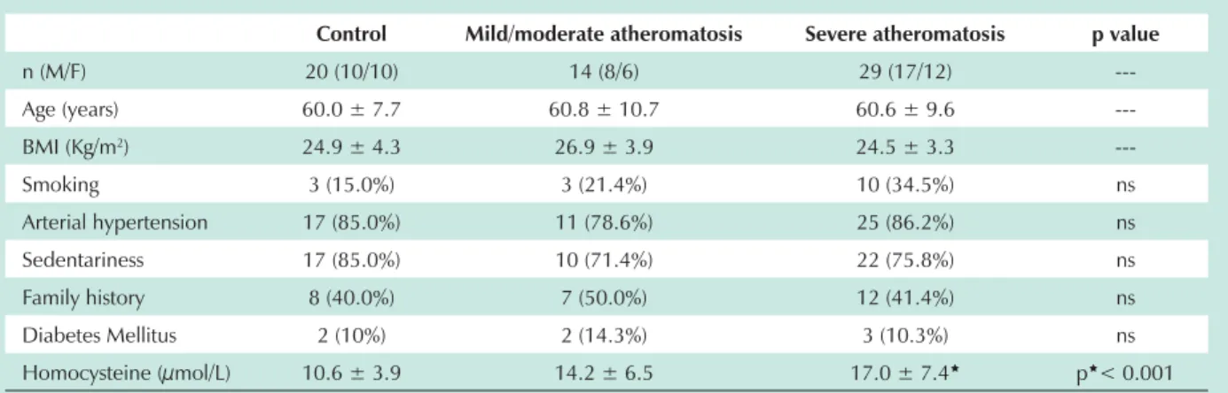
Table 1 characterizes the study groups with regard to gender, age, and BMI, as well as risk factors associated with CAD. It also shows the number of subjects and rate of a particular variable. No statistically significant differences were found in smoking, arterial hypertension, physical inactivity, family history, and diabetes mellitus among the three groups. No participant had overweight or obesity, suggesting that these subjects lacked the metabolic component.
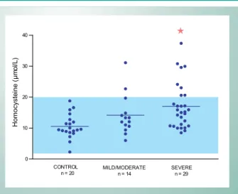
Homocysteine measurements are also presented in table 1, as means and respective standard deviations. A statistically significant difference was found between the control group and severe atheromatosis group (p < 0.001). Among the other groups, there were no significant differences in plasma homocysteine levels. Figure 1 shows the distribution of homocysteine values in the groups studied.
The incidence of MTHFR C677T mutation in the control, mild/moderate, and severe atheromatosis groups is shown in table 2, taking into account the presence or absence of mutation in homozygosis or heterozygosis. There was no statistically significant difference between groups C677T frequency in heterozygous or homozygous mutation.
Discussion
This cross-sectional cohort study assessed an intermediate-to-high risk population, since all selected subjects were referred for catheterization due to chest pain and presented demographic profile and risk factors for CAD, as shown in table 1. The three groups studied were homogeneous with respect to age, gender, and BMI. There were no significant differences in age, smoking, arterial hypertension, sedentariness, family history, and diabetes mellitus among the three groups (Table 1). This may be explained by the structure of the study model itself, in which, although patients in the control group did not have coronary atheromatosis, they had several risk factors for CAD (Table 1).
A number of landmark studies were published that correlate plasma homocysteine levels and CAD. Some have demonstrated an association between hyperhomocysteinemia and CAD incidence, while others reported lack of association between the two parameters20. These controversial results
involving homocysteine concentration and CAD may be explained by the fact that some factors that affect this amino acid’s plasma levels differ in each population. Homocysteine circulating levels may be affected by dietary habits, genetic factors, lifestyle, and race6. In the Brazilian population,
characterized by ethnic heterogeneity21, the results of
studies involving plasma homocysteine levels and CAD are equally controversial. It is well to point out that most authors consider hyperhomocysteine as an independent risk factor for CAD22. Table 1 shows that plasma homocysteine levels
were significantly higher in patients with severe atheromatosis, compared with the control group (p < 0.001). Yet, no statistically significant differences were found between the control group and the mild/moderate atheromatosis group, as well as between the mild/moderate atheromatosis group and severe atheromatosis group.
Folson et al14 suggested that elevated homocysteine levels
may be a consequence, not only a cause, of CAD. These authors also suggested that hyperhomocysteinemia may predict a poor prognosis, reflecting CAD severity. However, the hypothesis of increased homocysteine plasma levels as a result of atherosclerotic lesion was refuted by Bostom & Selhub23, because it is not supported by the epidemiological
evidence of several studies. This study’s findings indicate that if hyperhomocysteinemia did not contribute to worsen CAD in the assessed subjects, at least it was implicated in the atherosclerotic process. Questions remain, however, as to whether increased homocysteine is an aggravating factor for the disease or manifests itself with the development of the atherosclerotic process. Prospective studies with follow-up involving a large number of subjects with known hyperhomocysteinemia but no coronary disease are essential to authoritatively elucidate the relationship between hyperhomocysteinemia and CAD.
In the present study, the method used for homocysteine measurement advocates a reference value for this parameter ranging from 2 to 20 µmol/L. Also controversial among authors are the reference values for plasma homocysteine in the general population.
Some authors regard plasma homocysteine between 5
and 15 µmol/L24-27 as “normal”, while others believe that
the mean value in the general population is around 9 to 10 µmol/L28,29. Boushey et al4, in a meta-analysis involving 27
studies and more than four thousand patients, concluded that when homocysteine values are greater than 10 µmol/L, each 5µmol/L increment in circulating homocysteine concentration is associated with an 80% CAD risk in women and 60% in men. Currently, values equal to or greater than 10µmol/L are considered hyperhomocysteinemia and linked to increased risk for CAD30. In this study, 72% of the patients with severe
atheromatosis had plasma homocysteine within the reference range of the method used. Similar observation was made by Gravina-Taddei et al31, who analyzed elderly people with
and without coronary disease using a method similar to that of this study.
Among the 63 subjects that comprised the control, mild/ moderate atheromatosis and severe atheromatosis groups, the presence of homozygous C677T MTHFR mutation was found in only four subjects (Table 2), resulting in a 6.3% incidence. The frequency of heterozygous MTHFR C677T mutation was 44.4%, totaling 28 subjects (Table 2), all study subjects considered (n = 63). According to Arruda et al32, this mutation
prevalence in Brazilian Caucasians is approximately 10.3% and 54.2% in homozygosis and heterozygosis, respectively. Yet, this study (n = 63) was composed of 28.1% Caucasians, 22.8% blacks, and 49.1% mestizos (mulatto, dark skinned or others), with no statistically significant difference among the three groups for the different races.
The C677T mutation in the MTHFR gene was more frequent in subjects with severe atheromatosis (62.0%); yet, there was no significant difference between the severe atheromatosis group and the others. A possible explanation for this finding is that several factors are related to plasma homocysteine elevation, and since the number of participants evaluated in this study was insufficient to draw any conclusion in this respect, a multicenter, epidemiological study is needed.
Fig. 1 -Characterization of the study groups and homocysteine plasma levels
expressed as mean and standard deviation. Characterization by n (size), M (male gender) and F (female gender); age, BMI (body mass index), and plasma homocysteine, expressed as mean and standard deviation, and risk factor percentage in the studied group. A significant difference was found between the control group and the severe atheromatosis group for the homocysteine parameter, as indicated by the symbol (Ì). ns = nonsignificant.
0 10 20 30
40
CONTROL
n = 20 SEVEREn = 29 MILD/MODERATE
n = 14
H
o
mo
cyst
e
in
e
(
µ
m
o
l/L
Pisciotta et al33 obtained similar results when evaluating
subjects with hypercholesterolemia and CAD, with greater prevalence of mutation in patients with CAD, although with no significant difference relative to those without CAD.
The positive correlation observed between increased plasma homocysteine and CAD (r = 0.4191, p < 0.001) in the present study is in keeping with literature34. However, there
was no correlation between the presence of C677T mutation and high levels of homocysteine in the population studied. Although the severe atheromatosis group was found to have higher mutation incidence and significantly higher plasma homocysteine levels, no significant difference was observed when compared with the other groups. Other authors reported similar results35,36. Therefore, homocysteine elevation in these
cases should be explained by other mechanisms that were not examined in this study.
Among the CAD subjects, mild/moderate atheromatosis and severe atheromatosis groups considered, (n = 43); 22 had heterozygous mutation (51.1%) and three had homozygous mutation (6.9%), with no significant differences when compared with the control group. On the other hand, Almawi et al37 found higher frequency (71.9%) of heterogygous
mutation in patients with CAD and 45.5% in the control group, with a significant difference between both groups (p < 0.001). These authors also demonstrated a significant higher incidence of homozygous mutation (31.3%) in patients with severe atheromatosis, compared to the present study
(6.9%). The small sample size herein presented may have contributed to our data being discordant with that reported in the literature.
In this study, no correlation was found between mutation and coronary atheromatosis, all subjects from the mild/ moderate atheromatosis and severe atheromatosis groups considered (n = 43). Folsom et al14, in a prospective study
involving more than 15.000 participants, also demonstrated the lack of association between CAD and MTHFR C677T mutation. Likewise, Kölling et al38, in a case-control study
involving 2121 CAD patients and 617 non-CAD patients, demonstrated a lack of association between mutation and angiographically documented CAD.
Both studies mentioned above have demonstrated significantly elevated plasma homocysteine levels in CAD subjects, as compared with the control group, and these data are consistent with those of the present study.
Study limitations - Although the number of subjects evaluated was adequate for demonstrating a significant difference in homocysteine levels between the control group and severe atheromatosis group, studies published in the literature involved much large samples, especially to evaluate the association between C677T mutation and CAD. Another limiting factor was that the present study failed to take into account other key factors in homocysteine metabolism, such as deficiencies of other enzymes involved in this metabolism, dietary habits, and vitamin B12 and folic acid plasma levels
Control Mild/moderate atheromatosis Severe atheromatosis p value
n (M/F) 20 (10/10) 14 (8/6) 29 (17/12)
---Age (years) 60.0 ± 7.7 60.8 ± 10.7 60.6 ± 9.6
---BMI (Kg/m2) 24.9 ± 4.3 26.9 ± 3.9 24.5 ± 3.3
---Smoking 3 (15.0%) 3 (21.4%) 10 (34.5%) ns
Arterial hypertension 17 (85.0%) 11 (78.6%) 25 (86.2%) ns
Sedentariness 17 (85.0%) 10 (71.4%) 22 (75.8%) ns
Family history 8 (40.0%) 7 (50.0%) 12 (41.4%) ns
Diabetes Mellitus 2 (10%) 2 (14.3%) 3 (10.3%) ns
Homocysteine (µmol/L) 10.6 ± 3.9 14.2 ± 6.5 17.0 ± 7.4Ì pÌ< 0.001
Table 1 - Characterization of the study groups and homocysteine plasma levels expressed as mean and standard deviation. Characterization by n (size), M (male gender) and F (female gender); age, BMI (body mass index), and plasma homocysteine, expressed as mean and standard
deviation, and risk factor percentage in the studied group. A significant difference was found between the control group and the severe atheromatosis group for the homocysteine parameter, as indicated by the symbol (Ì). ns = nonsignificant
Control Mild/moderate atheromatosis Severe atheromatosis p value
n 20 14 29
---Heterozygous 6 (30%) 4 (28.6%) 18 (62.0%) ns
Homozygous 1 (5%) 1 (7.1 %) 2 (6.9%) ns
Nondetected 13 (65%) 9 (64.3%) 9 (31.1%) ns
Referências
1. Falk E, Zhou J, Moller J. Homocysteine and atherothrombosis. Lipids. 2001; 36 (Suppl. 1): S3-S11.
2. G u i l l a n d J C , Fa v i e r A , D e C o u r c y P G , G a l a n P, H e r c b e r g S . Hyperhomocysteinemia: an independent risk factor or a simple marker of vascular disease? Pathol Biol (Paris). 2003; 51:101-10.
3. Frosst P, Blom HJ, Milos R, Goyette P, Sheppard C, Matthews RG, et al. A candidate genetic risk factor for vascular disease: a common mutation in methylenetetrahydrofolate reductase. Nat Genet. 1995; 10: 111-3.
4. Boushey CI, Beresford SA, Omenn GS, Matulsky AG. A quantitative assessment of plasma homocysteine as a risk factor for cardiovascular disease. Probable benefits of increasing folic acid intakes. JAMA. 1995; 274: 1049-57.
5. Murphy-Chutorian D, Alderman EL. The case that hyperhomocysteinemia is a risk factor for coronary artery disease. Am J Cardiol. 1994; 73: 705-7.
6. Nehler MR, Taylor LM, Porter JM. Homocysteinemia as a risk factor for atherosclerosis: a review. Cardiovasc Pathol. 1997; 6: 1-9.
7. Nygärd O, Nordrehaug JA, Refsum HS. Plasma homocysteine levels and mortality in patients with coronary artery disease. N Eng J Med. 1997; 337: 230-6.
8. Graham IM, Daly LE, Refsum HM, Robinson K, Brattstrom LE, Ueland PM, et al. Plasma homocysteine as a risk factor for vascular disease. The European Concerted Action Project. JAMA. 1997; 277: 1775-81.
9. Puri A, Gupta OK, Dwivedi RN, Bharadway RP, Narain VS, Singh S. Homocysteine and lipid levels in young patients with coronary artery disease. J Assoc Physicians India. 2003; 51: 681-5.
10. Gupta M, Sharma P, Garg G, Kaur K, Bedi GK, Vig A. Plasma homocysteine: an independent or an interactive risk factor for coronary artery disease. Clin Chim Acta. 2005; 352: 121-5.
11. Soinio M, Marniemi J, Laakso M, Lehto S, Ronnemaa I. Elevated plasma homocysteine level is an independent predictor of coronary heart disease events in patients with type 2 diabetes mellitus. Ann Intern Med. 2004; 140: 94-100.
12. Kazemi MB, Eshraghian K, Omrani GR, Lankarani KB, Hosseini E. Homocysteine level and coronary artery disease. Angiology. 2006; 57: 9-14.
13. L a w r e n c e d e K o n i n g A B, We r s t u c k G H , Z h o u J, A u s t i n R C . Hyperhomocysteinemia and its role in the development of atherosclerosis. Clin Biochem. 2003; 36: 431-41.
14. Folson AR, Nieto FJ, McGovem PG, Tsai MY, Malinow MR, Eckfeldt JH, et al. Prospective study of coronary heart disease incidence in relation to fasting total homocysteine, related genetic polymorphism, and B vitamins. The Atherosclerotic Risk in Communities (ARIC) Study. Circulation. 1998; 98: 204-10.
15. Nikfardjam M, Graf S, Hornykewycz S. Homocysteine plasma levels in young patients with coronary artery disease. Relation to history of acute myocardial
infarctation and anatomical extent of disease. Thromb Res. 2001; 103 (Suppl. 1): 35-9.
16. Sociedade Brasileira de Cardiologia. III Diretrizes Brasileiras Sobre Dislipidemias e Diretrizes de Prevenção da Aterosclerose. Arq Bras Cardiol. 2001; 77 (Suppl. 3): 1-48.
17. Expert Committee on the Diagnosis and Classification of Diabetes Mellitus. Report of the Expert Committee on the diagnosis and classification of diabetes mellitus. Diabetes Care. 2003; 26 (Suppl. 1): S5-20.
18. Chobanian AV, Bakris GL, Black HR, Cushman WC, Green LA, Izzo JL Jr., et al. National High Blood Pressure Education Program Coordinating Committee. The Seventh Report of the Joint National Committee on prevention, detection, evaluation, and treatment of high blood pressure: The JNC 7 report. JAMA. 2003; 289: 2560-72.
19. National Committee for Clinical Laboratory Standards. Evaluation of precision performance of clinical chemistry devices. 2nd ed. Tentative Guideline. NCCLS Document EP5-T2, 1992.
20. Voutilainen S, Lakka TA, Hamelahti P, Lehtimaki T, Pousen HE, Salonen JT. Plasma total homocysteine concentration and risk of acute coronary events: the Kuopio Ischemic Heart Disease Risk Factor Study. J Intern Med. 2000; 248: 217-22.
21. Parra FC, Amado RC, Lambertucci JR, Rocha J, Antunes CM, Pena SD. Color and genomic ancestry in Brazilians. Proc Natl Acad Sci USA. 2003; 100: 177-82.
22. Chua S, Wu CJ, Chang HW, Hang CL, Chen CJ, Yang CH, et al. Impact of elevated plasma total homocysteine concentration on coronary atherosclerosis in Chinese patients with acute myocardial infarctation undergoing primary coronary intervention. Int Heart J. 2005; 46: 181-93.
23. Bostom A, Selhub J. Homocysteine and arteriosclerosis: subclinical and clinical disease associations. Circulation. 1999; 99: 2361-3.
24. Malinow MR, Bostom AG, Krauss RM. Homocysteine, diet, and cardiovascular diseases: a statement for healthcare professionals from the Nutrition Committee, American Heart Association. Circulation. 1999; 99: 178-82.
25. Christen W, Ajani U, Glynn R. Blood levels of homocysteine and increased risks of cardiovascular disease. Causal or casual? Arch Intern Med. 2000; 169: 422-34.
26. Girelli D, Martinelli N, Pizzolo F, Friso S, Olivieri S, Stranieri C, et al. The interaction between MTHFR 677 CoT genotype and folate status is a determinant of coronary atherosclerosis risk. J Nutr. 2003; 133: 1281-5.
27. Stangl V, Baumann G, Stangl K. Coronary atherogenic risk factor in women. Eur Heart J. 2002; 23: 1738-52.
28. Duell PB, Malinow MR. Homocysteinemia and risk of atherosclerosis: a clinical approach to evaluation and management. Endocrinologist. 1998; 8: 170-7.
29. Malinow M. Hyperhomocysteinemia. A common and easily reversible risk factor for occlusive atherosclerosis. Circulation. 1990; 81: 2004-6.
of the participants. These data would have enriched this study’s discussion.
Conclusion
The most important finding of this study was the association between hyperhomocysteinemia and coronary stenosis > 70%; yet, whether elevated plasma homocysteine worsens atherosclerosis or is a consequence remains to be established.
Acknowledgement
To Professor Ângela Maria Quintão Lana, PhD, for the
statistical analysis. To Fundação de Amparo à Pesquisa de Minas Gerais (FAPEMIG), RECOPE (FAPEMIG/FIEMG/ IEL-number 32082), Coordenação de Aperfeiçoamento de Pessoal de Nível Superior (CAPES), and the Conselho Nacional de Desenvolvimento Científico e Tecnológico (CNPq) for their support.
Supported by: Fundação de Amparo à pesquisa de Minas Gerais - FAPEMIG, CAPES e CNPq.
Potential Conflict of Interest
30. Yang F, Tan HM, Wang H. Hyperhomocysteinemia and atherosclerosis. Acta Physiol Sinica. 2005; 57: 103-14.
31. Gravina-Taddei CF, Batlouni M, Sarteschi C, Baltar VT, Salvarini NA, Bertolami MC, et al. Hiper-homocisteinemia como fator de risco para doença aterosclerótica coronariana em idosos. Arq Bras Cardiol. 2005; 85: 166-73.
32. Arruda VR, Siqueira LH, Gonçalves MS, von Zuben PM, Soares MC, Menezes R, et al. Prevalence of the mutation C677T in the methylenetetrahydrofolate reductase gene among distinct ethnic groups in Brazil. Am J Med Gen 1998; 78: 332-5.
33. Pisciotta L, Cortese C, Gnasso A, Liberatoscioli L, Pastore A, Mannucci L, et al. S. Serum homocysteine, methylenetetrahydrofolate reductase gene polymorphism and cardiovascular disease in heterozygous familial hypercholesterolemia. Atherosclerosis. 2005; 179: 333-8.
34. Dedoussis GY, Panagiotakos DB, Chrysohoou C, Pitsavos C, Zampelas A, Choumerianou D, et al. Effect of interaction between adherence to a Mediterranean diet and the methylenetetrahydrofolate reductase 677Co T mutation on homocysteine concentrations in health adults: the ATTICA
Study. Am J Clin Nutr. 2004; 80: 849-54.
35. Yilmaz H, Isbir S, Agachan B, Ergren A, Farsak B, Isbir T. C677T mutation of methylenetetrahydrofolate reductase gene and serum homocysteine levels in Turkish patients with coronary artery disease. Cell Biochem Funct. 2006; 24 (1): 87-90.
36. Girelli D, Friso S, Trabetti E, Olivieri O, Russo C, Pessotto R, et al. Methylenetetrahydrofolate reductase C677T mutation, plasma homocysteine, and folate in subjects from Northern Italy with or without angiographically documented severe coronary atherosclerotic disease: evidence for an important genetic-environmental interaction. Blood. 1998; 11: 4158-63.
37. Almawi WY, Ameen G, Tamim H, Finan RR, Irani-Hakime N. Factor V G1691A, prothrombin G20210A, and methylenetetrahydrofolate reductase [MTHFR] C677T gene polymorphism in angiographycally documented coronary artery disease. J Thromb Thrombolysis. 2004; 17: 199-205.