Mailing Address: Neuza Helena Moreira Lopes • Av. Dr. Enéas Carvalho de Aguiar, 44 - 05403-000 – São Paulo, SP - Brazil
E-mail: [email protected] Received on 07/11/05 • Accepted on 10/07/05
QT Interval Dispersion Analysis in Acute Myocardial
Infarction Patients: Coronary Reperfusion Effect
Neuza Helena Moreira Lopes, César Grupi, Cleberson H. Dina, Aécio F. T. de Gois, Ludhimila A. Hajjar, Beatriz Ayub, Carlos Eduardo Rochitte, José Antonio Franchini Ramires, Whady A. Hueb, Roberto Kalil
Instituto do Coração do Hospital das Clínicas – HCFMUSP - São Paulo, SP - Brazil
O
BJECTIVETo study the effect of early reperfusion of infarct-related artery on QT(∆QT) dispersion interval, as well as how valuable it is as a marker for coronary reperfusion and ventricular arrhythmias.
M
ETHODSOne hundred and six patients with reperfusion (WR) and 48 without reperfusion (WtR) who have received thrombolytic therapy in the acute phase of infarction were studied. ECG carried out on admission as well as on day 4 of patient’s course were analyzed. ∆QT – defi ned as the difference between maximum and minimum QT interval – was measured by 12-lead ECG.
RESULTS
The reperfusion group showed signifi cant ∆QT reduction – from 89.66±20,47ms down to 70.95±21.65ms (p<0.001). On the other hand, the group without reperfusion showed ∆QT signifi cant increase - from 81.27±20.52ms up to 91.85±24.66ms (p<0.001). Logistic regression analysis showed that reduction magnitude beween pre- and post-thrombolysis ∆QT was the independent factor to most effectively identify coronary reperfusion (OR 1.045, p<0.0001; CI 95%). No significant difference was found in dispersion measures when patients with ventricular arrhythmias were compared with those with no arryhthmias in the course of the fi rst 48 hours.
C
ONCLUSIONThe study shows that ∆QT is signifi cantly reduced in patients with acute myocardial infarction submitted to successful thrombolysis, and is increased in infarcted patients with closed artery. ∆QT reduction between the pre- and post-thrombolysis condition was a predictor for coronary reperfusion of those patients, and did not show correlation to ventricular arrhythmias.
KEY WORDS
The concept of QT interval dispersion (∆QT) defi ned as the difference between the longest and the shortest QT interval as measued by 12-lead electrocardiogram (EKG) was introduced by Day and Campbell in the early 1990’s.1 At a fi rst moment it was proposed as an electric instability index to represent the expression of regional physiological variation of myocardial excitability recovery2,3.
From then on, QT dispersion analysis has been accepted as the non-invasive method for the detection of ventricular repolarization heterogeneity, acting as a marker for arrythmogenesis especially in the presence of a ischemic substratum4.Additionally, some studies report its as prognostic for heart failure and hypertrophic cardiomyopathy5,6.
However, few reports are available in the literature on the effect of early reperfusion on ventricular polarization of acute myocardial infarction (AMI) patients. Animal studies have indicated that regional ischemia and reperfusion alter the duration of action potential and conduction velocity, thus leading to lower homogeneity in ventricular recovery7,8.Some studies have demonstrated that QT dispersion is higher in the early stage of acute myocardial infarction (AMI), reduced along time in successful trhombolysis cases, and with a possibility of being kept high in patients who have developed ventricular fi brillation 9-11.
With the purpose of investigating the effect of early reperfusion in the infarct-associated artery on QT dispersion interval, as well as how valuable it is for coronary reperfusion and ventricular arrhythmias, patients who have received thrombolytic therapy in the acute phase of infarction were studied.
METHODS
One hundred and fi fty-four (154) patients admitted to the Heart Institute at the University of São Paulo Clinics Hospital were studied retrospectively. All patients had been diagnosed with acute myocardial infarction (AMI), and had been submitted to thrombolysis in the period between 1989 and 1992. Patients were selected from a group of 220 consecutive patients who had received tissue plamsinogene activator thrombolysis (rt-PA) for drug effi cacy evaluation.
Criteria for acute myocardial infarction diagnosis were: elevation of segment ST equal to or higher than 0.2 mV in at least two leads in frontal plane, or equal to or higher than 0.3mV in two adjacent leads in horizontal plane, with or without the presence of Q waves longer than 40 m, oclusion of infarct-related artery (IRA) kept after 200 mcg nitroglycerin intracoronary infusion, and ckmb two times normal levels.
Intravenous administration of rt-PA was started at 100 mg in the fi rst fi ve hours after onset of symptoms. After 90 minutes of thrombolytic administration, new coronariography and new ventriculography were carried
out to investigate the level of IRA reperfusion. In the time frame between 24 and 48 hours coronariography was repeated for IRA patency to be assessed.
Exclusion criteria in the present study included: previous or acute atrial fi brillation (8 patients), His bundle branch blocks, or intraventricular conduction disorders (8 patients), EKG traces that did not allow satisfactory assessment (22 patients), and the presence of new occlusion in control catheterism in 48 hours (11 patients), or the absence of perfusion in the fi rst catheterism with patency in the second exam (10 patients).
Following reperfusion angiographic criteria and arterial patency maintenance, patients were divided into two groups:
• With Reperfusion (WR): 106 patients with re-perfusion immediately after thrombolysis and with angiographic patency maintenance of IRA 48 hours after the event. Mean time of reperfusion was 56.60±16.33 minutes.
• Without reperfusion (WtR): 48 patients without reperfusion after thrombolysis and with no angiographic patency of IRA 48 hours after the event.
Clinical criterion for reperfusion was considered to be the presence of early ckmb peak level within 12 hours after infusion of thrombolytic.
Ventricular arrhythmia diagnosis was based on the presence of ventricular fi brillation (VF) and/or sustained ventricular tachycardia (SVT), characterized by successive ectopic episodes longer than 30 seconds within the fi rst 48 hours after infarction.
EKG traces were carried out at patient’s admission, before infusion of thrombolytic and on day 4 of infarction course. All patients were submitted to standard 12-lead computerized EKG with analogic digital signal conversor, model 4745 Hewlett-Packard, with simultaneous acquisiton for 3 leads. The 12 conventional leads reported velocity of 25 mm/s and amplitude of 10 mm/Mv.
To improve measuring accuracy, traces were enlarged to double their size by a copying machine, with velocity at 50 mm/s and amplitude at 20mm/Mv. Traces were analyzed by a single observer, with no previous knowledge on the status of IRA patency.
The duration of complex QRS and RR intervals, QT and JT was measured manually for each of the 12 leads for two consecutive cycles through a Kurta IS/ONE digitalization table with 1,000 points per inch resolution, and software developed by the Heart Institute Computer Center.
Table 1 – Distribution of angiographic variables in groups with reperfusion (WR) and groups without reperfusion (WtR)
WR WtR
Variable N N P
AMI Related Arteries
Right Coronary 36 (33.96%) 22 (45.83%)
0.336
Circumfl ex 6 (5.66%) 4 (3.77%)
Anterior interventricular branch 62 (58.49%) 22 (45.83%)
Coronary Disease Compromising
Uniarterial 49 (46.22%) 22 (45.83%)
0.520
Two-artery 35 (33.01%) 13 (27.08%)
Three-artery 22 (20.75%) 13 (27.08%)
Left Ventricle Ejection Fraction 0.63 ± 0.11 0.61 ± 0.12 0.492
. ( . ) = n (%) . ± . = mean ± standard deviation; WR = Reperfusion Group; WtR = Group without reperfusion; AMI =acute myocardial infarction; p = signifi cance probability; % absolute and relative frequency.
0 10 20 30 40 50 60 70 80 90
G-WR G-WtR
Early Late
%
P<0.001 QT interval was corrected for heart rate following
Bazett’s formula: QTc = QT/square root of RR. QT (∆QT) and JT (∆JT) dispersion, as well as QTc (∆QTc) dispersion were defi ned as the differences between maximum and minimum values of intervals, expressed in milliseconds (ms).
Statistic evaluation - Classifi catory variables were distributed in tables, with absolute and relative frequencies in the two groups, with proportions compared by using chi-square test or Fisher test. Continuous variables such as date, ckmb, ejection fraction, and duration of intervals were compared by using Student t test (normal distribution), or Wilcoxon parametric test for abnormal distribution. p<0.05 values were considered signifi cant. The SAS statistic system was used.
RESULTS
Comparative analysis of clinical characteristics between WR and WtR groups (Table 1) showed no signifi cant difference regarding age 53.57+10.00 and 53.00+9.44 (p=0.875), predominance of male sex (p=0.902), site of acute myocardial infarction, being anterior wall 58.49% and 45.83%, and inferior wall 41.41% and 54.17%, respectively (p=0.144), average time between the onset of pain and thrombolysis 4.5+0.8 hours and
4.8+0.9 hours, respectively (p=0.124) and maximum levels of CKMB 116 IU and 93.5 IU (p=0.063). The WtR group reported a higher trend towards in-hospital mortality rate (14.58%) when compared to the WR group (4.72%) (p=0.05). No signifi cant difference was reported regarding the presence of VF or SVT in the acute phase (15.09% and 16.67%) (p=0.803), or total atrioventricular blocking (5.66% and 10.42% (p=0.32), in the WR and WtR groups, respectively. No signifi cant difference between the WR and WtR groups was reported regarding risk factors for coronary artery disease.
In the WR group early ckmb peak was reported in 76.42% of cases, against 27.08% (p<0.001) in the WtR group (Figure 1). Early peak reported specifi city at 72.92% and sensitivity at 76.42% for the detection of coronary reperfusion.
No significant difference was reported in the distribution of the different infarct-related arteries between the two groups, as there was no difference between patients’ atherosclerotic disease extension and ventricular function (Table 1).
Electrocardiographic variables -Measure results from RR intervals, QRS duration, maximum QT intervals, minimum QT intervals, maximum QTc, minimum QTc, maximum JT, and minimum JT obtained from the
reperfusion group did not show signifi cant relation when compared to those from the group without reperfusion.
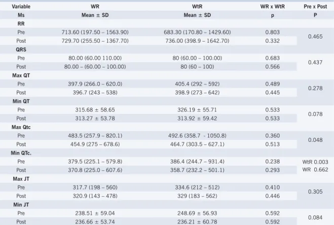
QRS duration, RR, QT, QTc and JT interval - No signifi cant difference was demonstrated in the groups, either for results from RR interval, QRS duration, maximum QT intervals, minimum QT intervals, maximum JT, and minimum JT obtained from EKG at admission (pre) and EKG on day 4 of condition course, after the administration of the thrombolytic (post). Maximum QT interval showed to be signifi cantly higher in pre-thrombolysis as compared to post-pre-thrombolysis in both groups (p=0.048), while minimum QTc interval showed to be higher in pre rather than post-thrombolysis in the WtR (p=0.003) (Table 2).
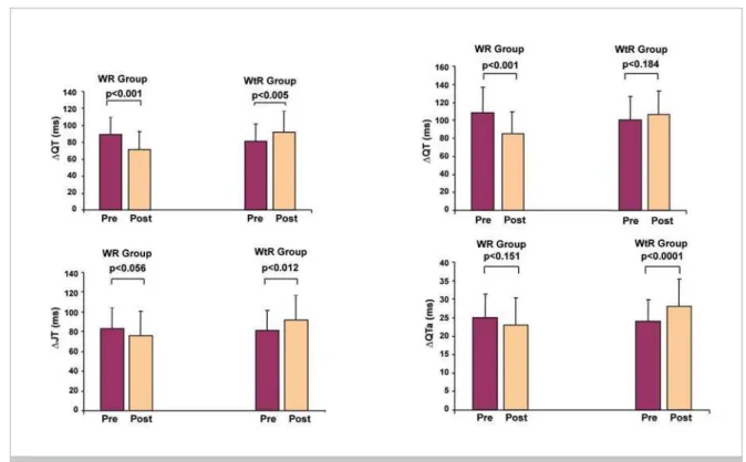
QT, JT and QTc interval dispersions pre- and post-thrombolysis - Before thrombolysis was carried out, values for dispersion parameters for QTc (∆QTc) and JT (∆JT) intervals did not show any difference between WR and WtR. The variable ∆QT showed to be higher in the WR group (89.66 + 20.47 ms), when compared to the WtR group (81.27 + 20.52 ms) (p=0.019). On day 4 after thrombolysis, values for dispersion parameters were signifi cantly lower in the WR group when compared to the WtR group, respectively: ∆QT 70.95+21.65 ms and 91.85 + 24.66 ms (p<0.001), ∆QTc 84.58 + 24.61 ms and 106.00 +26.48 ms (p<0.001); and ∆JT 77.75+24.43 ms and 92.19 + 24.67 ms (p=0.001). (Table 3).
Table 2 – Evaluation of electrocardiographic variables: comparative analysis between patients in groups with reperfusion and groups without reperfusion
Variable WR WtR WR x WtR Pre x Post
Ms Mean ± SD Mean ± SD p P
RR
Pre 713.60 (197.50 – 1563.90) 683.30 (170.80 – 1429.60) 0.803
0.465
Post 729.70 (255.50 – 1367.70) 736.00 (398.9 – 1642.70) 0.332
QRS
Pre 80.00 (60.00 110.00) 80 (60.00 – 100.00) 0.683
0.437
Post 80.00 – (60.00 – 100.00) 80 (60 – 100) 0.566
Max QT
Pre 397.9 (266.0 – 620.0) 405.4 (292 – 592) 0.489
0.278
Post 396.7 (243 – 538) 398.9 (273 – 642) 0.445
Min QT
Pre 315.68 ± 58.65 326.19 ± 55.71 0.533
0.078
Post 313.27 ± 53.78 313.92 ± 59.42 0.533
Max Qtc
Pre 483.5 (257.9 – 820.1) 492.6 (358.7 - 1050.8) 0.360
0.048
Post 454.9 (275 – 678.6) 464.7 (303.5 – 627.1) 0.513
Min QTc.
Pre 379.5 (225.1 – 579.8) 386.4 (244.7 – 931.4) 0.238 WtR 0.003
WR 0.662
Post 370.8 (225.0 – 607.6) 358.7 (232.2 – 501.1) 0.293
Max JT
Pre 317.7 (198 – 560) 334.6 (212 – 512) 0.410
0.305
Post 320.9 (143 – 478) 329 (183 – 562) 0.446
Min JT
Pre 238.51 ± 59.04 248.69 ± 56.93 0.592
0.084
Post 236.66 ± 53.74 236.21 ± 60.78 0.592
. ( . - . ) = median (minimum – maximum); . ± . = mean ± standard deviation; p = signifi cance probability; WR = Group with reperfusion; WtR
= Group without reperfusion.
Table 3 – QT interval dispersion in pre and post-thrombolysis groups
WR WtR WRxWtR
Parameter (ms) Mean± SD Mean ± SD p
Pre
∆QT 89.66 ± 20.47 81.27 ± 20.53 0.019
∆QTc 108.28 ± 28.37 99.63 ± 26.85 0.079
∆JT 83.42 ± 21.54 81,09 ± 20.28 0.528
Post
∆QT 70.95 ± 21.66 91.85 ± 24.66 <0.001
∆QTc 84.58 ± 24.62 106,00 ± 26.49 < 0.001
∆JT 77.75 ± 24.43 92.29 ± 24.67 0.001
Fig. 2 - QT, QTc, JT and QTA interval dispersion in the groups: pre- and post-thrombolysis. In WR patients signifi cant reduction was shown in
variables ∆QT (p<0.001), ∆QTc (p<0.001), and ∆JT (p=0.020). On the other hand, the WtR group showed signifi cant increase in variables ∆QT (p=0.005) and ∆JT (p=0.022). (Figure 2).
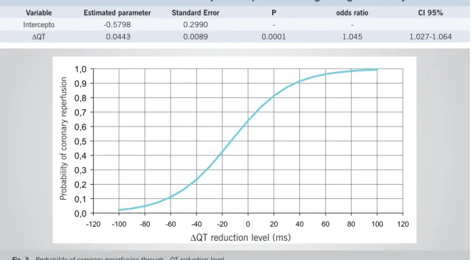
Predictive factors for reperfusion - Considering the signifi cant variables in the univariate analysis – pre- and post-thrombolysis ∆QT, ∆QTc and ∆JT, CKMB peak, Killip > 2 functional class for reperfusion – the logistic regression model was adjusted through stepwise selection procedure. After model adjustment, reduction of QT interval dispersion obtained after thrombolysis was selected as predictive factor for reperfusion, which is to say, the difference between pre and post-thrombolysis QT (dif ∆QT) (Table 4). Therefore, the higher the reduction of that parameter, the higher the probability of reperfusion, which is to say, for each ms of difference found between pre- and post-thrombolysis ∆QT, the chance of reperfusion will be increased 1.045 times (CI 95%; 1.027-1.064) (Figure 3). The sensitivity and specifi city of that model are 87.74% and 75%, respectively, for a 4 ms reduction, with accuracy level at 83.77.
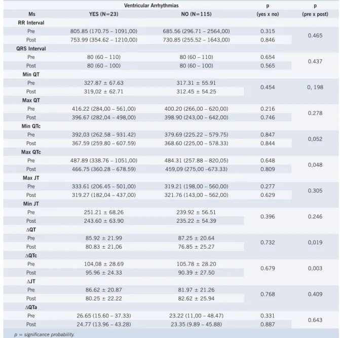
Ventricular arrhythmias and electrocardiographic variables - No significant difference was found in electrocardiographic measures, ∆QT included, when patients with or without ventricular arrythmias were compared in the course of the first 48 hours after infarction. Although the groups did not differ in regard do electrocardiographic characteristics, maximum QT, maximum QTc and maximum JT showed to be higher pre-thrombolysis when compared to post-thrombolysis in the ventricular arrhythmia group (Table 5).
DISCUSSION
The study shows increased dispersion in ventricular repolarization in the acute phase of myocardial infarction, assessed through QT, QTc and JT intervals dispersion measure. Additionally, reperfusion and the maintenance of infarct-related artery in patients submitted to thrombolytic therapy were associated to lower dispersion of QT, QTc and JT intervals when compared to patients without reperfusion.
Time dependent changes in QT interval have been demonstrated during the acute phase of infarction. ∆QT and duration increase take place within the fi rst 48 hours of infarction, and reach their peak on day 312,13.
Thrombolysis and consequent reperfusion can also affect ∆QT. At a first moment, reperfusion of ischemic myocardium increases the heterogeneity of ischemia-induced ventricular recovery due to ventricular repolarization immediately after reperfusion. But later on, patients reporting better reperfusion level in the infarct-related artery report lower ∆QT14.
Recently, Bonnemeier et al15 have demonstrated that patients submitted to angioplasty due to acute myocardial infarction who reported incomplete reperfusion showed changes in ventricular repolarization which affected QT dynamics.
Our results are in agreement with data found in the literature. In 1988, Cowan et al16 described higher post-myocardial infarction ∆QT in 42 patients when compared to a control group. Van de Loo17 demonstrated signifi cant
Table 4 – Predictive value of ∆QT as reperfusion parameter in logistic regression analysis
Variable Estimated parameter Standard Error P odds ratio CI 95%
Intercepto -0.5798 0.2990 -
-∆QT 0.0443 0.0089 0.0001 1.045 1.027-1.064
0,0 0,1 0,2 0,3 0,4 0,5 0,6 0,7 0,8 0,9 1,0
-120 -100 -80 -60 -40 -20 0 20 40 60 80 100 120
Probabilidade de re
p
erfusão
Redução ∆QT (ms)
Fig. 3 - Probability of coronary reperfusion through QT reduction level. phase in infarction recovery. In 1995, while comparing
∆QT in angina and infarction patients, Higman et al18 demonstrated that those patients reported ∆QTc reduction after infarction, thus suggesting that could be due to the reperfusion by thrombolysis with streptokinase.
However, those preliminary studies did not correlate the changes found in the ∆QT of infarcted patients with infarct-related artery reperfusion effect or lack of effect, as this study has done. In the present study, QT behavior both in acute phase and myocardial infarction recovery stage coincides with the early proposition of clinical and experimental studies, emphasizing that the period after infarction and infarct-related artery reperfusion level are relevant in determining ∆QT variation.
Additionally, the present study has demonstrated that all dispersion variables are increased in acute infarction patients, and recanalization and patency of related artery caused reduction in those variables when evaluated on day 4 after the event.
It was possible to demonstrate, through logistic regression analysis, that among the variables related to reperfusion, ∆QT reduction between pre-thrombolysis condition and day 4 of patient’s evolution was the parameter that best identifi ed the presence of coronary reperfusion.
Infarct-related artery patency was turned into the major
goal in therapeutic strategy for acute myocardial infarction patients, since it is known that early reperfusion and artery patency maintenance are responsible for mid and long-term mortality reduction in infarcted patients19,20 . Therefore, the interest in research for methods that sponsor early and non-invasive evaluation of coronary reperfusion and artery patency is of high relevance for their prognostic implication in infarction course. This was the fi rst study whose methodology included coronariographic evaluation pre-thrombolysis, at 90 minutes and at 48 hours after the use of rt-PA, which allowed the correlation of ∆QT variation with coronary reperfusion.
In the present study, the relation between higher occurrence of ventricular arrhythmias and QT dispersion magnitude was not obtained. Results in literature are contradictory, especially in what it refers to arrythmias in the acute phase. Studies based on different methodologies as well as the diffi culty in analyzing an event caused by multiple factors have resulted in controversial data.
Therefore, QT interval dispersion is signifi cantly reduced in patients with acute myocardial infarction submitted to successful thrombolysis, and is signifi cantly increased in infarcted patients with closed artery. The evaluation of QT dispersion reduction between the pre- and psot-thrombolysis condition is a predictor for coronary reperfusion of infarcted patients submitted to thrombolysis.
P
robability of coronar
y reper
fusion
Table 5 – Comparison of electrocardiographic variables following the occurrence of ventricular arrythmias
Ventricular Arrhythmias p p
Ms YES (N=23) NO (N=115) (yes x no) (pre x post)
RR Interval
Pre 805.85 (170.75 – 1091,00) 685.56 (296.71 – 2564,00) 0.315
0.465
Post 753.99 (354.62 – 1210,00) 730.85 (255.52 – 1643,00) 0.846
QRS Interval
Pre 80 (60 – 110) 80 (60 – 110) 0.654
0.437
Post 80 (60 – 100) 80 (60 – 100) 0.565
Mín QT
Pre 327.87 ± 67.63 317.31 ± 55.91
0.454 0, 198
Post 319,02 ± 62.71 312.45 ± 54.25
Max QT
Pre 416.22 (284,00 – 561,00) 400.20 (266,00 – 620,00) 0.216
0.278
Post 396.67 (282,04 – 498,00) 398.90 (243,00 – 642,00) 0.746
Min QTc
Pre 392,03 (262.58 – 931.42) 379.69 (225.22 – 579.75) 0.847
0,052
Post 367.59 (259.80 – 607.59) 368.60 (225,00 – 578.33) 0.844
Max QTc
Pre 487.89 (338.76 – 1051,00) 484.31 (257.88 – 820,05) 0.648
0,048
Post 466.75 (360.28 – 678.59) 459,09 (275,00 –673.33) 0.809
Max JT
Pre 333.61 (206.45 – 501,00) 319.21 (198,00 – 560,00) 0.277
0.305
Post 319.27 (182,04 – 437,00) 321.76 (143,00 – 562,00) 0.629
Min JT
Pre 251.21 ± 68.26 239.92 ± 56.51
0.396 0.246
Post 243.60 ± 63.90 235.22 ± 54.39
∆QT
Pre 85.92 ± 21.99 87.25 ± 20.64
0.732 0,019
Post 80.83 ± 21,06 76.85 ± 25.27
∆QTc
Pre 104,08 ± 28.69 105.78 ± 28.20
0.679 0,003
Post 95.96 ± 24.33 90.39 ± 27.50
∆JT
Pre 86.62 ± 20.87 81.97 ± 21.26
0.768 0.409
Post 80.25 ± 22.22 82.62 ± 25.94
∆QTa
Pre 26.65 (15.60 – 37.33) 23.22 (11,00 – 48.47) 0.331
0.643
Post 24.77 (13.96 – 43.28) 23.35 (9.89 – 45.88) 0.887
p = signifi cance probability.
R
EFERENCES1. Day CP, McComb JM, Campbell RW. QT dispersion: an indication of arrythmia risk in patients with long QT intervals. Br Heart J. 1990; 63: 342-4.
2. Merri M, Benhorin J, Alberti M, Locati E, Moss AJ. Electrocardiographic quantitation of ventricular repolarization. Circulation. 1989; 80:301-8.
3. Butrous GS, Dabbas N, Patel PR, Cochrane T, Camm AJ. Measurement of the QT interval. In: Butrous GS, Swartz PJ. Clinical aspects of ventricular repolarization. London: Farrand Press; 1989: 41-8.
4. Tomassoni G, Pisano E, Gardner kMW, Natale A. QT prolongation and dispersion in myocardial ischemia and infarction. J Electrocardiol. 1998; 30: 187-90.
5. Day CP, McComb JM, Campbell RW. QT dispersion: an indication of arrythmia risk in patients with long QT intervals. Br Heart J. 1990; 63: 342-4.
6. Dristas A, Gilligan D, Nihoyannopoulus P, Oakley CM. Amiodarone reduces QT dispersion in patients with hypertrophic cardiomyopathy. Intern J Cardiol. 1992; 36: 345-9.
7. Han J, Millet D, Chizzonitti B, Moe GK. Temporal dispersion of recovery of excitability in atrium and ventricle as a function of heart rate. Am Heart J. 1996; 71: 481-7.
8. Kurz RW, Xiao-Lin R, Franz MR. Increased dispersion of ventricular repolarization and ventricular tachyarrhythmias in the globally
Potencial Confl ict of Interest
ischaemic rabbit heart. Eur Heart J. 1993; 14: 1561-71.
9. Moreno FL, Villanueeva T, Karagounis LA, Anderson JL. Reduction in QT interval dispersion by successful thrombolytic in acute myocardial infarction. Circulation. 1994; 90: 94-100.
10. Sahu P, Lim PO, Rana BS, Struthers AD. QT dispersion in medicine: electrophysiological Holy Grail or fool’s gold? QJM 2000; 93 (7): 423-31.
11. Kleber AG, Janse MJ, Van Capelle FJL, Durrer D. Mechanisms and time course of S-T and T-Q segment changes during acute regional myocardial ischaemia in the pig heart determined by extracellular and intracellular recording. Circ Res. 1978; 42: 603-13.
12. Glancy JM, Garrat CJ, Bono DP. Dynamics of QT dispersion during myocardial infarction and ischemia. Intern J Cardiol. 1996; 57: 55-60.
13. Higham PD, Furniss S, Campbell RWF. QT dispersion and components of the QT interval in ischaemia and infarction. Br Heart J. 1995; 73: 32-6.
14. Sporton SC, Taggart P, Sutton PM, Walker JM, Hardman. Acute ischaemia: a dynamic infl uence on QT dispersion. Lancet. 1997; 349: 306-9.
15. Bonnemeier H, Wiegand KH, Bode F, Hartmann F, et al. Impact of infarct-related artery fl ow on QT dynamicity in patients undergoing direct percutaneous coronary intervention for acute myocardial infarction. Circulation. 2003; 108: 2979-86.
16. Cowan JC, Yusoff KM, Amos PA, Gold AE, Bourke JP, Tansuphaswadikul S, Campbell RWF. Importance of lead selection in QT interval measurement. Am J Cardiol. 1988; 61: 83-7.
17. Van de Loo A, Arendts W, Hohnloser SH. Variability of QT dispersion measurements in the surface electrocardiogram in patients with acute myocardial infarction and in normal subjects. Am J Cardiol. 1994; 74: 1113-8.
18. Higham PD, Campbell RWF. QT dispersion. Br Heart J. 1994; 71: 508-10.
19. .AIMS Trial Study Group. Long-term effects of intravenous anistreplase in acute myocardial infarction. Final report of the AIMS study. Lancet. 1990; 335: 427-31.