Clinical and neurophysiological
investigation of a large family with
dominant Charcot-Marie-Tooth type
2 disease with pyramidal signs
Eduardo Luis de Aquino Neves1, Fernando Kok2
ABSTRACT
Charcot-Marie-Tooth (CMT) disease is a hereditary neuropathy of motor and sensory impairment with distal predominance. Atrophy and weakness of lower limbs are the first signs of the disease. It can be classified, with the aid of electromyography and nerve conduction studies, as demyelinating (CMT1) or axonal (CMT2). Objective: Clinical and neurophysiological investigation of a large multigenerational family with CMT2 with autosomal dominant mode of transmission. Method: Fifty individuals were evaluated and neurophysiological studies performed in 22 patients. Results: Thirty individuals had clinical signs of motor-sensory neuropathy. Babinski sign was present in 14 individuals. Neurophysiological study showed motor-sensory axonal polyneuropathy. Conclusion: The clinical and neurophysiological characteristics of this family does not differ from those observed with other forms of CMT, except for the high prevalence of Babinski sign.
Key words: Charcot-Marie-Tooth disease, CMT2, axonal hereditary neuropathy.
Investigação clínica e neurofisiológica de família com doença de Charcot-Marie-Tooth tipo 2 com sinais piramidais
RESUMO
A doença de Charcot-Marie-Tooth (CMT) é uma neuropatia hereditária de acometimento sensitivo e motor de predomínio distal. Atrofia e fraqueza em membros inferiores são os primeiros sinais da doença. Pode ser classificada, com auxílio da eletroneuromiografia, em desmielinizante (CMT1) ou axonal (CMT2). Objetivo: Investigação clínica e neurofisiológica de família com portadores de CMT2 de herança dominante. Método: Foi feita avaliação neurológica de 50 indivíduos e eletroneuromiografia em 22 pacientes. Resultados: Trinta indivíduos tinham sinais clínicos de neuropatia sensitivo-motora. Sinal de Babinski estava presente em 14 indivíduos. A eletroneuromiografia demonstrou polineuropatia axonal sensitiva e motora. Conclusão: As características clínicas e neurofisiológicas desta família não se diferem das observadas em outras formas de CMT, exceto pela alta prevalência de sinal de Babinski.
Palavras-chave: doença de Charcot-Marie-Tooth, CMT2, neuropatia hereditária axonal.
Correspondence
Eduardo Luis de Aquino Neves Av. Gonçalo Prado Rollemberg 211 / sala 507
49015-230 Aracaju SE - Brasil E-mail: eduardoaquinoneves@ hotmail.com
Received 13 January 2011 Received in final form 2 February 2011 Accepted 10 February 2011
1Neurologist and Neurophysiologist from the Postgraduate Program in Neurology, Department of Neurology, University of
São Paulo, São Paulo SP, Brazil; 2Associate Professor in Neurology, Department of Neurology, School of Medicine, University
of São Paulo, São Paulo SP, Brazil.
Charcot-Marie-Tooth (CMT) disease is one of the most common genetically de-termined neurological disorder, with an estimated prevalence of 37/100,000 indi-viduals1. It is characterized by impairment
of peripheral nerves function, with distal
predominance and a highly variable clin-ical course.
According to the nature of periph-eral nerve injury, CMT disease can be divided into two main groups:
Other classiications are based on transmission pattern and genetic basis.
CMT disease causes progressive weakness and at-rophy, initially in distal muscles of lower limbs and later reaching the upper limbs, with foot deformities, loss of sensitivity, and reduced tendon relexes3.
Especially among individuals afected by demyelin-ating neuropathy (CMT1), it is frequent to ind atrophy of the distal third of the legs, giving the appearance of an inverted champagne glass and of pes cavus. he prox-imal muscles are rarely afected3. Cranial nerve
involve-ment is rare, but there are descriptions of families with vocal cord paresis and deafness associated with CMT4,5.
Reduction and even abolition of the relexes are the rule, although in some forms of CMT it is possible to find brisk relexes and even the presence of Babinski sign3,4,6-8.
It is not common for individuals with CMT to present subjective sensory symptoms or neuropathic
pain9. he sensory abnormalities are primarily due to a
more pronounced loss of myelinated thick ibers (Aα)1.
Sense for vibration and pain are the irst to be changed. More rarely, additional signs can be found such as ac-tion tremor, optic atrophy, deafness, pupillary abnormal-ities, and foot ulcers4,10. In some individuals and families,
pyramidal signs, as brisk tendon relexes and Babinski sign, without spasticity, might be detected3,4,8,10.
Electromyography (EMG) and nerve conduction studies (NCS) allow differentiation of demyelinating (CMT1) from axonal (CMT2) neuropathies: in the irst, the conduction velocity is less than 38 m/s, while in the second it is over 38 m/s2,3,11-14. here are also
interdiate forms, with motor conduction velocities of the me-dian between 30 and 40 m/s15.
According to the type of inheritance, CMT 1 is of au-tosomal dominant inheritance, and CMT2 is transmitted as a dominant or recessive character1,4,16. he forms of
inheritance linked to the X are known as CMTX, and the autosomal-recessive demyelinating forms are called CMT4. CMT5 is used to refer to CMT with spasticity.
For CMT1, ive loci and the genes associated with them have been located and identiied: about 70 to 80%
of cases are caused by duplication of the PMP22 gene,
and 5 to 10% are caused by mutation in the MPZ gene1.
To date, 13 loci are known to be associated with
CMT2, and only 9 genes have been identiied so far4,17.
Four of the 13 loci are related to forms of recessive in-heritance and some have been identified in only one family. CMT 2A is the most prevalent form of axonal CMT, being responsible for approximately 20% of ax-onal CMT17. It is caused by mutation of the MFN2 gene,
located on the 1p36 chromosome, which encodes the GTPase mitofusin, involved in mitochondrial fusion4,17.
his study aims to investigate, from the clinical,
neu-rophysiological, and genetic point of view, a family living in the municipality of Tobias Barreto, situated 180 km from Aracaju, Sergipe State, Brazil.
METHOD
Clinical evaluation
After completion of the heredogram and identifi-cation of possible individuals afected by CMT among family members, a clinical neurological evaluation was performed. All participants of the study signed a con-sent form, approved by the Institutional Committee for Ethics in Research.
Neurophysiological studies
EMG was performed using Viking Quest (Nicolet) 9.0 equipment. he records were obtained following a conventional protocol as reported by Oh and cols.18,
sur-face electrodes and stimulator were used for NCS. he action potentials amplitudes for motor nerves were mea-sured from the peak of the negative wave to the baseline, and for sensory nerves from peak-to-peak. Temperature were controlled, and never less than 33°C.
Application of the Charcot-Marie-Tooth Neuropathy Score
The Charcot-Marie-Tooth Neuropathy Score
(CMTNS), developed by Shy and cols.19 were used to
as-sess disease severity. his score combines motor and sen-sory manifestations with data obtained from NCS. he maximum possible score is of 36 points and patients with less than 10 points are classiied as mildly afected, with between 11 and 20 points as moderately compromised and with more than 21 points as severely afected19.
RESULTS
hirth-ive individuals with CMT were recognized, and 30 completed clinical evaluation which allows in-clusion in this study. Age at ascertainment varied from 3 to 75 years, and age of onset varied greatly. Four pa-tients with abnormal indings at clinical examination did not have complaints. Frequent falls was the the irst rec-ognized abnormality in cases with onset at childhood. In adolescents and adults, inability to run was usually seen as an early manifestation. Neuropathic pain was never reported.
This family presents a form of CMT with auto-somal dominant transmission, as can be observed in the heredogram (Figure). The presence of father-to-child transmission excludes an X-linked transmission.
Table 1. Clinical data of CMT2 family.
Patient (years)Age (years) AmbulationOnset Atrophy Pes cavus Babinski sign Tibialis anterior weakness Achilles relex Sensitivity*
II-2 75 40 yes no no no present reduced
II-14 74 53 yes legs and hands yes yes absent reduced
III-9 46 14 yes hands and legs yes yes absent reduced
III-10 48 12 no legs and hands no bilateral yes exalted reduced
III-12 51 10 yes hands and feet no yes absent reduced
III-14 36 14 yes forearms and legs yes yes present reduced
III-25 47 30 yes hands and feet no yes present reduced
III-35 41 30 yes hands and feet no bilateral yes present reduced
III-39 44 16 yes legs yes yes absent reduced
III-41 45 7 with aid legs and hands yes yes absent reduced
III-49 43 37 yes no no yes present reduced
III-51 37 5 yes no yes yes brisk reduced
IV-1 23 15 yes no yes yes present reduced
IV-5 18 13 yes hands and legs yes bilateral yes absent reduced
IV-8 28 9 yes legs yes yes absent reduced
IV-13 13 5 yes feet and hands yes bilateral yes absent reduced
IV-14 12 7 yes legs and hands no right yes absent reduced
IV -18 19 19 yes no yes right yes present reduced
IV-20 13 8 yes hands and legs yes bilateral yes present reduced
IV-31 11 9 yes legs yes yes absent reduced
IV-32 16 10 yes legs yes bilateral yes present reduced
IV-39 7 6 yes hands and feet yes left yes present reduced
IV-44 11 6 yes legs and hands yes bilateral yes brisk reduced IV-50 13 6 yes legs and hands yes bilateral yes brisk reduced IV-52 11 8 yes legs and hands yes bilateral yes absent reduced
IV- 54 20 7 yes feet and hands yes yes absent reduced
IV-57 14 12 yes hands and feet yes right yes present reduced
IV-59 10 5 yes hands and feet yes left yes present reduced
V- 3 6 3 yes no no no absent
V-4 3 3 yes no no yes present
TA: tibialis anterior; CMT2: Charcot-Marie-Tooth type 2; *Sensitivity surface-painful (pinprick) and deep-vibration.
Figure. Heredogram of family with CMT 2.
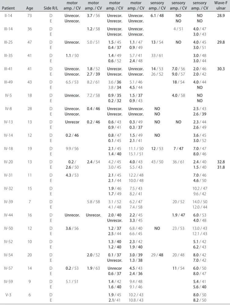
Table 2. Neurophysiological data of patients with CMT 2.
Patient Age Side R/L
Median motor amp. / CV
Ulnar motor amp. / CV
Peroneal motor amp. / CV
Tibialis motor amp. / CV
Median sensory amp. / CV
Ulnar sensory amp. / CV
Sural sensory
amp. / CV Wave-F ulnar
II-14 73 D
E Unrecor.Unrecor. 3.7
/56 Unrecor.
Unrecor. Unrecor.Unrecor. 6.1
/ 48 NO
NO NONO 28.9
III-14 36 D
E 1.2
/58 Unrecor.
Unrecor. Unrecor.Unrecor.
4 / 51 4.0 / 47
3.0 / 43
III-25 47 D
E Unrecor.
5.0 / 51 1.5 /45
0.4 / 37 1.1
/ 47
0.9 / 49 13
/ 54 NO 4.0 / 45
3.0 / 51 29.8
III-35 41 D
E 1.1
/ 50 1.4 / 49
0.6 / 52
5.7 / 41
2.4 / 48
33 / 61 3.0 / 48
3.0 / 44
III-41 41 D
E Unrecor.Unrecor. 1.8
/52
2.7 / 39 Unrecor.Unrecor. Unrecor.Unrecor. 14
/ 53 26 / 52 7.0
/ 56
9.0 / 57 2.0 / 46
2.0 / 42 30.3
III-49 43 D
E
6.5 / 53 8.2 / 61 3.6 / 36
3.8 / 34
5.1 / 46
4.5 / 44 18
/ 54 4.0 / 44
NO
IV-5 18 D
E Unrecor.
7.2 / 58 0.9 / 35 0.2 / 32 1.5
/ 37
0.9 / 43 4.0
/ 58 NO NO
IV-8 28 D
E Unrecor.Unrecor. 0.4
/ 46 Unrecor.
Unrecor. Unrecor.Unrecor. NONO 2.5
/ 43
2.6 / 39
IV-13 13 D
E Unrecor 0.2
/ 46 0.6 /43
0.9 /41 0.3 /49
0.3 / 37 NO NO 2.3
/ 44
2.6 / 49
IV-14 12 D
E 0.2
/ 46 0.8 /47
0.1 /45 1.5 /49
2.1 /41 NO 3.6
/ 45
3.0 / 52
IV-18 19 D
E
9.9 / 56 2.1 /45
1.4 / 40
11.1 / 50 15.1 / 51 12
/ 53 7 / 47 7.0 / 47
8.0 / 46
IV-20 13 D
E 0.2 /
2.6 / 50 2.4
/54 4.2 / 45 3.0 / 45 4.0
/ 43 5.5 / 43
43 / 50 36 / 61 2.4 / 40
1.5 / 40 32.831.8
IV-31 11 D
E 4.3
/53 2.1 /45
2.1 /44
12.2 / 48
10.0 / 48 7.0
/ 46
4.6 / 50
IV-32 15 D
E 1.9
/46
1.7 /49
7.5 / 43 8.2 / 41
10.2 / 47 9.6 / 42
IV-39 7 D
E
5.8 / 58 3.1 / 52 4.1 / 48
6.2 / 47 7.4 / 58
20 / 52 14.0 / 50 12.0 / 44
IV-44 16 D
E Unrecor. Unrecor. 2.0
/ 40 Unrecor. 2.2
/ 45
3.3 / 45 1.9
/ 47 6.0 / 53
4.0 / 48
IV-50 12 D
E 3.6
/56 1.2 / 37 2.5 /44
6.8 / 40
6.6 / 45 NO
23 / 53 13.0 / 43 12.1 / 43
IV-52 10 D
E 1.3
/ 40 1.2 / 40 2.3
/ 42
1.9 / 40 5.1
/ 42
6.2 / 43
IV-54 20 D
E 2.0
/52 0.1 / 37 Unrecor. 3.0
/ 39 1.3 / 38
29 / 48 20 / 48 8.0 / 42
7.0 / 42
IV-57 14 D
E 0.2
/53 1.9 /63 Unrecor 0.6 / 37 4.5
/43
2.4 / 36 11
/ 54 6.0 / 50
8.0 / 47
IV-59 9 D
E
5.1 / 51 1.4 /42
1.6 / 40
9.4 / 48
9.1 / 46 5.4
/ 41
5.6 / 40
V-3 6 D
E 1.9
/45
2.1/41
10.2 / 43
10.8 / 43 8.0
/ 50
8.2 / 50
ambulate with an walker. he siblings IV-13 and IV-14 had a very unstable gait, but could walk without assis-tance; they are nephews of patient III-10, and all afect individuals in this branch of the kindred had a more se-vere form of CMT, associated with pyramidal signs.
Pyramidal signs, characterized by the presence of Babinski sign with or without brisk relexes, was present in 14 patients. None of the subjects presented spasticity. Lower limbs atrophy was seen in 23 patients (76%) and hands atrophy in 19 individuals (63%). he atrophy was restricted to the distal third of the legs and to the hands, except for patients III-10, III-14, 8, 44, 52, IV-57, and II-14, who also had forearm atrophy , and pa-tients IV-13 and IV-14, who had forearms and arms at-rophy. Pes cavus was present in 21 patients (70%) and lat feet were observed in six individuals. Paresis of the tibi-alis anterior was the most consistent clinical abnormality, present in 28 patients (93%). In all patients, with excep-tion of individuals V-4 and V-3, who where too young to give consistent answers, a reduction of supericial and deep sensitivity in the distal portions of the lower limbs were detected. Pain and vibration hypoesthesia were also detected in ingers of 10 patients. hirteen individual had bilateral abolition of the Achilles relexes and four pa-tients (IV-44, III- 51, IV-50, and III-10) presented brisk relexes. Seven patients (IV-13, IV-14, IV-44, IV-5, IV-57, III-14, and III-10) had brisk patellar relexes .
Neurophysiological studies – EMG and NCS were performed in 22 individuals and the data obtained are summarized in Table 2. All 22 patients had neurophys-iological changes consistent with predominantly motor axonal neuropathy and greater distal involvement, except for patient IV-39. he most consistent neurophysiolog-ical inding was reduced amplitudes of compound motor action potentials of the motor nerves (CMAPs). he pe-roneal nerves showed the greatest reductions in CMAPs. Motor conduction velocity was slightly reduced in only 10 of the 22 patients. The lowest conduction velocity of the median nerve was 46 m/s. In the study of sory conduction, a reduction of the amplitudes of
sen-sory nerve action potentials (SNAPs) was also the most common inding, especially in the sural nerves. EMG with concentric needle proved to be abnormal in the 12 patients in which it was performed. he most consis-tent inding was reduction in the recruitment pattern of motor units, seen in the short extensor muscles of the ingers and tibialis anterior.
CMTNS application – he CMTNS was applied in 21 patients and results can be seen in Tables 3 and 4. he
Table 3. Charcot-Marie-Tooth Neuropathy Score in 21 patients with CMT2.
Patient Age Score disease (years)Duration of
II-14 75 18 35
III-10 48 26 36
III-14 36 22 22
III-25 47 16 17
III-35 41 12 11
III-41 45 21 37
III-49 43 6 6
IV-5 18 13 5
IV-8 28 16 19
IV-13 13 25 8
IV-14 12 23 5
IV-18 19 11 Not known
IV-20 13 12 5
IV-32 16 9 6
IV-39 7 6 1
IV-44 11 17 5
IV-50 13 11 7
IV-52 11 14 3
IV-54 20 10 13
IV-57 14 14 2
IV-59 10 9 5
Mean age: 25.6 years; Average score: 14.8 points; CMT 2: Charcot-Marie-Tooth type 2.
Table 4. Disease duration and Charcot-Marie-Tooth Neuropathy Score.
Generation II Generation III Generation IV Gereration V
Number of patients 2 10 16 2
Mean age 43.33 years 14.63 years
Age range 36 to 48 years 7 to 28 years
Mean age of onset 46.5 years 17.5 years 9.06 years 3 years Range of age of onset 40 to 50 years 5 to 37 years 5 to 19 years 3 years
Mean duration of disase 21.5 years 7 years
CMTNS mean 17.16 13.91
patients showed variation from 6 to 26 points. Five were mildly afected (score up to 10), 11 were moderately com-promised (score between 11 and 20), and ive had a severe compromise (score >21). he mean score in generation III was 17.16 points (range 6 to 26 points). In genera-tion IV, the mean was 13.9 points (range 6 to 25 points).
DISCUSSION
In the last two decades several loci and genes related
to CMT2 have been recognized20. We conducted a
clin-ical and neurophysiologclin-ical study of a multigenerational family with 35 individuals afected by an autosomal dom-inant form of CMT2, which is probably the largest kin-dred ever reported in Brazil.
Thirty individuals with CMT from completed the clinical study. We observed an earlier onset and larger severity of the disease at later generations, a fact already observed by other authors21-23. Assessment of disease
se-verity by CMTNS shows that ten individuals of genera-tion IV already have high scores. Generagenera-tion IV, with a mean of 7 years of disease, has in the CMTNS a mean of 13.9 points (moderate incapacity). In generation III, with a mean of 21.5 years of disease, CMTNS mean scorer was of 17.1 points. his inding could be a result of an ascertainment bias, caused by an increased awareness about the disease. he assessment of individuals of gen-erations V and VI might help establish this issue.
Neuropathic pain and other positive sensory symp-toms have not been fully investigated in most families
studies of CMT1. Germignani et al.9, showed that
ap-proximately 71% of the individuals had positive sensory symptoms, including neuropathic pain. In contrary, none of the individuals evaluated in this family complained of neuropathic pain. When present, sensory complaints were most often consistent with late onset nociceptive pain, and probably arising from osteo-muscular defor-mities. as suggested by Padua et al.24.
Trophic changes seen in the feet, legs, and hands of individuals from this family are typical of CMT disease.
he presence of pes cavus, so characteristic of CMT
dis-ease, was absent in one-third of the patients.
In some individuals, tendon relexes were abolished and in others were brisk. The presence of pyramidal signs, with Babinski sign and hyperrelexia seen in sev-eral individuals of this family, has been described in other families with CMT3,8,25,26. What diferentiates CMT2 with
pyramidal signs from CMT V (or HMSN V) is the ob-ligate presence of spasticity in the latter. Vucci et cols. found CMT with pyramidal signs in 9% of the families that they have studied, and demonstrated that the pres-ence of CMT with Babinski sign was genetically distinct
from all known forms of CMT226. In the present family,
we observed pyramidal signs in 46 % of the individuals.
hose individual had usually higher scores at CMTNS, which could not be explained by central nervous system involvement , but probably by a more marked progres-sion of atrophy, afecting proximal muscles of the upper and lower limbs.
he neurophysiological study was consistent with an axonal motor-sensory neuropathy. Reduction of the am-plitudes of motor action potentials in peroneal nerves was the earlier and more frequent inding. his study showed no singularities when compared to other fami-lies with CMT2, but provided information allowed de-termination of CMTNS, which allowed rating severity of disease. he CMTNS seeks to assess precisely the ax-onal loss that the disease eventually determines over time in both the distal and proximal upper and lower limbs.. he application of the score is fast and able to be held in all locations where the clinical neurological evaluation and neuroconduction study can be done, with no need for more sophisticated equipment, such as quantitative sensory testing (QST).
To conclude, in this study, we studied 30 individuals of a multigenerational family with autosomal dominant CMT2, which was associated in 46 % of cases with py-ramidal signs. Neurophysiological study was typical of an axonal compromise. It was also remarkable that dis-ease severity was higher and age of symptoms onset ear-lier in the more recent generation. A genetic study is un-derway to better characterize the molecular basis of this condition.
REFERENCES
1. Klein CJ, Dyck PJ. HMSN II (CMT2) and miscellaneous inherited system at-rophies of nerve axon: clinical-molecular genetic correlates. In: Dyck PJ, Thomas PK (Eds). Peripheral Neuropathy. Fourth Edittion. Philadelphia: Elsevier Saunders, 2005:1717-1752.
2. Davis CJF, Bradley WG, Madrid R. The peroneal muscular atrophy syndrome: clinical, genetic, electrophysiological and nerve biopsy indings and classi-ication. J Genet Hum 1978;26:311-349.
3. Harding AE, Thomas PK. The clinical features of hereditary motor and sensory neuropathy types I and II. Brain 1980; 103: 259-280.
4. Barisic N, Claeys KG, Sirotkovic-Skerlev M, et al. Charcot-Marie-Tooth disease: a clinic-genetic confrontation. Ann Hum Genet 2008;2:416-441. 5. Verhoeven K, Claeys KG, Züchner S, et al. MFN2 mutation distribution and
genotype/phenotype correlation in Charcot-Marie-Tooth type 2. Brain 2006; 129:2093-2102.
6. Chung KW, Kim SB, Park KD, et al. Early onset severe and late-onset mild Charcot-Marie-Tooth disease with mitofusin 2 (MFN2) mutations. Brain 2006;129:2103-2118.
7. Bienfait HME, Baas F, Koelman JHTM, et al. Phenotype of Charcot-Marie-Tooth type 2. Neurology 2007;68:1658-1667.
8. Zhu D, Kennerson ML, Walizada G, Züchner S, Vance JM, Nicholson GA. Charcot-Marie-Tooth with pyramidal signs is genetically heterogeneous: families with and without MFN2 mutations. Neurology 2005;65:496-497. 9. Gemignani F, Melli G, Alieri S, Inglese C, Marbini A. Sensory manifestations
in Charcot-Marie-Tooth disease. J Peripher Nerv Syst 2004;9:7-14. 10. Lawson VH, Graham BV, Flanigan KM. Clinical and electrophysiologic
features of CMT2A with mutations in the mitofusin 2 gene. Neurology 2005; 65:197-204.
neu-ropathy type 2A: novel mutations in the mitofusin 2 gene (MFN2). BMC Med Genet 2006;7:53.
13. Kimura J. Electrodiagnosis in diseases of nerve and muscle: principles and pratice. Ed. 3. Oxford University Press, 2001.
14. Muglia M, Zappia M, Timmerman V, et al. Clinical and genetic study of a large Charcot-Marie-Tooth type 2ª family from southern Italy. Neurology 2001;56:100-103.
15. Sevilla T, Jaijo T, Nauffal D, et al. Vocal cord paresis and diaphragmatic dysfunction are severe and frequent symptoms of GDAP1-associated neu-ropathy. Brain 2008;131:3051-3061.
16. Harding AE, Thomas PK. Genetic aspects of hereditary motor and sensory neuropathy (types I and II). J Med Genet 1980;17:329-336.
17. Züchner S, Vance JM. Molecular genetics of autosomal-dominant axonal Charcot-Marie-Tooth disease. Neuromolecular Med 2006;8:63-74. 18. Oh J. Clinical electromyography: nerve conduction studies. 2nd Ed. Williams
& Wilkins, Baltimore 1993.
19. Shy ME, Blake J, Krajewski K, et al. Reliability and validity of the CMT neuropathy score as a measure of disability. Neurology 2005;64:1209-1214.
20. Nicholson GA. The dominantly inherited motor and sensory neuropathies: clinical and molecular advances. Muscle Nerve 2006;33:589-597. 21. Kovach MJ, Campbell KC, Herman K, et al. Anticipation in a unique family
with Charcot-Marie-Tooth syndrome and deafness: delineation of the clin-ical features and review of the literature. Am J Med Genet 2002;108:295-303. 22. Steiner I, Gotkine M, Steiner-Birmanns B, et al. Increased severity over gener-ations of Charcot-Marie-Tooth disease type 1A. J Neurol 2008;255:813-819. 23. Marques W, Hanna MG, Marques SR, Sweeney MG, Thomas PK, Wood NW.
Phenotypic variation of a new PO mutation in genetically identical twins. J Neurol 1999;246:596-599.
24. Padua L, Cavallaro T, Pareyson D, Quattrone A, Vita G, Schenone A+ Italian CMT QoL Study Group. Charcot-Marie-Thooth and pain: correlations with neurophysiological , clinical, and disability indings. Neurol Sci 2008;29: 193-194.
25. Gemignani F, Marbini A. Charcot-Marie-Tooth disease (CMT): distinctive phenotypic and genotypic features in CMT type 2. J Neurol Sci 2001;184:1-9. 26. Vucic S, Kennerson M, Zhu D, Miedema E, Kok C, Nicholson GA. CMT with