Primary progressive aphasia
patients evaluated using difusion
tensor imaging and voxel based
volumetry-preliminary results
Fábio Pascotto de Oliveira1, Jaderson Costa da Costa2,Sabine Possa Marroni3, Ana Maria Marques da Silva4,
Sthefanie Hansen Barreiro5, Fábio Kunihiro Maeda6,
Mirna Wetters Portuguez7
ABSTRACT
There are individuals who have a progressive language deficit without presenting cognitive deficits in other areas. One of the diseases related to this presentation is primary progressive aphasia (PPA). Objective: Identify by means of diffusion tensor imaging (DTI) and measurements of cortical volume, brain areas that lead to dysphasia when presenting signs of impaired connectivity or reduced volume. Method: Four patients with PPA were evaluated using DTI, and measurements of cortical volumes in temporal areas. These patients were compared with two normal volunteers. Results: There is a trend to a difference in the number and volume of related fibers between control group and patients with PPA. Comparing cortical volumes in temporal areas between groups yielded a trend to a smaller volume in PPA patients. Conclusion: Patients with PPA have a trend to impairment in cortical and subcortical levels regarding relevant areas.
Key words: anomia, primary progressive aphasia, DTI, tractography, arcuate fasciculus, language
Avaliação de pacientes com afasia primária progressiva com imagem tensor de difusão e volumetria baseada em voxels
RESUMO
Existem indivíduos que apresentam deterioração progressiva da linguagem sem apresentar déficits em outros domínios cognitivos; estes pacientes em geral apresentam afasia primária progressiva (APP). Objetivo: Identificar, pela imagenologia, áreas cerebrais que quando sofrem atrofia, ou quando seu sistema de conexões apresenta alterações, levam a disfasia. Método: Foram avaliados quatro pacientes com APP, utilizando-se ressonância magnética com as técnicas de imagem por tensores da difusão (DTI) e medida do volume cortical em áreas de interesse previamente definidas. Estes pacientes foram comparados com um grupo controle constituído por dois voluntários. Resultados:
Redução da média dos volumes dos giros temporais esquerdos no grupo de pacientes, em relação ao grupo controle, e alterações quanto ao volume e número de fibras nos tratos correspondentes. Conclusão: Pacientes com APP têm tendência a apresentar danos tanto ao nível cortical quanto subcortical.
Palavras-chave: anomia, afasia primária progressiva, DTI, tratografia, volume cerebral, fascículo arqueado, linguagem.
Correspondence
Fábio Pascotto de Oliveira Av. Fernando Ferrari 1067 97050-801 Santa Maria RS - Brasil E-mail: [email protected]
Received 12 February 2010
Received in final form 21 December 2010 Accepted 3 January 2011
1Neurologist, HUSM, Santa Maria RS, Brazil; 2Professor of Neurology, Director Brain Institute, PUCRS, Porto Alegre RS, Brazil; 3Unit of Neuropsychology Hospital São Lucas, PUCRS, Porto Alegre RS, Brazil; 4Professor of the School of Physics, PUCRS,
Porto Alegre RS, Brazil; 5Medical Physicist, NIMed, PUCRS, Porto Alegre RS, Brazil; 6Physics Student, NIMed, PUCRS, Porto
Language is one of the pillars of human intellect, and it is deined as the ability to communicate. It involves as-sociating sounds and symbols with signiicant concepts and allow us to describe the external environment and abstract thoughts1. Currently, one of the most accepted
theories about language suggests it is processed in two pathways the ventral and the dorsal2. his model
pro-poses that the ventral pathway, which involves structures in portions of the upper and middle temporal lobe, is involved in signal processing for understanding spoken language. Moreover, the dorsal pathway, which involves structures of the posterior region of the frontal lobe, pos-terior dorsal temporal lobe, and parietal operculum, is involved in translating acoustic signals to articulatory language representations in the frontal lobe. he latter route would be essential for the development of language and its normal production3.
hus oral communication occur as follows, irst the speech would be examined in the auditory cortex bilat-erally in the supratemporal plane, and processed in pho-nological and representative levels involving the middle portion of the posterior superior temporal sulcus bilat-erally. Subsequently the system splits towards the dorsal and ventral pathway; the dorsal pathway have dominance in the left hemisphere and the ventral pathway is repre-sented bilaterally and has a weak dominance in the left hemisphere. he anatomical areas involved in these path-ways are not known perfectly but the approximate loca-tions are described below.
he dorsal pathway, which has a strong dominance in the left hemisphere involves in its posterior aspect an area near the sylvian issure in the temporoparietal junction, proposed as a sensory-motor interface. And in its anterior aspect it would involve the anterior part of the insula, the inferior frontal gyrus and also the pre-motor cortex2.
he ventral pathway, which has weak dominance in the left hemisphere being organized bilaterally, involves the middle temporal gyrus and inferior temporal sulcus. he most posterior areas of the ventral pathway would correspond to a lexical interface, which would link the phonological information with semantics3.
At the subcortical level it has been proposed to the inferior fronto-occipital fasciculus a semantic function, since it was observed that after intraoperative stimula-tion semantic paraphasias were noticed, regardless of the point in which this pathway was stimulated4.
The arcuate fasciculus is classically linked to lan-guage, and it has been demonstrated that stimulation in diferent parts of this structure generates diferent ex-pressions including phonemic paraphasias, and disor-ders that afect the formation of phonological words5.
Currently it is recognized that the arcuate fasciculus that
connects the parietal and temporal regions of the frontal lobe is composed of various subcomponents. Besides the classic arcuate fasciculus connecting Wernicke’s area and Broca’s area directly, there is an indirect route that runs parallel and lateral to the arcuate fasciculus6, and this
in-direct route connects the frontal lobe with the parietal lobe, and the temporal lobe with the parietal lobe.
Knowing the way that language is processed, we can infer that people with speech disorders present some involvement of the related structures. Those who de-velop a slow and progressive decline in language func-tion without cognitive deicits in other domains, usually have a disease called primary progressive aphasia (PPA)7.
he diagnosis of PPA can now be performed in any patient in which the progressive loss of language con-stitutes the most salient aspect of the clinical condition, when it is caused by a neurodegenerative disease8. he
damage to language can be luent or nonluent and may or may not interfere with word recognition. Memory for recent events recognition of familiar faces and ob-jects are preserved but when tested orally may seem ab-normal. Small changes in personality and behavior can occur, but usually do not interfere with the patient’s life. For the diagnosis of PPA the aphasia has to be the most salient deficit and the major cause of impaired daily living activity for approximately two years.
his study aims to determine in patients with PPA which structures in cortical or subcortical levels are in-volved in language deterioration. For this purpose we performed brain magnetic resonance (MR) examina-tions; measured cortical volumes and used diffusion tensor imaging (DTI) in areas involved in speech.
METHOD
Subjects
Four right handed, patients with diagnosis of PPA were included aged 59-82 (mean 69.25). his diagnosis was conirmed by at least two neurologists according to previously published criteria9 and neuropsychological
tests. Exclusion criteria were patients or controls that had any condition that could simulate PPA, or had a his-torical of a previous neurological disease.
hese patients were compared with two age-matched health volunteers aged 65.
All subjects gave written informed consent and the study was approved by the Internal Review Board.
Image acquisition
MR was performed on a 1.5 T scanner (Siemens Avanto 76×18 HQ).
mm2; flip angle 90; pixel spacing 1.25×1.25×2.5; slice
thickness 2.5 mm.
he pulse sequences used for the morphometric anal-yses weretwo three-dimensional, T1-weighted [magneti-zation prepared rapidgradient echo (MP-RAGE)] scans, with the following parameters:repetition time, 2400 ms; echo time, 3.61 ms; inversion time,1000 ms; lip angle, 8°; matrix, 192×192; ield of view,192. Each scan took 7 minutes and 42 seconds.
Image analysis
MR data were transferred to an of-line computer for further analysis. Tractography was applied to the DTI data to reconstruct white matter tracts by successively following the path of preferred direction of water dif-fusion when fractional anisotropy (FA) is higher than a selected threshold. Using MedINRIA software ver-sion 1.9.0 (http://www-sop.inria.fr/asclepios/software/ MedINRIA) difusion tensors were calculated from all voxels within the brain and iber tracts were calculated by connecting adjacent voxels with similar principal ei-genvectors, using a threshold FA value of 0.2. Only ibers with lengths of >10 mm were included. hese parameters are similar to those used by others who applied a iber assignment by continuous tracking algorithm. Regions of interest were drawn in each brain on axial slices by a single coder. Regions of interest were drawn according published DTI atlas10. We investigated bilaterally the
ar-cuate fasciculus, inferior fronto-occiptal fasciculus and the inferior longitudinal fasciculus. Variables obtained for statistical comparison included the fractional anisot-ropy, the volume of ibers, and the number of ibers in each of the structures analysed.
Cortical volume was studied using IBASPM11, which
is a fully automated, easy volumetric method (http:// thomaskoenig.ch/lester/ibaspm.htm). Volume mea-surement with IBASPM software is based on an indi-vidual brain atlas masked using an automated anatom-ical labeling. his procedure consists of the following ive
steps: [1] Segmentation: The MR image is segmented into gray matter, white matter, and cerebral spinal luid. [2] Normalization: he MR image is normalized to the MNI space to obtain the spatial transformation matrix; using this transformation matrix the gray matter is trans-formed to MNI space. [3] Automatic labeling: each nor-malized individual gray matter voxel is labeled based on an atlas of predeined 116 segmented structures. [4] At-lasing: Individual atlases are created from each subject’s MR image. he individual atlases are reversely normal-ized, and the gray matter image in native space is masked using its individual 116 brain structures. [5] Volume sta-tistic: For all individual atlases, the volumes of 116 pre-deined brain structures are calculated. When the voxel values of the gray matter image exceed the voxel values of the white matter and CSF images, the voxel is included for volume calculation.
Measurements were obtained in mm3 using this
method. Structures studied included the superior tem-poral gyri, medial temtem-poral gyri, inferior temtem-poral gyri, and the supramarginal gyri12.
Statistical analysis
It was applied the nonparametric Mann-Whitney test for comparing several variables between cases and controls. A nonparametric test was chosen because the sample is small, not allowing us to ensure the assump-tions necessary for a parametric test for comparison of means. We used also the nonparametric Wilcoxon test to compare variables measured between the left and right hemisphere. he Wilcoxon test was used in this situa-tion because we are comparing two measurements in the same individuals,a fact that leads us to use a test for related samples.
RESULTS
A visual comparison between patients and controls demonstrates diferences observed mainly in the left ar-cuate fasciculus, the left fronto-occipital and left inferior
Table 1. P value for the Mann-Whitney* test to compare number of ibers between PPA group and control group.
LAF LIFF LFLI RAF RIFF RFLI
0.481 0.064 0.159 0.355 0.481 0.165
*P values for bilateral test; LAF: left arcuated fascilus; LIFF: left inferior fronto-occiptal fasciculus; LFLI: left inferior longitudinal fasciculus; RAF: right arcuate fasciculus; RIFF: right inferior fronto-occiptal fasciculus; RFLI: right inferior longitudinal faciculus.
Table 2. P value for the Mann- Whitney* test to compare tract volume between PPA group and control group.
LAF LIFF LFLI RAF RIFF RFLI
0.064 0.064 0.165 0.643 0.165 0.165
longitudinal fasciculus. In Figures 1 and 2 are shown the inferior longitudinal fasciculus in a patient and in a con-trol respectively
When anisotropy was assessed using DTI, we could not ind signiicant diference between the PPA group and the control group when FA was analyzed (P=0.24). However when compared the number of fibers and volume of ibers there is a trend to a signiicant difer-ence as shown in Tables 1 and 2.
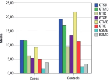
he cortical volumes for studied areas are shown in Figure 3. It can be seen in this graphic the clear dispro-portion between groups for all cortical areas except the right supramarginal gyrus (Table 3).
he comparison of cortical volumes between areas of both hemispheres for each group is shown in Table 4. In this comparison there is a trend to a diference regarding supramarginal and superior temporal gyri.
DISCUSSION
Fiber tracts serve as barriers to free water difusion in the brain. his barrier is the reason for the high an-isotropy of white matter when compared to gray matter, in which the diffusion of water is almost isotropic13.
here are many ways of measuring anisotropy, but one
Fig 1. Patient’s left inferior longitudinal fasciculus. Fig 2. Control left inferior longitudinal fasciculus.
Table 3. P values for the Mann-Whitney* test for comparison between cortical volumes between groups.
GTSD GTMD GTID GTSE GTME GTIE GSME GSMD
0.355 0.355 0.355 0.064 0.064 0.064 0.355 0.643
*P values for bilateral test; GTSD: right superior temporal gyrus; GTMD: right medium temporal gyrus; GTID: right inferior temporal gyrus; GTSE: left superior temporal gyrus; GTME: left medium temporal gyrus; GTIE: left inferior temporal gyrus; GSME: left supramarginal gyrus; GSMD: right supramarginal gyrus.
Table 4. P values for the Wilcoxon for comparison of cortical volumes between hemispheres.
Individuals Number GTSE × GTSD GTME × GTMD GTIE × GTID GSMD × GSME
Cases 4 0.068 0.465 0.465 0.068
Controls 2 0.180 0.180 0.180 0.180
*P values for bilateral comparison; GTSD: right superior temporal gyrus; GTMD: right medium temporal gyrus; GTID: right inferior temporal gyrus; GTSE: left superior temporal gyrus; GTME: left medium temporal gyrus; GTIE: left inferior temporal gyrus; GSME: left supramarginal gyrus; GSMD: right supramarginal gyrus.
25,00
20,00
15,00
10,00
5,00
0,00
Med
ia
Cases Controls
GTSD GTMD GTID GTSE GTME GTIE GSME GSMD
GTSD: right superior temporal gyrus; GTMD: right medium temporal gyrus; GTID: right inferior temporal gyrus; GTSE: left superior temporal gyrus; GTME: left me-dium temporal gyrus; GTIE: left inferior temporal gyrus; GSME: left supramarginal gyrus; GSMD: right supramarginal gyrus.
Fig 3. Mean cortical volume in mm3 for cases (left side) and con-trols (right side).
mol-ecules in these tissues increases, reducing the direction-ality of the water, thus decreasing the value of the FA14.
Probabilistic tractrography uses the anisotropy of water and tract direction to generate probabilistic maps of connectivity between brain regions and it may draw i-bers into the gray matter15. he number of ibers in each
tract must be understood as a quantitative measure of connectivity between anatomical locations, as deter-mined by the chosen region of interest (ROI)16. he loss
of iber loss by axonal degeneration secondary to neu-ronal injury occur after three months17.
One well known limitation of DTI resides in ibber crossings, since there are brain areas in which many i-bers with diferent directions intersect in the same voxel, which typically has dimensions of a few cubic millime-ters, but within this area can go up to thousands of axons. In these areas probably this method may have limited use. Another limitation of DTI is that the val-idation of a tract evidenced by this method is based on prior anatomical knowledge, making it diicult to inter-pret the absence of a fascicle, or the presence of a tract in place other than the usual18. Despite the limitations of
DTI, it is the only method for the investigation of brain ibers in vivo14.
Evaluation of the white matter integrity using FA has been used in several studies For each disease tracts that may justify patient’s condition are studied. In amyo-trophic lateral sclerosis research is mainly the pyramidal tract19, in Alzheimer’s disease the main focus of research
is the parahippocampal white matter20. his study
as-sessed the arcuate fasciculus bilaterally, the longitudinal fasciculus and inferior fronto-occipital fascicle, because in the existing literature these are subcortical structures most involved in spoken language21.
In this study the values of FA are very similar for cases and controls, however when compared the number and volume of iber tracts, mainly in the left hemisphere between groups it is perceived that patients have a lower number of ibers and a decrease in volume tract volume as shown in Table 1 and 2. Considering what was pre-viously mentioned this could suggest that initially there is a cortical damage with subsequent loss of subcor-tical structures. It can be inferred that a reduction in the number of ibers can occur in more advanced stages of disease. here is evidence from studies in patients with amyotrophic lateral sclerosis showing that patients with rapid progression of the disease have decreased mea-sures of brain connectivity in the pyramidal tract when compared to controls, and it is suggested that this should occur by degeneration of the motor tract15.
his study demonstrated diferences of the average volume between patients and controls in the three tem-poral gyri studied, and also that there is an imbalance of
cortical volume in the patient group when comparing ce-rebral hemispheres in the superior temporal and supra-marginal gyri. his is in line with the literature showing a reduction in left perisylvian areas in patients with PPA22.
hese cortical volumetric changes are consistent with the idea presented above in which there must be cortical cell death, at irst, with subsequent changes of subcor-tical networks.
We would like to highlight the indings of the DTI on the volume and number of ibers of the left inferior fronto-occipital fascicle that was disproportionately af-fected in the PPA group. his tract when disabled seems to cause semantic paraphasias, which are errors about the meaning of the word. These findings suggest that this tract is related is the semantic stream of the ventral pathway of language21.
When compared hemispheres in PPA group, cortical volume of the supramarginal gyri and the superior tem-poral gyri appear to be reduced in the left hemisphere. his could be explained because the superior temporal gyri are the initial point for both routes of language the ventral and dorsa3,23.
he ventral pathway, which processes language has a weak dominance in the left hemisphere, then a lesion in this area would not afect comprehension so importantly. On the other hand, the supramarginal gyrus is a part of the dorsal pathway; this pathway has a strong dominance in the left hemisphere and is involved in the articulation of speech2. his explains a more pronounced deicit in
speech production.
Our study sought to evaluate the circuit of language in patients with primary progressive aphasia using voxel based measures and difusion tensor imaging obtained by magnetic resonance imaging of the brain. Although the study sample was small relecting low frequency of PPA data show that patients with this disorder have a tendency for impairment in cortical and subcortical level.
REFERENCES
1. Price CJ. The anatomy of language: contributions from functional neuro-imaging. J Anat 2000;197(Pt 3):335-359.
2. Hickok G, Poeppel D. The cortical organization of speech processing. Nat Rev Neurosci 2007;8:393-402.
3. Shalom DB, Poeppel D. Functional anatomic models of language: assem-bling the pieces. Neuroscientist 2008;14:119-127.
4. Duffau H, Gatignol P, Mandonnet E, Peruzzi P, Tzourio-Mazoyer N, Ca-pelle L. New insights into the anatomo-functional connectivity of the se-mantic system: a study using cortico-subcortical electrostimulations. Brain 2005;128:797-810.
5. Dufau H, Capelle L, Sichez N, et al. Intraoperative mapping of the subcor-tical language pathways using direct stimulations: an anatomo-functional study. Brain 2002;125:199-214.
6. Catani M, Jones DK, Ffytche DH. Perisylvian language networks of the human brain. Ann Neurol 2005;57:8-16.
7. Ingles JL, Fisk JD, Passmore M, Darvesh S. Progressive anomia without semantic or phonological impairment. Cortex 2007;43:558-564. 8. Mesulam MM. Primary progressive aphasia: a 25-year retrospective.
9. Rogalski E, Mesulam M. An update on primary progressive aphasia. Curr Neurol Neurosci Rep 2007;7:388-392.
10. Catani M, Thiebaut de Schotten M. A difusion tensor imaging tractography atlas for virtual in vivo dissections. Cortex 2008;44:1105-1132.
11. Alemán-Gómez YM-GL, Valdés-Hernandesbn P. IBASPM: Toolbox for au-tomatic parcellation of brain structures CD-Rom in NeuroImage 27 vol. Florence, Italy; 2006.
12. Tae WS, Kim SS, Lee KU, Nam EC, Kim KW. Validation of hippocampal volumes measured using a manual method and two automated methods (FreeSurfer and IBASPM) in chronic major depressive disorder. Neuroradi-ology 2008;50:569-581.
13. Ciccarelli O, Catani M, Johansen-Berg H, Clark C, Thompson A. Difusion-based tractography in neurological disorders: concepts, applications, and future developments. Lancet Neurol 2008;7:715-727.
14. Johansen-Berg H, Behrens TE. Just pretty pictures? What diffusion tractography can add in clinical neuroscience. Curr Opin Neurol 2006;19: 379-385.
15. Ciccarelli O, Behrens TE, Altmann DR, et al. Probabilistic difusion tractog-raphy: a potential tool to assess the rate of disease progression in amyo-trophic lateral sclerosis. Brain 2006;129:1859-1871.
16. Lindenberg R, Renga V, Zhu LL, Betzler F, Alsop D, Schlaug G. Structural
integrity of corticospinal motor ibers predicts motor impairment in chronic stroke. Neurology 2010;74:280-287.
17. Rutgers DR, Fillard P, Paradot G, Tadie M, Lasjaunias P, Ducreux D. Difusion tensor imaging characteristics of the corpus callosum in mild, moderate, and severe traumatic brain injury. AJNR Am J Neuroradiol 2008;29:1730-1735. 18. Pierpaoli C, Barnett A, Pajevic S, et al. Water difusion changes in Wallerian
degeneration and their dependence on white matter architecture. Neuro-image 2001;13:1174-1185.
19. Aoki S, Iwata NK, Masutani Y, et al. Quantitative evaluation of the pyra-midal tract segmented by difusion tensor tractography: feasibility study in patients with amyotrophic lateral sclerosis. Radiat Med 2005;23:195-199. 20. Rogalski EJ, Murphy CM, deToledo-Morrell L, et al. Changes in parahip-pocampal white matter integrity in amnestic mild cognitive impairment: a difusion tensor imaging study. Behav Neurol 2009;21:51-61.
21. Duffau H. The anatomo-functional connectivity of language revisited. New insights provided by electrostimulation and tractography. Neuropsy-chologia 2008;46:927-934.
22. Gorno-Tempini ML, Dronkers NF, Rankin KP, et al. Cognition and anatomy in three variants of primary progressive aphasia. Ann Neurol 2004;55:335-346. 23. Saur D, Kreher BW, Schnell S, et al. Ventral and dorsal pathways for