XXX
Raphe obscurus neurons participate in
thermoregulation in rats
Neurônios do núcleo obscuro da rafe participam da termorregulação em ratos
Melissa Araújo Ulhoa1, Nyam Florencio da Silva1, José Guilherme Pinheiro Pires2,3, Henrique de Azevedo
Futuro Neto1,2,4
he thermoregulatory mechanism constitutes a complex and integrated system. he “main controller” is the hypothal-amus, which comprises many diferent sub-areas; it receives inputs either from temperature sensitive neurons within the hypothalamus itself to monitor core temperature or from re-ceptors in the skin which monitor the changes in external temperature. Nevertheless, the inal drive or command to the vascular smooth muscle is not controlled by the hypo-thalamus but by a complex neuronal circuitry that comprises brainstem and spinal cord nuclei1.
In rats, non-evaporative heat loss occurs mainly through the tail2, the blood vessels vasodilate when the
hypothalam-ic preopthypothalam-ic area is warmed3. he eferent signals for this
re-sponse originate in heat-sensitive neurons, and the descend-ing pathway passes through the medial forebrain bundle4.
Blood low to the rat tail is determined by the level of activ-ity in its sympathetic postganglionic vasoconstrictor ibers5.
hese are supplied by preganglionic sympathetic neurons sit-uated mainly in the intermediolateral cell column of the irst and second lumbar segments6,7.
1Departamento de Morfologia, Centro de Ciências da Saúde, Universidade Federal do Espírito Santo (UFES), Vitória ES, Brazil; 2Curso de Medicina da Faculdade Brasileira (UNIVIX), Vitória ES, Brazil;
3Faculdade de Medicina, Centro Universitário do Espírito Santo (UNESC), Colatina ES, Brazil;
4Escola Superior de Ciências da Saúde da Santa Casa de Misericórdia de Vitória (EMESCAM), Vitória ES, Brazil.
Correspondence: José Guilherme Pinheiro Pires; Departamento de Morfologia, Centro de Ciências da Saúde, Universidade Federal do Espírito Santo; Avenida Marechal Campos 1.468; 29042-751 Vitória ES - Brasil; E-mail: [email protected]
Support: Research supported by Conselho Nacional de Desenvolvimento Cientíico e Tecnológico (CNPq) and Fundação de Amparo à Pesquisa do Espírito Santo (FAPES).
Conflict of interest: There is no conlict of interest to declare.
Received 29 June 2012; Received in inal form 20 October 2012; Accepted 29 October 2012. ABSTRACT
In mammalian, several evidences suggest that central serotonin participates in thermoregulation. Nucleus raphe obscurus (NRO), a serotonergic nucleus, has been recognized to be the source of generation of various hemodynamic patterns in different behavioral conditions, but its involve-ment in thermoregulation is unclear. In the present study, extracellular action potentials of NRO neurons were recorded in anesthetized rats, which were submitted to cold and warm stimuli in the tail. The iring rate of the neurons was compared before and after each stimulation. It was found that 59% of the neurons submitted to a cold stimulus trial had a signiicant increase in their iring frequency, while 48% of the neurons submitted to warm stimulation trial were inhibited. The opposite responses in neuronal activity of NRO units to cooling or heating suggest that these cells are involved in producing the homoeothermic vascular adaptations secondary to changes in cutaneous temperature in the rat tail.
Key words: nucleus raphe obscurus, serotonin, serotonergic neurons.
RESUMO
A termorregulação em mamíferos envolve a participação da serotonina. O núcleo obscuro da rafe (NRO), que é serotoninérgico, participa do controle autonômico, mas seu envolvimento na termorregulação é incerto. Neste estudo, registramos potenciais de ação extracelulares de neurônios do NRO em ratos anestesiados nos quais a cauda foi submetida a estímulos de calor ou frio. A frequência de disparo dos neurônios foi comparada antes e depois dos estímulos. O grupo controle não apresentou modiicação da frequência de disparo, enquanto que 59% dos neurônios registrados em ani-mais submetidos a estímulo de frio tiveram sua frequência aumentada. Por outro lado, 48% dos aniani-mais submetidos a estímulo de calor tiveram sua frequência de disparo diminuída. As respostas opostas da frequência de disparo em neurônios de animais submetidos à estimulação com frio e calor sugere que estes neurônios estejam envolvidos na geração de respostas hemodinâmicas, que são coerentes com a termorregulação nesta espécie.
Palavras-Chave: núcleo obscuro da rafe, serotonina, neurônios serotoninérgicos.
DOI: 10.1590/0004-282X20130010
250 Arq Neuropsiquiatr 2013;71(4):249-253
hese spinal cord sympathetic neurons are strongly mod-ulated by neurons located at the brainstem and suprasegmen-tal areas, particularly the raphe neurons8. he raphe nuclei are
a group of structures distributed in the midline of the brain-stem from the midbrain to the caudal pole of the medulla9.
hese nuclei have the highest density of serotonergic neurons in the central nervous system10. Classically the caudal raphe
nuclei (magnus, pallidus and obscurus) are involved in several physiological and pathological processes. hese nuclei have been implicated in homeostatic circuitry and regulate life-sustaining respiratory and thermoregulatory networks11. A
re-cent study found that caudal raphe nuclei are related to pro-cessing thermogenesis, cardiovascular and gastric functions, by neuronal connections from the hypothalamus12.
Regarding thermoregulation, a multiple-input system operates within the spinal cord, raphe nuclei and locus subcoe-ruleus, all involved in generating aferent thermal signals and modulating eferent vasomotor thermoregulatory responses1,13.
Projections from medullary raphe cells to the thoracolumbar intermediolateral cell column are implicated in the sympa-thetic control of physiological functions such as brown adipose metabolism and cutaneous vasoconstriction, therefore these cells are implicated in temperature control14.
he main serotonergic nuclei involved in thermoregu-lation are raphe pallidus (rostral pole) and raphe magnus. Raphe pallidus and magnus nuclei receive projections from the dorsomedial hypothalamus and warm-sensitive preoptic neurons, and contains spinally projecting premotor neurons that provide the excitatory drive to spinal circuits controlling the activity of thermogenic efectors and heat loss control-ling cutaneous vasoconstriction1. Dense projections of these
nuclei have been described to the spinal cord neurons that innervate the vessels of the tail of the rat, which is a major organ of heat loss in this species. Sympathetic premotor neu-rons controlling the tail circulation are located in the rostral medullary raphe15-21.
On the other hand, nucleus raphe obscurus (NRO) has been implicated in generating sympathetic patterns of ac-tivity in diferent behavioral situations as paradoxical sleep22
and in response to nociceptive stimulation23. herefore this
nucleus has the ability to mediate hemodynamic changes in diferent behavioral situations, so it is reasonable to specu-late its participation in the vasomotor adaptations induced by thermoregulatory mechanisms. Hence, the present work was designed to evaluate the participation of NRO neurons in thermoregulatory functions in the rat.
METHODS
he experiments were performed on anesthetized, spontaneously breathing, male Wistar rats of 250–300 g body weight (n=24 overall; 4 animals in the time-matched
control group and 10 animals per experimental group), ob-tained from the breeding stock of the Universidade Federal do Espírito Santo (UFES). All procedures were conducted in ac-cordance with the Biomedical Research Guidelines for Care and Use of Laboratory Animals, as stated by the Federation of the Brazilian Societies of Experimental Biology (FeSBE). he experimental protocol was approved by the Animal Use Committee at Escola Superior de Ciências da Santa Casa de Misericórdia (nº 021/2007 — CEUA/EMESCAM, Vitória, ES, Brazil). Anesthesia was induced with halothane and main-tained with urethane (1.2 g/kg, i.v.) and supplementary dos-es of urethane administered as required. A tracheotomy was performed in all animals and respiratory frequency was con-tinuously monitored. he femoral artery was cannulated for the measurement of blood pressure by means of a pressure transducer (Viggo-Spectramed, P23XL) and the heart rate was electronically derived from the blood pressure signal us-ing a rate meter (Biotach, Gould 13-64616-66). hese param-eters were continuously monitored. he left femoral vein was cannulated for drug administration. Rectal temperature was maintained between 37–37.5°C with a thermostatically controlled heating blanket (Harvard).
he animal’s head was positioned in a stereotaxic head holder (Stoelting). he dorsal surface of the medulla was ex-posed by a drilled hole for electrode insertions. We employed stainless steel electrodes, etched electrolytically and insu-lated with resin except for the tip (stainless steel needles, Darning nº 8; 4.5 cm length, 0.1 cm diameter and 50 μm tip diameter). Extracellular action potentials were recorded with an AC ampliier (NL 104, NeuroLog, Digitimer) connected to a high-impedance headstage (NL 100). he recording elec-trodes were positioned in the following stereotaxic coordi-nates, midline -11.8 mm AP and 8.0 to 8.5 mm from brain-stem surface24. he ampliied signals were iltered (NL 126:
low pass ilter 5 to 20 Hz, high pass ilter 900 to 1,000 Hz, with a 60 Hz band notch), connected to an audio ampliier, were processed using a spike trigger (NL 200) and a rate me-ter (NL 256) for subsequent analysis. he processed data was digitalized (Biopac MP 100, acquisition frequency 2,000 Hz) and stored on a hard disk.
Neurons at the rostral raphe nuclei, which include the nu-cleus obscurus, are mainly but not exclusively serotonergic. Serotonergic raphe neurons were described as displaying ir-ing rates inferior than 10 Hz, while higher frequencies are as-sociated to iring of non-serotonergic neurons. Since we were interested in the efects of 5-hydroxytrypyamine (5-HT) neu-rons, we limited our analysis to the efects of temperature on NRO cells iring at 10 Hz or less25.
cold and hot baths up to the moment of the stimulation, so as to assure maintenance of a constant temperature. In a time-matched control group (n=4 animals), a rubber condom at room temperature was applied to the tail, in a similar protocol.
Stimulation time was of 30 seconds and intervals be-tween stimuli were at least of 5 minutes. he iring rates of the neurons were compared before and after each stimula-tion, a new trial was only initiated after the iring rate of the unit returned to pre-trial values. Each experimental session did not last more than 120 minutes.
At the end of the recording period the position of the stainless steel electrode was marked by passing a DC current of 200 µA for 30 seconds (stainless steel electrode anode); in this manner a small deposit of iron was made at the stimu-lus site. At the end of the experiment the animals were killed, by a lethal anesthetic injection (urethane, i.v.), and the brain-stem was removed and placed in 1% potassium ferrocyanide in 10% formaldehyde saline for 7 days, so that the ferrocy-anide would cause the conversion of the iron deposit to an identiiable Prussian Blue spot. Brainstem 60 µm thick fron-tal sections were cut with a freezing microtome (Ernst Leitz, Wetzlar, Germany) and stained with neutral red. Individual maps were drawn for each experiment.
All data are expressed as mean±standard error mean (SEM). Unless otherwise stated, comparisons of the chang-es in the parameters were carried out by means of a one-way ANOVA of repeated measures, followed by a multiple comparison test (Tukey). To discard the possibility of a chance inding, regarding the iring frequency distribution, it was applied a χ2 test. Diferences were considered
signii-cant at p<0.05.
RESULTS
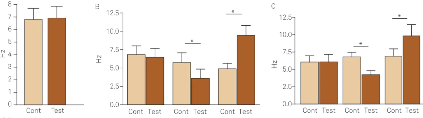
A total number of 51 cells were recorded in 24 animals. In the time-matched control group (n=4), 6 cells were re-corded and the basal iring rates varied from 2.7 to 9.0 Hz with a mean of 6.2±0.9 Hz. In the experimental groups (n=20), i.e., submitted to thermal stimulation, 45 cells were recorded and the basal iring rate varied from 0.3 to 10.0 Hz with a mean of 6.1±0.4 Hz.
Time-matched control group
In four animals, we stimulated the rat tail with a con-dom with water at room temperature, therefore no ther-mal stimuli were applied but just a mild tactile stimulation (Fig 1A). he basal iring rate of the 6 cells varied from 2.7 to 9.0 Hz, with a mean of 6.2±0.9 Hz. he mild tactile stimula-tion did not induce any signiicant changes to the basal iring rate of these units, varying from 3.7 to 10.0 Hz with a mean of 6.9±0.9 Hz (p>0.05). Fig 2 depicts representative sections with the plotted recording sites.
Neuronal firing rate modifications due to cold stimulation of the rat tail
In 10 animals, the basal iring rate of the 22 cells sub-mitted to cold stimulation of the rat tail varied from 0.3 to 10.0 Hz with a mean of 5.4±0.6 Hz (Fig 1B). Of the total num-ber of cells; 4 (18%) showed no response to the stimulus, with the control iring rate varying from 4.3 to 10.0 Hz with a mean of 6.5±1.2 Hz, and after cold stimulation the iring rate of the units was: 3.7 to 9.5 Hz with a mean of 6.5±1.2 Hz (p>0.05). Five cells (23 %) decreased its control iring rate, varying from 2.7 to 9.3 Hz with a mean of 5.7±1.3 Hz, and after cold stimulation the iring rate of the units was: 1.0 to 7.7 Hz with a mean of 3.6±1.2 Hz (p<0.01). Whilst 13 (59 %) increased its control iring rate, varying from 0.3 to 10.0 Hz with a mean of 4.9±0.7 Hz, and after cold stimulation the iring rate of the units was: 2.3 to 15.0 Hz with a mean of 9.5±1.3 Hz (p<0.01). Fig 2 depicts representative sections with the plotted recording sites.
Neuronal firing rate modifications due to warm stimulation of the rat tail
In 10 animals, the basal iring rate of the 23 cells sub-mitted to warm stimulation of the rat tail varied from 2.7 to 10.0 Hz with a mean of 6.6±0.5 Hz (Fig 1C). Of the total number of cells; 6 (26%) showed no response to the stimu-lus, with the control iring rate varying from 3 to 8 Hz with a mean of 6.1±0.9 Hz, and after warm stimulation the iring rate of the units was: 2.7 to 9.3 Hz with a mean of 6.1±0.9 Hz (p>0.05). Eleven cells (48%) decreased its control iring rate, varying from 3 to 10 Hz with a mean of 6.8±0.7 Hz, and af-ter warm stimulation the iring rate of the units was: 1.7 to 8.0 Hz with a mean of 4.2±0.6 Hz (p<0.001). Whilst 6 (26%) increased its control iring rate, varying from 2.7 to 10.0 Hz with a mean of 6.9±1.1 Hz, and after cold stimulation the iring rate of the units was: 3.3 to 16.0 Hz with a mean of 9.8±1.7 Hz (p<0.001). Fig 2 depicts representative sections with the plotted recording sites.
he basal iring rate distribution of neurons in the two ex-perimental groups, i.e. warm or cold stimulation, were com-pared with a χ2 test and no signiicant diferences were
ob-served in the distributions, implying that the two neuronal populations recorded from are similar.
DISCUSSION
hermoregulation is a complex relex mechanism that is controlled mainly by temperature responsive neurons in the anterior hypothalamus and preoptic area. Nevertheless, this system has its inal pathway in brainstem nuclei that control vasomotricity1. To our knowledge, this is the irst
report of the involvement of NRO in this brainstem thermo-regulatory circuitry.
252 Arq Neuropsiquiatr 2013;71(4):249-253
Serotonergic neurons in the caudal and rostral raphe nuclei discharge steadily in a state-dependent manner and share common pharmacological properties26. Nevertheless,
there are diferences in the mean iring rate of dorsal and cau-dal raphe nuclei neurons, being the iring rate of caucau-dal nu-clei neurons higher than of the dorsal raphe nunu-clei neurons25.
he iring rates distribution of the population of cells record-ed from in the present work is compatible with the expectrecord-ed behavior of caudal raphe nuclei neurons.
Cutaneous nociceptors and thermal receptors are struc-turally similar; they are both free axon terminals and origi-nate in similar neurons in the dorsal root ganglion. heir pro-jection to the spinal cord are mainly circumscribed in the irst and second spinal laminas where they make synaptic connections with second order neurons that project to spe-ciic thalamic nuclei via anterolateral ascending system27.
herefore, there are great similarities between the nocicep-tive and thermal neural processes starting at the receptor level, projections on the spinal cord, ascending pathways and even in its projection on the primary somatosensory cortex cortical area27. In this work, special care was taken so as not
to stimulate thermal sensitive nociceptors instead of true thermal sensitive terminals involved in thermoregulation.
As stated above, tail skin cooling in these experiments was within the non-noxious range. Such stimulation elicited an increase in the spontaneous activity of the majority of cells tested (59%). Tail skin heating experiments, conducted as pre-viously stated, induced a decrease in the spontaneous activ-ity of the majoractiv-ity of cells tested (48%). he opposite respons-es in neuronal activity of raphe obscurus units to cooling or heating suggest that these cells are involved in producing the homeothermic relevant vascular adaptations secondary to changes in cutaneous temperature in the rat tail.
he present results demonstrate a signiicant number of neurons that have its basal activity modiied by thermal stim-ulation. his may be reinforced by the fact that caudal raphe nuclei have been implicated in the induction of a variety of
speciic patterns of hemodynamic responses, such as those found in desynchronized sleep22 and nociception23.
he “serotonin syndrome” is a rare disorder of 5-HT ex-cess, with indings of hyperthermia, shivering, seizures, coma or even death, which can acutely result from seroto-nergic drug interactions28. his association between 5-HT
excess in the central nervous system and hyperthermia is possibly consistent with our indings showing that NRO neurons, which are serotonergic, play a role in thermoregu-lation. However, the precise mechanisms involved remains to be established.
Fig 2. Representative sagittal, coronal sections and
microphotography of the rat brainstem, displaying electrode track (Tr) and recording sites plotted from four animals.
: increase in unitary discharge rate due to warm stimulation; : decrease in unitary discharge rate due to warm
stimulation; O: no response; Δ: increase in unitary discharge rate due to cold stimulation; : decrease in unitary discharge rate due to cold stimulation; NRM: nucleus raphe magnus; NRO: nucleus raphe obscures; NRP: nucleus raphe pallidus; pyx: pyramidal tract. Representative sections modiied from Paxinos and Watson24.
Tr
NRM pyx
NRP
100 µm
NRO
NRP pyx NRO
Obex Tr Tr Tr
Fig 1. Firing rate responses of nucleus raphe obscurus neurons to thermal stimulation in the rat tail. (A) Time-matched control group; (B) effect of cold stimulation in the rat tail; (C) effect of warm stimulation in the rat tail. In (B and C), cells were separated according to their response proiles to thermal stimulation; i.e.,no response (left double columns), reduction (middle double columns) or increase in iring rate (right double columns). Cont = unitary basal iring rate. Test = unitary iring rate after thermal challenge.
8
7
6
5
4
3
2
1
0 A
Hz
Cont Test Cont Test Cont Test Cont Test 0.0
2.5 5.0 7.5 10.0 12.5
*
* B
H
z
Cont Test Cont Test Cont Test 0.0
2.5 5.0 7.5 10.0 12.5
*
* C
H
1. M ur . etue t f t
Ar ut u t u m eulatu rtuo utal
u al pat wayf t m eulat f u . J pl Phy
2011;110:1137-1149.
2. G uJ. T m e f t at at. Phy s v
1990;47:963-991.
3. J, La u eM. t u ut t m eulat
fftf pr tarm u e uat. Phy s v 1979;23:723-732.
4. K u a, Yu ujiwara M, u T. t m
u t f t m eulat mt ut . m J Phy
1994;267(1 Pt 2):R283-288.
5. O’Leary DS, Johnson JM, Taylor WF. Mode of neural control mediating rat tail vasodilatation during heating. J Appl Physiol 1985;59:1533-1538.
6. Rathner JA, McAllen RM. Differential control of sympathetic drive to the rat tail artery and kidney by medullary premotor cell groups. Brain Res 1999;834:196-199.
7. Smith JE, Jansen AS, Gilbey MP, Loewy AD. CNS cell groups projecting to sympathetic outlow of tail artery: neural circuits involved in heat loss in the rat. Brain Res 1998;786:153-164.
8. Coote J! F t t, L e ur.T uv m utf t u u
u u ut u t uf ru u tt u e uhr u " .s u #$ % &' (:277-$ (.
#. T ),s , W g F.T u f t utm
ut t.* . mt eaphu t ct t u eu al u .J m #+&114:161-187.
10. swk M ! Abtt .
, utt t
valuat uf u u e
t u eu u u tu gic pr t u f m t m
ft uto u f xt u x t u f t u uu
t ut m u y pr t u eu u, tutf mt
u m eu .s u # #&% o15-%.
11. Ray RS, u),s t , t . Impair at u
t mat ut u t tue u u u t u .
r u &' ' 'o +'; +( .
. s t ! Patt uM!r tt uG M !M u, Z ue.* u t
t udal r
-u u uv
utmal, car
,
u
e t ut t u eulat u . t ms % &123:(; %+.
'. M u r , Nakamura K. ut u al pat way f
t meulat u . Fr uts ;16:;((.
(. Nakamura K,M ur .ut ff ut t waym t u e u
u ev ympat tt m e u u u t .
Am J Phy e * ut e mA y ;&# o ; '+ .
%. rmt JE, Ju uS,G y MP, Lwy AD.r e t u e
tympat tic outlow of tail artery: neural circuits involved in heat
loss in the rat. Brain Res 1998;786:153-164.
16. Tanaka M, Nagashima K, McAllen RM, Kanosue K. Role of the medullary raphé in thermoregulatory vasomotor control in rats. J Physiol 2002;540:657-664.
17. Tanaka M, Ootsuka Y, McKinley MJ, McAllen RM. Independent vasomotor control of rat tail and proximal hairy skin. J Physiol 2007;582:421-433.
18. Nakamura K, Matsumura K, Hübschle T, et al. Identification of sympathetic premotor neurons in medullary raphe regions mediating fever and other thermoregulatory functions. J Neurosci 2004;24:5370-5380.
19. Ootsuka Y, Blessing WW, McAllen RM. Inhibition of rostral medullary raphé neurons prevents cold-induced activity in sympathetic nerves to rat tail and rabbit ear arteries. Neurosci Lett 2004;357:58-62.
20. Ootsuka Y, Blessing WW. Inhibition of medullary raphé/parapyramidal neurons prevents cutaneous vasoconstriction elicited by alerting stimuli and by cold exposure in conscious rabbits. Brain Res 2005;1051:189-193.
21. Ootsuka Y, McAllen RM. Interactive drives from two brain stem premotor nuclei are essential to support rat tail sympathetic activity. Am J Physiol Regul Integr Comp Physiol 2005;289:R1107-1115.
22. Futuro-Neto HA, Coote JH. Changes in sympathetic activity to heart and blood vessels during desynchronized sleep. Brain Res 1982;252:259-268.
23. Dantas MA, Có W, Futuro-Neto HA. Responses of neurons of the nucleus raphe obscurus to noxious stimuli. Braz J Med Biol Res 1990;23:923-926.
24. Paxinos G, Watson C. The rat brain in stereotaxic coordinates. 6th ed. San Diego: Elsevier, 2007. p. 1-166.
25. Futuro Neto HA, Dantas MA, Gilbey MP. A comparison of the effects of eserine sulfate on the activity of medullary raphe neurons in the anesthetized rabbit and rat. Braz J Med Biol Res 1994;27:1445-1454.
26. Jacobs BL, Azmitia EC. Structure and function of the brain serotonin system. Physiol Rev 1992;72:165-229.
27. Chéry-Croze S. Painful sensation induced by a thermal cutaneous stimulus. Pain 1983;17:109-137.
28. Sternbach H. The serotonin syndrome. Am J Psychiatry 1991;148:705-713. References