217
Embolism caused by metallic mercury: a case report
Radiol Bras 2007;40(3):217–219
Case Report
EMBOLISM CAUSED BY METALLIC MERCURY: A CASE REPORT*
João Paulo Kawaoka Matushita1
, Roberto Márcio A. Peixoto2
, Hilton Muniz Leão Filho2 , Guilherme Carvalho Missiaggia2
, Ricardo Saraiva Dias2
, Wilson Campos Tavares Junior2 , Cristina Sebastião Matushita3
, José Nelson Mendes Vieira4
A case of a male, 29-year-old patient who, in a suicide attempt, has self-administered a 2 ml injection of metallic mercury into his left arm is reported. Radiological studies have shown the presence of several me-tallic spheres in the subcutaneous tissue of the olecranal fossa and along the veins of the left arm, with dissemination to lungs, heart, liver, spleen and brain.
Keywords: Embolism; Mercury; Radiology; Computed tomography.
Embolismo por mercúrio metálico: relato de caso.
Relata-se um caso de um paciente do sexo masculino, de 29 anos de idade, que na tentativa de auto-exter-mínio injetou 2 ml de mercúrio industrial no antebraço esquerdo. O estudo radiológico mostrou várias esfe-ras metálicas no subcutâneo da fossa olecraniana e ao longo das veias do braço esquerdo, com dissemina-ção para pulmões, coradissemina-ção, fígado, baço e encéfalo.
Unitermos: Embolismo; Mercúrio; Radiologia; Tomografia computadorizada. Abstract
Resumo
* Study developed in the Department of Radiology and Imag-ing Diagnosis of Hospital das Clínicas, Faculdade de Medicina da Universidade Federal de Minas Gerais (UFMG), Belo Hori-zonte, MG, Brazil.
1. PhD, Associate Professor at Faculdade de Medicina da Universidade Federal de Minas Gerais (UFMG), Full Professor of Radiology and Imaging Diagnosis, Course of Physiotherapy at Faculdade Biológicas da Saúde do Centro Universitário Newton Paiva, Belo Horizonte, MG, Brazil.
2. MDs, Residents in the Service of Imaging Diagnosis at Hospital das Clínicas, Faculdade de Medicina da Universidade Federal de Minas Gerais (UFMG), Belo Horizonte, MG, Brazil.
3. Graduate Student of Medicine at Faculdade de Medicina da Universidade do Vale do Sapucaí (Univás), Pouso Alegre, MG, Brazil.
4. MD, Radiologist, Professor at Faculdade de Medicina da Universidade Federal de Minas Gerais (UFMG), Belo Horizonte, MG, Brazil.
Mailing address: Prof. Dr. João Paulo Kawaoka Matushita. Rua Caetés, 530/216, Centro. Belo Horizonte, MG, Brazil, 30120-080. E-mail: [email protected]
Received April 12, 2005. Accepted after revision July 6, 2005.
INTRODUCTION
Reports on metallic mercury injection are unusual in the literature, and embolism caused by these metallic particles is rare. Most frequent causes are intravenous injec-tions defined as suicide attempts(1,2) or a
complication resulting from drugs abuse(3).
Injection of mercury by accident may oc-cur because of injuries from a broken ther-mometer(4,5).
Subcutaneous administration(6,7) or
vas-cular migration following injection(2,3,8)
may result in granulomas and abscesses in the site.
Systemic signs of toxicity by mercury are not found immediately; however, four cases have been found in the literature with
systemic manifestation after a subcutane-ous injection(9,10).
CASE REPORT
Male, 29-year-old patient, born in Divi-nópolis, MG, Brazil, who, in January/1988, with 16 years of age, tried to suicide with a subcutaneous auto-injection of 2 ml me-tallic mercury into his left forearm. Eight hours post-injection, he presented an creased tenderness in the region of the in-jection, with “tugging” and loss of coordi-nation in the trunk and in the correspondent upper limb lasting for about three days.
He sought medical assistance in the fol-lowing day, by the doctor did not believe him and no laboratory test or study was requested. Some weeks later, the patient presented with an inflammatory foreign-body-type reaction at the site of the injec-tion which had to be surgically drained for three times, eliminating a solid, silvery material resembling sand grains. The pa-tient developed anorexia with a 10 kg weight loss in few weeks. Clinically, he complained of excessive sudoresis, somno-lence, and metallic taste in the mouth. As regards family antecedents, the patient re-ported suicidal ideas after his parents´ sepa-ration, when he was eight years old.
On physical/neurological examination, the patient presented intentional tremor, with upper spastic paraparesis at left,
shuf-fling walk and hyperreflexia of lower limbs. On clinical examination of the left arm, firm, tumor-like lesions covered by erythemato-violaceous and smooth skin could be observed. One of these lesions presented an elastic consistency, with fluc-tuation in the cubital fossa. Among the le-sions, there was a 5 cm-long and 1 cm-wide fibrotic cord. His left upper limb extension was impaired.
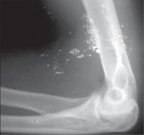
Radiological studies demonstrated: left
forearm radiograph – multiple spheres
with a metallic density subcutaneously clustered in the olecranal fossa, and other, sparse along the veins of the correspondent arm (Figure 1); chest radiographs – several spheres with a metallic density, bilaterally
218
Matushita JPK et al.
Radiol Bras 2007;40(3):217–219
sparse throughout the pulmonary paren-chyma and heart shadow (Figure 2); plain
abdominal radiograph – some spherical
structures with a metallic density in the liver, spleen and abdominal vessels (Figure 3); brain computed tomography (without infusion of hydrosoluble iodinated contrast agent) – small sphere with a metallic den-sity on the left cerebellar hemisphere (Fig-ure 4).
Laboratory tests showed normal levels of urea, creatinine, bilirubin as well as nor-mal hemogram values. Measurements of 24-hour urinary mercury excretion showed the following values: May/1990, 81µg/l; July/1990, 71µg/l; February/1993, 340µg/ l; August/2000, 60.7 µg/l (reference value: up to 5 µg/l; biological tolerance limit: 50 µg/l). Electroneuromiography performed in July/2001 was normal.
On July 13, 1989, the patient was suc-cessfully submitted to surgical excision of the lesion in the left upper limb, with skin graft. The anatomopathological report on the surgical specimen described:
macro-scopy – irregular fragment of fibrotic
tis-sue measuring 10 cm × 4,6 cm × 4 cm, with areas covered by ulcerated skin, pres-ence of fragments of fibrotic tissue in pel-lets of inorganic mercury; microscopy – dermis in granulation tissue and intense histiocytic reaction, with microabscesses forming granulomas; blackened, round
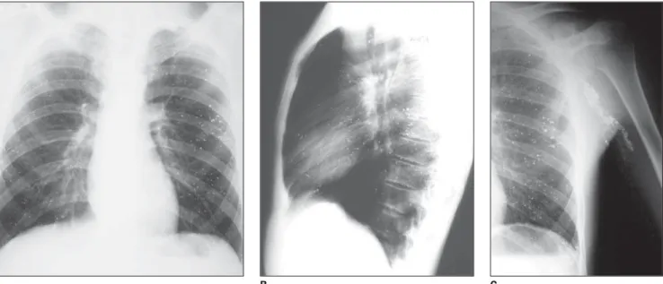
Figure 2. Posteroanterior (A) and lateral (B) chest radiographs showing several, bilateral, sparse spherical metallic densities throughout the pulmonary paren-chyma and in the mediastinal shadow. Anteroposterior left hemithorax radiograph (C) showing other metallic, spherical structures along the axillary vein and homolateral lung.
A B C
Figure 3. Plain abdominal radiograph showing spherical, metallic bodies in the topography of the liver, spleen and abdominal vessels.
Figure 4. Non-contrast-enhanced computed to-mography of the skull identifying a small metallic sphere in the left cerebellar hemisphere.
structures varying in size, lymph nodes presenting follicles with enlarged centers and signs of phagocytosis. Diagnosis: Chronic granulomatous inflammation sec-ondary to foreign body. Lymph nodes in reactional status. Sinusal histiocytosis.
DISCUSSION
Subcutaneous deposition of metallic mercury with formation of granulomas
occurs because of leakage during the at-tempt of intravascular injection(3,11–13).
Accidents may occur with arterial pressure monitors with a liquid mercury manometer connected to an intra-arterial needle. Ra-diologically, mercury emboli present as small metallic densities in soft tissues of extremities, distal to the site of the manom-eter needle insertion(14).
at-219
Embolism caused by metallic mercury: a case report
Radiol Bras 2007;40(3):217–219
tempts(1,2) and thermometers break(4,5).
Ra-diological findings in patients with metal-lic mercury emboli in the vascular system identify multiple small, spherical metallic densities in the drainage pathways at the site of insertion, and also in one or both lungs.
Probably, the most difficult differential radiological diagnosis would be between mercury emboli through the pulmonary artery which may occur by venous way, and intrabronchial mercury, which may be as-pirated in case of a thermometer break in-side the oral cavity. Identification of mer-cury inside abdominal vessels, in the right ventricle apex, or in subcutaneous tissue of extremities suggests the arterial pathway as route of entry, and identification of
intra-intestinal mercury suggests the metal inges-tion.
REFERENCES
1. Johnson HR, Koumides O. Unusual case of mer-cury poisoning. Br Med J 1967;1:340–341. 2. Roden R, Fraser-Moodie A. Self-injection with
mercury. Injury 1993;24:191–192.
3. Netscher DT, Friedland JA, Guzewicz RM. Mer-cury poisoning from intravenous injection: treat-ment by granuloma resection. Ann Plast Surg 1991;26:592–596.
4. Rachman R. Soft-tissue injury by mercury from a broken thermometer. A case report and review of the literature. Am J Clin Pathol 1974;61:296–300. 5. Tanner DC, Branch M, Schreiner RD, Morgan HJ. Subcutaneous deposition of elemental mercury. J Tenn Med Assoc 1988;81:698–699. 6. Hill DM. Self-administration of mercury by
sub-cutaneous injection. Br Med J 1967;1:342–343. 7. Gebauer K, Connor B. Cutaneous mercury granu-loma. Australas J Dermatol 1991;32:129–132.
8. Hannigan BG. Self-administration of metallic mercury by intravenous injection. Br Med J 1978; 2:933.
9. Kumar A, Jain R, Sawhney S, Goel AK, Chattopa-dhyay K. Intravenous administration of metallic mercury with homicidal intent. J Assoc Physi-cians India 1992;40:640–641.
10. Schulz E, Beskind H. Systemic deposition of metallic mercury. J Pediatr 1960;57:733–737. 11. Oliver RM, Thomas MR, Cornaby AJ, Neville E.
Mercury pulmonary emboli following intrave-nous self-injection. Br J Dis Chest 1987;81:76– 79.
12. Berger RL, Madoff IM, Ryan TJ. Mercury embo-lization during arterial pressure monitoring. J Thorac Cardiovasc Surg 1967;53:285–287. 13. Esau P. Flooding of pulmonary circulation with
mercury. Deutsche Ztschr f. Chir 1924;188:221– 233.