XI
Radiol Bras. 2009 Jul/Ago;42(4):XI–XII
Pedro José de Santana Júnior1
, Kim-Ir-Sen Santos Teixeira2
, Pedro Paulo Teixeira e Silva Torres3 , Renato Tavares Daher1
, Patrícia Karla Vilarinho Santana4
, Ana Caroline Vieira Aurione5
Study developed at Departamento de Radiologia e Diagnóstico por Imagem do Hospital das Clínicas da Universidade Federal de Goiás (UFG), Goiânia, GO, Brazil. 1. MD, Resident, Department of Radiology and Diagnostic Imaging – Hospital das Clínicas da Universidade Federal de Goiás (UFG), Goiânia, GO, Brazil. 2. PhD, Associate Professor and Head, Department of Radiology and Diagnostic Imaging – Hospital das Clínicas da Universidade Federal de Goiás (UFG), Goiânia, GO, Brazil. 3. MD, Radiologist, Trainee at Unit of Magnetic Resonance Imaging, Department of Diagnostic Imaging – Santa Casa de Misericórdia de São Paulo, São Paulo, SP, Brazil. 4. MD, Secretaria Municipal de Saúde de Goiânia, Goiânia, GO, Brazil. 5. Graduate Student, Faculdade de Medicina da Universidade Federal de Goiás (UFG), Goiânia, GO, Brazil. Mailing address: Dr. Pedro José de Santana Júnior. Rua T-36, nº 3485, ap. 104, Setor Bueno. Goiânia, GO, Brazil, 74223-050. E-mail: [email protected]
0100-3984 © Colégio Brasileiro de Radiologia e Diagnóstico por Imagem
Which is your diagnosis?
•
Qual o seu diagnóstico?
Santana Jr PJ, Teixeira KISS, Torres PPTS, Daher RT, Santana PKV, Aurione ACV. Which is your diagnosis? Radiol Bras. 2009;42(4):XI–XII.
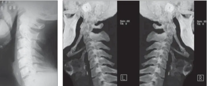
A male, 41-year-old patient, agricultural worker, complaining of pain in the right shoulder and in the whole cervical region besides facial pain in the region of the temporomandibular joint. At clinical exami-nation, a hardened bulging mass was felt at palpation of the right anterior pillar of the tonsillar fossa in association with limited mouth opening. The patient underwent radiography and multidetector computed tomography of the skull base.
Figure 3. Computed tomography image with 3D reconstruction of the cervical spine.
Figure 2. Computed tomography image with 3D reconstruction of the cervical spine.
XII Radiol Bras. 2009 Jul/Ago;42(4):XI–XII Images description
Figures 1, 2 and 3. Bilateral stylohyoid ligament calcification.
Diagnosis: Eagle syndrome.
COMMENTS
Eagle syndrome corresponds to a set of symptoms including recurrent facial and cervical pain (with or without irradiating to the ear or mastoid region), dysphagia, sen-sation of a foreign body in the throat, sia-lorrhea, glossalgia, dysphonia, recurrent headache, carotidynia, vertigo, visual per-turbation and restricted neck motion. This disease was first described in 1937 by the otolaryngologist Watt W. Eagle, and is di-rectly associated with the styloid process elongation or stylohyoid ligament calcifi-cation. Incidence is higher in women, at the fourth decade of life, with no preference for unilateral or bilateral presentation(1).
The temporal bone styloid process cor-responds to an osseous projection measur-ing approximately 25 mm in length, located posteriorly to the pharynx, between the internal and external carotid arteries. The set formed by the styloid process, stylooid ligament and the small horn of the hy-oid bone corresponds to the stylohyhy-oid complex or apparatus whose embryologi-cal origin is the Reichert’s cartilage of the second brachial arch(2).
The etiology of the styloid apophysis elongation (> 30 mm) still remains un-known. There are several theories explain the etiopathology of the disease such as id-iopathic, congenital alteration resulting from the persistence of one of the
precur-sory cartilages, or ossification of the styloid ligament(3).
Most patients with elongated styloid process are asymptomatic, and the diagno-sis can only be achieved through imaging studies. The incidence of this abnormality in the general population ranges between 4% and 28%, and only 4% to 10.3% of the patients in this group are symptomatic(2).
Differential diagnoses for Eagle syn-drome include: cervical arthritis, temporo-mandibular joint disorders, otitis, mastoidi-tis, sialadenimastoidi-tis, sialolithiasis, esophageal diverticulosis, temporal arteritis, myofas-cial pain, chronic pharyngotonsillitis, unerupted or impacted third molar, hemic-rania, histamine headache, pharynx tumor, tumor in the base of the tongue, trigeminal neuralgia, glossopharyngeal, upper laryn-geal and sphenopalatine tumors(1).
Elongated styloid apophysis is sus-pected through clinical examination with palpation of the tonsillar fossa, and evi-denced by imaging methods on facial, lat-eral, anteroposterior and oblique views. Computed tomography is the method of choice for this evaluation. Considering the variable degree of calcification and vari-ability in the stylohyoid complex presenta-tions, some authors have developed a ra-diographic classification system describing the stylohyoid complex as elongated, pseudoarticulated or segmented, the sec-ond one correspsec-onding to the computed tomography images shown in the present case. Computed tomography plays a criti-cal role in the diagnosis of Eagle syndrome for allowing images acquisition in the axial, sagittal and coronal planes, as well
as multiplanar and 3D reconstructions. Thus, this method demonstrates not only bone structures and calcified components — likewise radiographic studies —, but also their relationship with the other adja-cent structures, overcoming the conven-tional radiology limitations(3).
Although the surgical therapy (styloid apophysis resection) is more effective, in the present case a conservative manage-ment was adopted, utilizing anti-inflamma-tory drugs, with a good outcome till the present moment(4,5).
The presentation of a case like this is important because of the scarcity of simi-lar reports in the radiological literature, in spite of the dental and otolaryngological lit-erature. Considering that Eagle syndrome is included in the spectrum of differential diagnoses of several cervicofacial diseases, the knowledge about this entity is critical for a correct diagnosis and consequent therapeutic approach(5).
REFERENCES
1. Savranlar A, Uzun L, Uður MB, et al. Three-di-mensional CT of Eagle’s syndrome. Diagn Interv Radiol. 2005;11:206–9.
2. Chiang KH, Chang PY, Chou ASB, et al. Eagle’s syndrome with 3-D reconstructed CT: two cases report. Chin J Radiol. 2004;29:353–7. 3. Sá ACD, Zardo M, Paes Junior AJO, et al.
Alon-gamento do processo estilóide (síndrome de Eagle): relato de dois casos. Radiol Bras. 2004; 37:385–7.
4. Tiago RSL, Marques Filho MF, Maia CAS, et al. Síndrome de Eagle: avaliação do tratamento ci-rúrgico. Rev Bras Otorrinolaringol. 2002;68:196– 201.