XI
Radiol Bras. 2009 Nov/Dez;42(6):XI–XIII Marcelo Souto Nacif1
, Karen Amaral do Vabo2
, Telmo Pimentel do Vabo3
, Ronaldo Maselli de Pinheiro Gouvêa4
, Ricardo Ebecken5
, Alair Augusto Sarmet Moreira Damas dos Santos6
Study developed at the Imaging Center of Hospital de Clínicas de Niterói (HCN) and at Department of Radiology – Universidade Federal Fluminense (UFF), Niterói, RJ, Brazil. 1. Professor at Centro Universitário Serra dos Órgãos (Unifeso), Teresópolis, RJ, in charge of the Unit of Cardiac MRI at Hospital de Clínicas de Niterói (HCN), Niterói, RJ, Brazil. 2. MD, Radiologist, Hospital de Clínicas de Niterói (HCN), Niterói, RJ, Brazil. 3. Sub-chief of Imaging Center of Hospital de Clínicas de Niterói (HCN), Niterói, RJ, Brazil. 4. Post-graduation in Radiology and Imaging Diagnosis, Instituto de Pós-Graduação Médica Carlos Chagas (IPGMCC), Niterói, RJ, Brazil. 5. Head for Division of Endoscopy, Hospital de Clínicas de Niterói (HCN), Niterói, RJ, Brazil. 6. Titular Professor, Course of Post-Graduation in Radiology and Imaging Diagnosis at Instituto de Pós-Graduação Médica Carlos Chagas (IPGMCC), Coordinator for Imaging Center of Hospital de Clínicas de Niterói (HCN), Niterói, RJ, Brazil. Mailing address: Dr. Marcelo Souto Nacif. Rua Tavares de Macedo, 136, ap. 1503, Bloco A, Icaraí. Niterói, RJ, Brazil. E-mail: [email protected] / www.msnacif.med.br
0100-3984 © Colégio Brasileiro de Radiologia e Diagnóstico por Imagem
Which is your diagnosis?
•
Qual o seu diagnóstico?
Nacif MS, Vabo KA, Vabo TP, Gouvêa RMP, Ebecken R, Santos AASMD. Which is your diagnosis? Radiol Bras. 2009;42(6):XI–XIII.
Female, 66 year-old patient complain-ing of stomach pain. On January 27, 2007, the patient presented a sudden, dull ache in the epigastric region, with no irradiation or trigger factor that improved with the use of symptomatic medication. The patient re-mained asymptomatic for approximately one month, when the epigastric pain re-turned upon ingestion of fatty food, but now with higher intensity, demanding emergency treatment. She denied vomiting, choluria, acholia or fever. Investigation on an out-patient basis was indicated for the suspicion of hepatitis, and the patient was submitted to abdominal ultrasonography and computed tomography.
One week later, the patient presented a new episode of epigastric pain followed by vomiting, choluria and acholia, and on February 28, 2007 she was admitted to Hospital de Clínicas de Niterói, RJ, Brazil. During her hospital stay, the patient re-ported improvement of the algic symp-tomatology with null diet and underwent
magnetic resonance cholangiography. The only relevant fact in the patient’s previous history was a cholecystectomy ten years ago. At clinical examination, the only find-ing was jaundice (2+/4+). Laboratory tests demonstrated cholestasis indicators with
Figure 2. A,B,C,D: Upper abdominal magnetic resonance cholangiography.
A B C D
high levels of bilirubin (remarkably direct bilirubin), TGO and TGP, as well as alka-line phophatase and gamma-GT. Also, CA-19-9 serum levels were high. Serum amy-lase and lipase levels were normal.
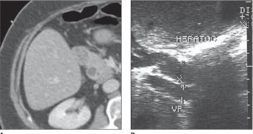
Figure 1.A: Contrast-enhanced abdominal computed tomography B: Upper abdominal ultrasonography.
XII Radiol Bras. 2009 Nov/Dez;42(6):XI–XIII
Images description
Figure 1. A: Contrast-enhanced ab-dominal computed tomography B: Upper abdominal ultrasonography. The sono-graphic image (impaired by the excessive presence of intestinal gas) and the CT im-age demonstrate dilatation of the extrahe-patic biliary tract. No abnormality is ob-served in the pancreatic duct and head.
Figure 2. A,B,C,D: Upper abdominal magnetic resonance cholangiography. A polypoid mass is observed in the major papilla of the duodenum, in association with choledocal dilatation with poorly fac-eted calculi. On the reconstructed image, distal interruption of the irregular lumen, neither abrupt nor filiform, is observed.
Diagnosis: Adenoma in the hepatopan-creatic ampulla.
COMMENTS
Adenomas are the most common tu-mors of the hepatopancreatic ampulla (0.04–0.12% of necropsies), being less fre-quently found than malignant lesions in this structure. Other benign tumors such as lipomas, hamartomas, fibromas and neuro-genic tumors may be found in this region. Typically, these tumors do not present any sex predilection, and mean age of patients is about 60 years(1,2).
These tumors may achieve 4 mm to 7 cm in size and, although extremely small tumors may be asymptomatic, 75% of pa-tients are symptomatic at the moment of the diagnosis(1,3,4).
The most frequent symptoms are the following: non-specific abdominal pain and jaundice (respectively 75% and 70%), besides digestive hemorrhage (50%). Laboratory tests demonstrate a cholestatic pattern of jaundice, with high levels of bi-lirubin – remarkably direct bibi-lirubin –, gamma-GT and alkaline phosphatase. In-termittent episodes of pancreatitis are not infrequent and high CA 19-9 serum levels may be observed(5–7).
Frequently, imaging findings demon-strate coexistence with biliary lithiasis, and a partial distal obstruction leading to a “double lumen” pattern (dilatation of the main biliary and pancreatic tract lumen), which has not been found in the present case. Most commonly, defective duodenal
filling is observed as a single finding which is extremely non-specific in this region, and, also, the utilization of intravenous contrast media (either gadolinium or io-dine) is quite controversial in the differen-tiation between malignant and benign tu-mors of this region(4,8).
Considering that adenomas, particularly the villous ones, present a high maligniza-tion rate, a radical therapy must be consid-ered for these patients (6,8).
In summary, the imaging findings of the present case demonstrate a picture of dila-tation of extrahepatic biliary ducts, with preserved main pancreatic ducts. Other finding is defective filling of the chole-docal lumen compatible with the presence of poorly faceted and probably cholesterol-rich calculi (which cannot be seen at com-puted tomography). Additionally, a regular, “polypoid” mass is observed in the area of the hepatopancreatic ampulla leading to defective duodenal lumen filling, and de-termining a poorly regular transition with the choledocal lumen (neither abrupt nor filiform). Such mass measures approxi-mately 3 cm, representing an argument against a non-neoplastic disease of this
structure such as odditis (up to 1.5 cm) fre-quently secondary to calculi impaction.
Thus, the suspicion of neoplastic lesion was raised, notwithstanding the finding of “primary” calculi (generally cholesterol-rich and moulded by the choledocal lumen) be poorly expected in periampullary malig-nant diseases which generally present a bad prognosis and extremely short survival. It is important to remember that residual cal-culi are generally faceted and usually cause complications within up to three years fol-lowing cholecystectomy.
The patient was submitted to endo-scopic retrograde cholangiopancreato-graphy with biopsy and excision of the le-sion (Figure 3).
Final considerations
As demonstrated in the present case, di-agnostic suspicion may be raised even be-fore histopathological examination by means of noninvasive methods in associa-tion with clinical and laboratory data in the search of a complete semiology of periam-pullary lesions, facilitating the therapeutic guidance. Adenoma in the hepatopan-creatic ampulla must be considered by the
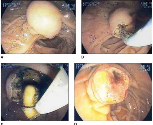
Figure 3. A,B,C,D: Endoscopic retrograde cholangiopancreatography demonstrating enlargement of the major duodenal papilla. Papillotomy was performed, with extraction of poorly faceted, brownish calculi, and extensive biopsy of the lesion.
D C
XIII
Radiol Bras. 2009 Nov/Dez;42(6):XI–XIII
radiologist in the differential diagnosis of lesions in this region.
REFERENCES
1. Buck JL, Elsayed AM. Ampullary tumors: radio-logic-pathologic correlation. Radiographics. 1993; 13:193–212.
2. Guibaud L, Bret PM, Reinhold C, et al. Bile duct obstruction and choledocholithiasis: diagnosis with MR cholangiography. Radiology. 1995;197: 109–15.
3. Kim MJ, Mitchell DG, Ito K, et al. Biliary
dilata-tion: differentiation of benign from malignant causes – value of adding conventional MR imag-ing to MR cholangiopancreatography.Radiology. 2000;214:173–81.
4. Asbach P, Klessen C, Kroencke TJ, et al. Magnetic resonance cholangiopancreatography using a free-breathing T2-weighted turbo spin-echo se-quence with navigator-triggered prospective ac-quisition correction. Magn Reson Imaging. 2005; 23:939–45.
5. Schindera ST, Merkle EM. MR cholangiopan-creatography: 1.5T versus 3T. Magn Reson Im-aging Clin N Am. 2007;15:355–64.
6. Morita S, Suzuki K, Machida H, et al. Prospec-tive trial of a navigator setting under left hepatic lobe on magnetic resonance cholangiopancrea-tography using a free-breathing prospective ac-quisition correction technique. Magn Reson Im-aging. 2008;26:841–6.
7. Kim JH, Kim MJ, Chung JJ, et al. Differential di-agnosis of periampullary carcinomas at MR im-aging. Radiographics. 2002;22:1335–52.