Accuracy of sonography and hysteroscopy in
the diagnosis of premalignant and malignant
polyps in postmenopausal women
Acurácia da ultrassonografia e da histeroscopia no diagnóstico de
pólipos endometriais pré-malignos e malignos na pós-menopausa
sirlEi siani morais1aarão mEndEs Pinto-nEto1 luCia Costa-Paiva1
Abstract
PURPOSE: To evaluate the accuracy of sonographic endometrial thickness and hysteroscopic characteristics in predicting malignancy in postmenopausal women undergoing surgical resection of endometrial polyps. METHODS: Five hundred twenty-one (521) postmenopausal women undergoing hysteroscopic resection of endometrial polyps between January 1998 and December 2008 were studied. For each value of sonographic endometrial thickness and polyp size on hysteroscopy, the sensitivity, speciicity, positive predictive value (PPV) and negative predictive value (NPV) were calculated in relation to the histologic diagnosis of malignancy. The best values of sensitivity and speciicity for the diagnosis of malignancy were determined by the Receiver Operating Characteristic (ROC) curve. RESULTS: Histologic diagnosis identiied the presence of premalignancy or malignancy in 4.1% of cases. Sonographic measurement revealed a greater endometrial thickness in cases of malignant polyps when compared to benign and premalignant polyps. On surgical hysteroscopy, malignant endometrial polyps were also larger. An endometrial thickness of 13 mm showed a sensitivity of 69.6%, speciicity of 68.5%, PPV of 9.3%, and NPV of 98% in predicting malignancy in endometrial polyps. Polyp measurement by hysteroscopy showed that for polyps 30 mm in size, the sensitivity was 47.8%, speciicity was 66.1%, PPV was 6.1%, and NPV was 96.5% for predicting cancer. CONCLUSIONS: Sonographic endometrial thickness showed a higher level of accuracy than hysteroscopic measurement in predicting malignancy in endometrial polyps. Despite this, both techniques showed low accuracy for predicting malignancy in endometrial polyps in postmenopausal women. In suspected cases, histologic evaluation is necessary to exclude malignancy.
Resumo
OBJETIVO: Avaliar a acurácia da espessura endometrial ecográica e características histeroscópicas em predizer malignidade em mulheres na pós-menopausa submetidas à ressecção cirúrgica de pólipos endometriais. MÉTODOS: Quinhentos e vinte e uma (521) mulheres na pós-menopausa submetidas à ressecção histeroscópica de pólipo endometrial entre janeiro de 1998 e dezembro de 2008 foram incluídas no estudo. Para cada valor de espessura endometrial ecográica e tamanho dos pólipos na histeroscopia, a sensibilidade, a especiicidade, valor preditivo positivo (VPP) e valor preditivo negativo (VPN) foram calculados em relação ao diagnóstico histológico de malignidade. Os melhores valores de sensibilidade e especiicidade para o diagnóstico de malignidade foram determinados pela curva Receiver Operating Characteristic (ROC). RESULTADOS: O diagnóstico histológico identiicou a presença de pré-malignidade ou malignidade em 4,1% dos casos. A espessura endometrial medida por ultrassonograia em casos de pólipos malignos foi maior quando comparado com pólipos benignos e pré-malignos. Na histeroscopia os pólipos malignos também foram maiores. A espessura endometrial de 13 mm mostrou uma sensibilidade de 69,6%, especiicidade de 68,5%, VPP de 9,3% e VPN de 98% para predizer malignidade em pólipo endometrial. A medida do pólipo por histeroscopia mostrou que para pólipos de 30 mm de tamanho, a sensibilidade foi de 47,8%, a especiicidade foi de 66,1%, VPP foi de 6,1% e VPN foi de 96,5% para predizer o câncer. CONCLUSÕES: A espessura endometrial ultrassonográica mostrou uma maior acurácia que a avaliação histeroscópica do tamanho do pólipo para predizer malignidade nessas lesões endometriais. Apesar disso, ambas as técnicas não mostraram boa acurácia para excluir a necessidade de fazer a avaliação histológica dos casos suspeitos.
Study carried out at Universidade Estadual de Campinas – Unicamp – Campinas (SP), Brasil.
1Departamento de Tocoginecologia, Faculdade de Ciências Médicas, Universidade Estadual de Campinas – Unicamp – Campinas
(SP), Brasil.
Keywords
Polyps/surgery Endometrium/pathology Endometrium/ultrasonography Hysteroscopy/methods Biopsy
Palavras-chave
Pólipos/cirurgia Endométrio/patologia Endométrio/ultrassonograia
Histeroscopia/métodos Biópsia
Correspondence Lucia Costa-Paiva Department of Obstetrics and Gynecology, Faculdade de Ciências Médicas, Universidade Estadual de Campinas – Unicamp P.O. Box 6111 CEP: 13083-970 Campinas (SP), Brazil
Received 05/22/2012
Accepted with modiications 05/14/2013
Introduction
With the routine use of ultrasound for the investi-gation of abnormal uterine bleeding or postmenopausal bleeding over the last 20 years, the diagnoses of endo-metrial thickening and endoendo-metrial polyps have become more frequent. The prevalence of endometrial polyps ranges from 10 to 40% in women with abnormal uterine
bleeding1-5. Furthermore, this disorder is found in up to
12% of asymptomatic women during routine gynecologic examinations6,7.
In postmenopausal women with risk factors asso-ciated with endometrial cancer, such as advanced age, obesity, hypertension, diabetes, tamoxifen use, or vaginal bleeding, hysteroscopic polypectomy has been adopted as a routine treatment.
The value of sonographic endometrial thickness, which allows us to predict malignant focal endometrial lesions with a higher level of diagnostic accuracy, remains controversial. Currently, postmenopausal patients with an
endometrial thickness ≥5 mm are referred for
endome-trial biopsy performed via uterine curettage or surgical hysteroscopy, particularly when they exhibit associated vaginal bleeding. Hysteroscopy has revealed the presence of endometrial polyps in up to 74.3% of the patients
with an endometrial thickness >12 mm8. A search in the
literature found no other studies that have assessed the value of ultrasound for the prediction of malignancy in focal endometrial lesions.
It is well known that the malignancy rate associated with endometrial polyps is low. In a meta-analysis
per-formed by Lee et al.9, it was determined that malignant
endometrial polyps were present in 0.8 to 8% of the patients, depending on the population studied and the
methods used for diagnosis and resection10-17.
In postmenopausal women, there is a direct rela-tionship between the size of endometrial polyps and the existence of atypical hyperplasia and endometrial
carcinoma. A study performed by Rahimi et al.15
deter-mined that polyps measuring >1.5 cm carry a 3.6-fold higher risk of malignancy compared with lesions with a smaller diameter.
Methods
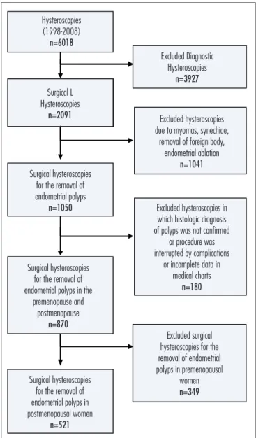
From January 1998 to December 2008, 6,018 hyste-roscopies were performed in the Prof. Dr. José Aristodemo Pinotti Women’s Hospital – CAISM/Unicamp. From this total, 3,927 diagnostic hysteroscopies and 2,091 surgical hysteroscopies were excluded due to myomas, synechiae, removal of a foreign body, and endometrial ablation, resulting in 1,050 hysteroscopies performed, during this period, to remove endometrial polyps. From these,
180 hysteroscopies in which the histologic diagnosis of endometrial polyps was not conirmed, or the procedure was interrupted by complications or incomplete data in medical charts were excluded. Finally, we excluded 349 cases in which the procedure was performed to remove endometrial polyps in premenopausal women, resulting in 521 surgical hysteroscopies performed to remove polyps in postmenopausal women, the subject of our study (Figure 1).
The clinical, pathological, and sonographic data were obtained from medical chart review. This study included 521 postmenopausal women with or without abnormal bleeding who had previously received an ultrasound diagnosis of endometrial polyps, based on indings of focal endometrial thickening associated with the presence of a vascular pedicle. Diagnostic hysteroscopy was performed by using a Karl Storz hysteroscope with optical systems
Figure 1. Flow chart.
Hysteroscopies (1998-2008)
n=6018
Excluded Diagnostic Hysteroscopies
n=3927
Surgical L Hysteroscopies
n=2091 Excluded hysteroscopies
due to myomas, synechiae, removal of foreign body,
endometrial ablation
n=1041
Excluded hysteroscopies in which histologic diagnosis of polyps was not confirmed
or procedure was interrupted by complications
or incomplete data in medical charts
n=180
Excluded surgical hysteroscopies for the removal of endometrial polyps in premenopausal
women
n=349
Surgical hysteroscopies for the removal of endometrial polyps in postmenopausal women
n=521
Surgical hysteroscopies for the removal of endometrial polyps in the
premenopause and postmenopause
n=870
Surgical hysteroscopies for the removal of endometrial polyps
of 2.8 mm. For distension of the uterine cavity, CO2 or saline solution was used. Through an evaluation of the endocervical canal, endometrial surface, vascularization, tubal ostia, the presence of endometrial polyps, myomas or synechiae were observed.
Surgical hysteroscopy with the patient under spinal anesthesia was performed using a 10 mm Karl Storz re-sectoscope. A glycine 1.5% solution was used to distend the uterine cavity. Evaluation of the endocervical canal and endometrial cavity was performed. Resection of en-dometrial polyps was performed with loop electrocautery that relied on a monopolar electrical current.
Pathologists from the Department of Pathologic Anatomy of the Unicamp Medical School analyzed the endometrial samples obtained using hematoxylin and eosin staining. Polyps were classiied as benign, non-atypical or atypical simple glandular hyperplasia, non-atypical or atypical complex glandular hyperplasia, and malignant.
This study was designed according to recommen-dations from the questionnaire Quality Assessment of
Diagnostic Accuracy Studies (QUADAS)18, and approved
by the Research Ethics Committee of FCM/Unicamp under number 769/2009.
Statistical analysis
Statistical analysis was performed by measurement of rates, means, and standard deviations. The sensitivity, speciicity, positive predictive value (PPV), and negative predictive value (NPV) were calculated for different measures of endometrial thickness and polyp size upon hysteroscopy. Histologic diagnosis was used as the gold standard and the cut-off point (the point with the hi-ghest sensibility and speciicity) was established by the methodology for the Receiver Operating Characteristic (ROC) curve. For statistical analysis, polyps were grouped according to histologic diagnosis into benign (benign polyps, non-atypical simple hyperplastic and non-atypical complex) or premalignant and malignant (atypical simple, atypical complex hyperplastic and carci-nomatous), and the prevalence ratios and their respective conidence intervals were calculated. The signiicance level was set at 5%. The SAS program version 9.2 was used for these estimates.
Results
Five hundred and twenty-one (521) postmenopausal women, with a mean age of 57.5 years (±10.6), were stu-died. Mean time since menopause was 12.4 years. There was a sonographic diagnosis of endometrial thickening (>5 mm) in 89.8% of cases. Histologic diagnosis iden-tiied the presence of premalignancy or malignancy in 4.1% of cases.
Sonographic measurement of the endometrial thick-ness in postmenopausal women undergoing hysteroscopic polypectomy revealed that mean thickness was 11.5 mm in benign polyps, 10.5 mm in premalignant polyps, and 17.4 mm in malignant polyps (p=0.002). Of the 16 malignant cases evaluated, 2 had endometrial thickness less than 5 mm on sonographic measurement (Table 1).
On surgical hysteroscopy, the median size of the benign polyps was 21.5 mm, premalignant polyps 24.3 mm, and malignant polyps 26.3 mm (p=0.003) (Table 1).
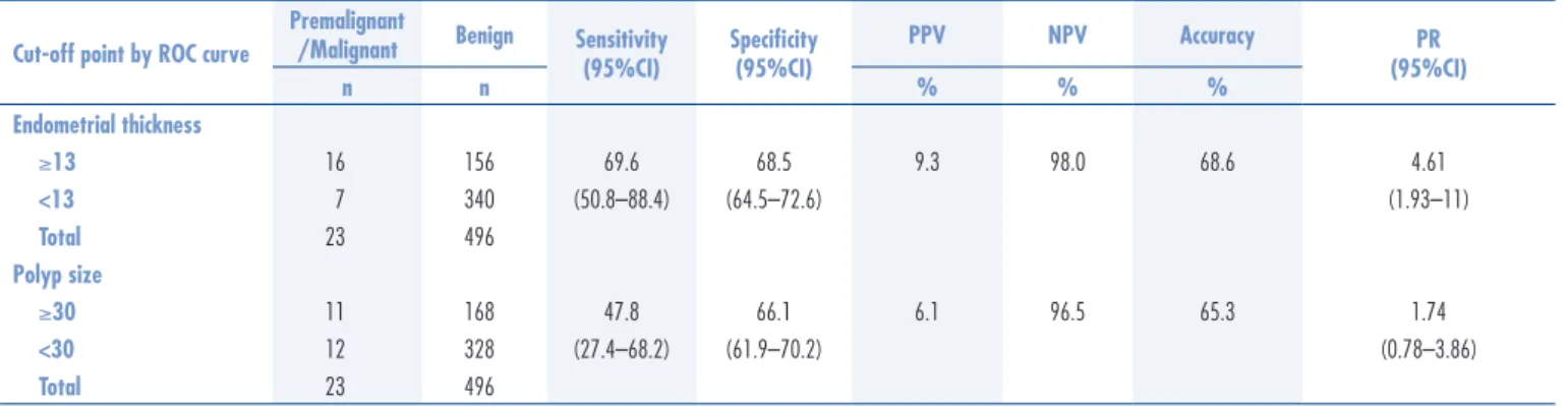
To predict malignancy, the sensitivity, speciicity, positive predictive value and negative predictive value were calculated for each value of sonographic endometrial thickness between 2 and 20 mm, resulting in a diagnostic accuracy of 68.6%. The best cut-off point established by the ROC curve was 13 mm, showing a sensitivity of 69.6%, speciicity of 68.5%, PPV of 9.3%, and NPV of 98%. On hysteroscopy, diagnostic accuracy for polyp size was 65.3% and the best cut-off point was 30 mm by the ROC curve, with a sensitivity of 47.8%, speciicity of 66.1%, PPV of 6.1%, and NPV of 96.5% (Table 2 and Figure 2).
With the purpose of determining the risk of ma-lignancy in endometrial polyps according to a group of different risk factors, malignancy risk was calculated in correlation with the presence or absence of postmenopausal bleeding, endometrial thickness greater or less than 13 mm and polyp size larger or smaller than 30 mm. For patients with vaginal bleeding, endometrial thickness less than 13 mm and polyps smaller than 30 mm, the prevalence ratio showed a risk of malignancy of 14.41 (95%CI 1.85–112.57), while for endometrial thickness
Endometrial thickness
Benign n=495 %
Premalignant n=08
%
Malignant n=16
%
p-value*
<5 mm 9 12 12
5.1 to 10 mm 46 37 –
10.1 to 15 mm 27 37 37
15.1 to 20 mm 10 12 18
>20 mm 6 – 31
Mean±SD 11.5±8.2 10.5±4.7 17.4±8.5 0.002
Polyp size
Benign n=498 %
Premalignant n=07
%
Malignant n=16
%
p-value*
<15 mm 38 28 25
15.1 to 20 mm 24 14 25
20.1 to 25 mm 2 – 6
25.1 to 30 mm 21 42 18
>30 mm 12 14 25
Mean±SD 21.5±13.9 24.3±11.3 26.3±13.2 0.003
Table 1. Sonographic endometrial thickness (n=519) and polyp size by surgical hysteroscopy (n=521) according to histologic diagnosis of endometrial polyp in postmenopausal women
greater than 13 mm and polyps larger than 30 mm, the risk was 32.71 (95%CI 3.94–271.84). In the absence of postmenopausal bleeding, the assessment of endometrial thickness associated with polyp size did not signiicantly increase the risk of malignancy.
Discussion
This study used histological diagnosis as the gold standard and assessed the accuracy of sonographic en-dometrial thickness and hysteroscopic characteristics in predicting malignancy in women undergoing hysteroscopic resection of endometrial polyps. The results revealed a diagnostic accuracy of 68.6% for endometrial thickness and 65.3% for polyp size.
Satisfactory diagnostic methods for the prediction of malignancy in focal endometrial lesions are still lacking, and a histological investigation is required in all suspected cases.
In the postmenopausal period, different cut-off values for endometrial thickness have been proposed to determine whether additional investigation is required, particularly in asymptomatic women who carry a lower risk of malignancy compared with women with genital
bleeding18-20. For focal endometrial lesions where the
adjacent endometrium has an atrophic pattern, these cut-off values are poorly deined. There is little informa-tion about the role of sonography as an exclusive method for predicting malignancy in endometrial polyps.
In the present study, ultrasound measurement of endometrial thickness showed that the mean endometrial thickness was greater with malignant polyps than with benign polyps. An endometrial thickness of 13 mm sho-wed the best sensitivity (69.6%) and speciicity (68.5%) in predicting malignancy in endometrial polyps.
According to Dreisler et al.6, ultrasound made it
possible to rule out the presence of benign focal endo-metrial lesions (polyps or submucous myomas) when the endometrial thickness was <2.8 mm, with a negative
predictive value of 98.5%. Grimbizis et al.21 observed
a sensitivity of 41.8% and a speciicity of 83.6% in the diagnosis of endometrial polyps using ultrasound exami-nation. The author was unable to discriminate between
benign, premalignant, and malignant focal lesions21.
In a study performed in the United Kingdom with 48,230 women undergoing transvaginal sonography to screen for endometrial cancer not associated with the presence of focal lesions, an endometrial thickness of 5.15 mm had a sensitivity of 80.5% and a speciicity of 86.2% in predicting a malignancy. When a cut-off value of 10 mm was used further to investigate malignancy,
the sensitivity was 54.1% and speciicity was 97.2%22.
Among the diagnostic methods for investigating endometrial disease, hysteroscopy has the highest diag-nostic eficacy. For hysteroscopic diagnosis of endometrial
polyps, a study performed by Cepni et al.23 showed a
sensitivity of 94% and a speciicity of 58%.
Few studies have evaluated the relationship between
the size of the polyp and the risk of malignancy15-17,24,25.
In this study, a size of 30 mm showed the best sensitivity (47.8%) and speciicity (66.1%) in predicting malignancy in endometrial polyps. A meta-analysis conducted to evaluate the oncogenic potential of endometrial polyps
Cut-off point by ROC curve
Premalignant
/Malignant Benign Sensitivity (95%CI)
Speciicity (95%CI)
PPV NPV Accuracy PR
(95%CI)
n n % % %
Endometrial thickness
≥13 16 156 69.6 68.5 9.3 98.0 68.6 4.61
<13 7 340 (50.8–88.4) (64.5–72.6) (1.93–11)
Total 23 496
Polyp size
≥30 11 168 47.8 66.1 6.1 96.5 65.3 1.74
<30 12 328 (27.4–68.2) (61.9–70.2) (0.78–3.86)
Total 23 496
Table 2. Accuracy of ultrasound and hysteroscopy in diagnosing malignancy in endometrial polyps in postmenopausal women (n=521)
PPV: positive predictive value; NPV: negative predictive value; PR: prevalence ratio
Figure 2. ROC curve for endometrial polyp size, endometrial stripe, and histologic diagnosis of malignancy
0 0,0 0,2 0,4 0,6 0,8 1,0
0,2 0,4 0,6 0,8
1-Speciicity
Polyps size Endometrial stripe
1
1. Anastasiadis PG, Koutlaki NG, Skaphida PG, Galazios GC, Tsikouras PN, Liberis VA. Endometrial polyps: prevalence, detection, and malignant potential in women with abnormal uterine bleeding. Eur J Gynaecol Oncol. 2000;21(2):180-3.
2. Clevenger-Hoeft M, Syrop CH, Stovall DW, Van Voorhis BJ. Sonohysterography in premenopausal women with and without abnormal bleeding. Obstet Gynecol. 1999;94(4):516-20. 3. Goldstein SR, Zeltser I, Horan CK, Snyder JR, Schwartz LB.
Ultrasonography-based triage for perimenopausal patients with abnormal uterine bleeding. Am J Obstet Gynecol. 1997;177(1):102-8. 4. Nagele F, O’Connor H, Davies A, Badawy A, Mohamed H, Magos
A. 2500 outpatient diagnostic hysteroscopies. Obstet Gynecol. 1996;88(1):87-92.
5. Van Bogaert LJ. Clinicopathologic indings in endometrial polyps. Obstet Gynecol. 1988;71(5):771-3.
6. Dreisler E, Stampe Sorensen S, Ibsen PH, Lose G. Prevalence of endometrial polyps and abnormal uterine bleeding in a Danish population aged 20-74 years. Ultrasound Obstet Gynecol. 2009;33(1):102-8.
7. Lieng M, Istre O, Sandvik L, Qvigstad E. Prevalence, 1-year regression rate, and clinical signiicance of asymptomatic endometrial polyps: cross-sectional study. J Minim Invasive Gynecol. 2009;16(4):465-71.
8. Schmidt T, Breidenbach M, Nawroth F, Mallmann P, Beyer IM, Fleisch MC, et al. Hysteroscopy for asymptomatic postmenopausal
women with sonographically thickened endometrium. Maturitas. 2009;62(2):176-8.
9. Lee SC, Kaunitz AM, Sanchez-Ramos L, Rhatigan RM. The oncogenic potential of endometrial polyps: a systematic review and meta-analysis. Obstet Gynecol. 2010;116(5):1197-205. 10. Martínez MA, Jou P, Nonell R, Cardona M, Alonso I, Vanrell JA.
Pólipos endometriales: riesgo de malignización y correlación clínico-anatómica. Prog Obstet Ginecol. 2004;47(11):506-10. 11. Antunes A Jr, Costa-Paiva L, Arthuso M, Costa JV, Pinto-Neto AM.
Endometrial polyps in pre- and postmenopausal women: factors associated with malignancy. Maturitas. 2007;57(4):415-21. 12. Savelli L, De Iaco P, Santini D, Rosati F, Ghi T, Pignotti E, et al.
Histopathologic features and risk factors for benignity, hyperplasia, and cancer in endometrial polyps. Am J Obstet Gynecol. 2003;188(4):927-31.
13. Lieng M, Istre O, Qvigstad E. Treatment of endometrial polyps: a systematic review. Acta Obstet Gynecol Scand. 2010;89(8):992-1002.
14. Baiocchi G, Manci N, Pazzaglia M, Giannone L, Burnelli L, Giannone E, et al. Malignancy in endometrial polyps: a 12-year experience. Am J Obstet Gynecol. 2009;201(5):462.e1-4. 15. Rahimi S, Marani C, Renzi C, Natale ME, Giovannini P, Zeloni R.
Endometrial polyps and the risk of atypical hyperplasia on biopsies of unremarkable endometrium: a study on 694 patients with benign endometrial polyps. Int J Gynecol Pathol. 2009;28(6):522-8.
References
in 10,552 patients identiied only eight studies that evaluated the association between polyp size and risk of
malignancy9. In four studies, larger polyps were directly
associated with a greater risk of malignancy. According
to Fernández-Parra et al.26, Goldstein et al.3, Shushan,
Revel and Rojansky27,and Gregoriou et al.28, polyp size
did not represent a risk factor for malignancy. The au-thors highlight that in this meta-analysis, polyp size was reported in different units of measurement (centimeters, millimeters, or grams) and data were not amenable; these factors hindered the analysis of this association.
The sensitivity of hysteroscopy for diagnosing any endometrial disease is 86%, whereas that of ultrasound is
54%22. For the diagnosis of endometrial hyperplasia and
endometrial cancer in the absence of focal lesions, the
sensiti-vity of hysteroscopy was 33% and the speciicity was 87%27.
Small uterine lesions or functional changes with polypoid pattern in the endometrium may result in failure to identify focal lesions during hysteroscopic evaluation. Furthermore, malignant endometrial neoplasms may coexist with indings of benign endometrial polyps. For this reason, resection of endometrial polyps accompanied by guided biopsy of the
adjacent endometrium is recommended in patients at risk29,30.
Data from the present study has shown that so-nographic endometrial thickness and hysteroscopic measurement of endometrial polyps have a low level of accuracy in predicting malignancy in focal lesions. These methods alone are not suficient to exclude the need for additional histological evaluation in suspec-ted cases. There are still no satisfactory diagnostic methods for identifying patients that should undergo more judicious surgical resection. On the basis of the literature, an individualized approach to the treatment of patients with endometrial polyps is recommended, considering the associated risk factors for each patient. Symptomatic postmenopausal women should undergo hysteroscopic polyp resection while asymptomatic postmenopausal women should receive individualized therapy based on polyp size, presence of risk factors for malignancy, general clinical conditions, and their
expectations from treatment31.
16. Ben-Arie A, Goldchmit C, Laviv Y, Levy R, Caspi B, Huszar M, et al. The malignant potential of endometrial polyps. Eur J Obstet Gynecol Reprod Biol. 2004;115(2):206-10.
17. Ferrazzi E, Zupi E, Leone FP, Savelli L, Omodei U, Moscarini M, et al. How often are endometrial polyps malignant in asymptomatic postmenopausal women? A multicenter study. Am J Obstet Gynecol. 2009;200(3):235.e1-6.
18. Whiting P, Rutjes AW, Reitsma JB, Bossuyt PM, Kleijnen J. The development of QUADAS: a tool for the quality assessment of studies of diagnostic accuracy included in systematic reviews. BMC Med Res Methodol. 2003;3:25.
19. Osmers R, Völksen M, Schauer A. Vaginosonography for early detection of endometrial carcinoma? Lancet. 1990;335(8705): 1569-71.
20. Seelbach-Göbel B, Rempen A, Kristen P. [Vaginal sonography of the endometrium in postmenopause. Initial results of a prospective study]. Gynakol Rundsch. 1991;31 (Suppl 2):253-5. German. 21. Grimbizis GF, Tsolakidis D, Mikos T, Anagnostou E, Asimakopoulos
E, Stamatopoulos P, et al. A prospective comparison of transvaginal ultrasound, saline infusion sonohysterography, and diagnostic hysteroscopy in the evaluation of endometrial pathology. Fertil Steril. 2010;94(7):2720-5.
22. Weigel M, Friese K, Strittmatter HJ, Melchert F. [Ultrasound assessment of the postmenopausal endometrium. Is measuring thickness adequate?] Ultraschall Med. 1994;15(3):117-21. German.
23. Cepni I, Ocal P, Erkan S, Saricali FS, Akbas H, Demirkiran F, et al. Comparison of transvaginal sonography, saline infusion
sonography and hysteroscopy in the evaluation of uterine cavity pathologies. Aust N Z J Obstet Gynaecol. 2005;45(1):30-5. 24. Jacobs I, Gentry-Maharaj A, Burnell M, Manchanda R, Singh N,
Sharma A, et al. Sensitivity of transvaginal ultrasound screening for endometrial cancer in postmenopausal women: a case-control study within the UKCTOCS cohort. Lancet Oncol. 2011;12(1):38-48. 25. Wang JH, Zhao J, Lin J. Opportunities and risk factors for premalignant
and malignant transformation of endometrial polyps: management strategies. J Minim Invasive Gynecol. 2010;17(1):53-8. 26. Fernández-Parra J, Rodríguez Oliver A, López Criado S, Parrilla
Fernández F, Montoya Ventoso F. Hysteroscopic evaluation of endometrial polyps. Int J Gynaecol Obstet. 2006;95(2):144-8. 27. Shushan A, Revel A, Rojansky N. How often are endometrial
polyps malignant? Gynecol Obstet Invest. 2004;58(4):212-5. 28. Gregoriou O, Konidaris S, Vrachnis N, Bakalianou K, Salakos N,
Papadias K, et al. Clinical parameters linked with malignancy in endometrial polyps. Climacteric. 2009;12(5):454-8.
29. Krampl E, Bourne T, Hurlen-Solbakken H, Istre O. Transvaginal ultrasonography sonohysterography and operative hysteroscopy for the evaluation of abnormal uterine bleeding. Acta Obstet Gynecol Scand. 2001;80(7):616-22.
30. Salim S, Won H, Nesbitt-Hawes E, Campbell N, Abbott J. Diagnosis and management of endometrial polyps: a critical review of the literature. J Minim Invasive Gynecol. 2011;18(5):569-81. 31. American Association of Gynecologic Laparoscopists. AAGL practice