2009; 59: 1: 67-73 CLINICAL REPORT
RESUMO
Sluminsky BG, Silva RC — Ocorrência de Lesão Pulmonar Aguda Relacionada com Transfusão (TRALI — Transfusion Related Acute LungInjury) em Pós-Operatório de Mastectomia com Reconstrução Microcirúrgica de Mama.
JUSTIFICATIVA E OBJETIVOS: Após sua descrição há mais de 20 anos, a TRALI — transfusion related acute lung injury — tornou-se, nos Estados Unidos e na Inglaterra, a principal causa de morbi-dade e mortalimorbi-dade relacionada com transfusão sanguínea. Por não existirem dados confiáveis com relação à sua epidemiologia no Brasil, seu diagnóstico é difícil, pois seu quadro clínico é variado e não há dados laboratoriais específicos. Sendo assim, os relatos de casos tornam-se importantes. É o primeiro relato dessa reação transfusional, neste situação cirúrgica, indexado na base de dados LILACS.
RELATO DO CASO: Paciente do sexo feminino, 36 anos, subme-tida à mastectomia com reconstrução microcirúrgica de mama sob anestesia geral. Logo após o término da transfusão de concentrado de hemácias, na sala de recuperação pós-anestésica, evoluiu com insuficiência respiratória, não necessitando reintubação traqueal. Foi realizado tratamento de suporte em unidade de terapia intensiva após serem descartadas outras hipóteses diagnósticas. Evoluiu bem, recebendo alta hospitalar no quarto dia de pós-operatório, sem seqüelas.
CONCLUSÕES: Ressalta-se a importância da realização crite-riosa de transfusão sanguínea, pois, apesar da transmissão de do-enças ser rara, a ocorrência de TRALI é muito freqüente, contudo subestimada pela diversidade de hipóteses diagnósticas. Por isso
é salutar o conhecimento e divulgação dessa doença, sobretudo em nosso meio.
Unitermos: CIRURGIA, Plástica: mamaplastia; COMPLICAÇÕES: le-são pulmonar aguda; SANGUE: Transfule-são.
SUMMARY
Sluminsky BG, Silva RC — Transfusion-Related Acute Lung Injury (TRALI) after Mastectomy with Microsurgical Breast Reconstruction.
BACKGROUND AND OBJECTIVES: After its description more than 20 years ago, TRALI – Transfusion Related Acute Lung Injury – be-came the main cause of transfusion-related morbidity and mortality in the United States and England. Since reliable data on its epide-miology in Brazil are not available, the difficulty to diagnose, varied clinical presentation, and absence of specific laboratory data, case reports are important. This is the first report of this transfusion reac-tion indexed in the LILACS data base.
CASE REPORT: A 36-year old female underwent mastectomy with microsurgical breast reconstruction under general anesthesia. Immediately after the transfusion of one unit of packed red blood cells in the post-anesthetic recovery room, she developed respi-ratory failure, which did not require reintubation. Supportive treat-ment was instituted in the intensive care unit after other diagnostic hypotheses were ruled out. She had a favorable evolution and was discharged from the hospital without sequelae.
CONCLUSION: The importance of judicious blood transfusion is emphasized since, although disease transmission is rare, TRALI is not, but it is underestimated due to the diversity of diagnostic hypotheses. Therefore, the knowledge of this disorder and its dissemination, especially in our country, is important.
Key Words: BLOOD: Transfusion; COMPLICATIONS: acute lung in-jury; SURGERY, Plastic: mammoplasty.
INTRODUÇÃO
Após sua descrição há mais de 20 anos, a TRALI — transfusion related acute lung injury — tornou-se, nos Estados Unidos e na Inglaterra, a principal causa de morbidade e mortalidade relacionada com transfusão sanguínea 1,2. Contudo, não há
dados confiáveis com relação à sua epidemiologia, sobretu-do no Brasil. A TRALI pode ser de difícil diagnóstico devisobretu-do à
Ocorrência de Lesão Pulmonar Aguda Relacionada com
Transfusão (TRALI — Transfusion Related Acute Lung Injury)
em Pós-Operatório de Mastectomia com Reconstrução
Microcirúrgica de Mama*
Transfusion-Related Acute Lung Injury (Trali) after Mastectomy
with Microsurgical Breast Reconstruction
Beatriz Garcia Sluminsky, TSA1, Ranger Cavalcante da Silva, TSA2
*Recebido do (Received from) Serviço de Anestesiologia do Hospital Vita Curitiba, Curitiba, PR
1. Co-Responsável pelo CET do Hospital de Clínicas da UFPR e Aneste-siologista dos Hospitais Vita Curitiba
2. Mestre em Medicina e Cirurgia pela UFPR; Co-Responsável pelo CET do Hospital de Clínicas da UFPR; Anestesiologista e Coordenador do Serviço de Anestesiologia do Hospital Vita Curitiba
Apresentado (Submitted) em 25 de março de 2008 Aceito (Accepted) para publicação em 27 de outubro de 2008
Endereço para correspondência (Correspondence to): Dra. Beatriz Garcia Sluminsky
Rua Reinaldo Hecke, 453/301–A — Abranches 82210-300 Curitiba, PR
E-mail: [email protected]
clínico citado, o diagnóstico de TRALI pode ser confundido com ALI (lesão pulmonar aguda ou acute lung injury) por sepse, trauma, aspiração de conteúdo gástrico, coagulação intravascular disseminada (CIVD) ou lesão pulmonar por ventilação mecânica.
Com relação à fisiopatologia, não há consenso estabeleci-do, apesar de estarem implicados anticorpos leucocitários HLA classe I (antígeno leucocitário humano) e HLA classe II presentes no sangue do doador. Menos de 10% dos ca-sos são de anticorpos de antígenos leucocitários presentes no sangue do receptor 6. Daí a necessidade de comunicar
o banco de sangue para que tome as providências devidas com relação a outros hemocomponentes do doador envolvi-do, que porventura possam ainda existir no estoque. Junto à alta prevalência de anticorpos HLA, nos produtos sanguí-neos implicados na TRALI, está a elevada prevalência de multíparas doadoras, sensibilizadas previamente por antí-genos fetais. A despeito da aparente associação entre a sensibilização prévia durante a gravidez e TRALI, sangue com taxas de sensibilização HLA de até 26,7% em 332 do-adoras multíparas não foi implicado no desenvolvimento de TRALI 3. Estudos recentes indicaram que há necessidade
de concordância antígeno e anticorpo, bem como alguma especificidade de anticorpos com relação às combinações. No presente relato o doador era masculino, hígido, com san-gue totalmente compatível (ABO e RH) e outros hemocompo-nentes de sua doação tinham sido transfundidos em outros pacientes previamente sem intercorrências (uma unidade de plasma fresco congelado e uma de plaquetas). Os da-dos do paciente em questão, assim como a complicação advinda da transfusão, foram lançados no banco de dados da hemovigilância, recentemente.
O principal diagnóstico diferencial é com o edema agudo de pulmão, apesar das características radiológicas sugerirem edema não-cardiogênico na maioria das vezes, como no caso em questão, a exclusão de causa cardíaca é premente. O tratamento da TRALI é de suporte, com necessidade de in-tubação e ventilação mecânica em até 72% dos casos 5.
Quando não há necessidade de intubação as medidas in-cluem oxigênio suplementar. A hipotensão arterial pode ser tratada com administração de fluidos ou, nos casos refratá-rios, com administração de vasopressores. A recomendação de administrar fluidos pressupõe a exclusão de sobrecarga hídrica e o edema agudo pulmonar de origem cardiogênica. Quando não há sobrecarga hídrica, o uso de diuréticos não é recomendado. O uso de corticosteróides é empírico, sem dados que suportem ou refutem o seu uso 5.
Quando comparada com a ALI, a TRALI tem mortalidade baixa (6% a 10%) sem comprometimento tardio da função pulmonar 5. No caso apresentado, apesar da precocidade
e rápida progressão dos sintomas a paciente não necessi-tou de intubação traqueal, o que possibilinecessi-tou rápida e com-pleta recuperação da função pulmonar.
O presente relato ressaltou a importância da realização cri-teriosa de transfusão sanguínea, pois os riscos inerentes a
esse procedimento vão muito além da transmissão de doen-ças virais, o risco mais temido, ainda que muito raro na atua-lidade, em países com elevado índice de desenvolvimento humano. Por outro lado, a ocorrência de TRALI é muito alta e notoriamente subestimada pela diversidade de hipóteses diagnósticas, justificando sua divulgação, sobretudo em nosso meio, no qual o anestesiologista está diretamente envolvido nas transfusões de sangue.
Transfusion-Related Acute Lung Injury
(Trali) after Mastectomy with
Microsur-gical Breast Reconstruction
Beatriz Garcia Sluminsky, TSA, M.D.; Ranger Cavalcante da Silva, TSA, M.D.
INTRODUCTION
After its description more than 20 years ago, TRALI – Trans-fusion-Related Acute Lung Injury – became the main cause of transfusion-related morbidity and mortality in the United States and England 1,2. However, since reliable data on its
epidemiology in Brazil are not available, the difficulty to diag-nose, varied clinical presentation, and absence of specific laboratory data, case reports are important. The authors re-port a case of TRALI in a patient who underwent a mastec-tomy with microsurgical breast reconstruction. This is the first report of this transfusion reaction indexed in the LILACS data base.
CASE REPORT
This is a 36 years old female, ASA I, scheduled for a mas-tectomy with microsurgical reconstruction of the breast. Du-ring the pre-anesthetic evaluation, done the day before, the patient stated she had undergone two breast surgeries under general anesthesia for removal of nodes without in-tercurrences. She was taking clonazepam for two months and denied using any other medication, smoking, or alcohol, as well as any systemic diseases. The physical exam was normal. Laboratory exams showed Hb 12.7 g.dL-1 and MCV
37.6%; all other results were normal. The electrocardiogram showed altered right bundle branch conduction and the chest X-ray was normal. Since the surgery was expected to be leng-thy, significant bleeding was a possibility and the red blood cell count was in the lower normal limit, two units of packed-red blood cells were requested.
and intraoperative losses were compensated by the admi-nistration of 2,500 mL of NS and 500 mL of hetastarch. At the end of the surgery, blood for the determination of Hb and MCV was drawn, and the patient was extubated without in-tercurrences. She was transferred to the post-anesthetic recovery room (PARR) with blood pressure 100 x 70 mmHg, HR 84 bpm, RR 12 bpm, which were similar to preoperative parameters. She had a SpO2 99% with O2 via a face mask, which was maintained in the PARR. Approximately 20 minutes after admission to the PARR, the results of her blood work revealed Hb 8 mg.dL-1 and MCV 24%. Although the patient
remained hemodynamically stable without any complaints it was decided to transfuse one unit of PRBCs because she was actively bleeding through the drain and to avoid trans-fusion in the room.
The PRBC was transfused in approximately 25 minutes and, immediately after the transfusion, the patient developed dyspnea, tachypnea (30 bpm), and progressive reduction of SpO2, down to 82% with 6 L.min-1 of O
2 via face mask. On
aus-cultation, she had rare crackles and rales bilaterally, more prominent on the bases. A chest X-ray revealed interstitial infiltrate and diffuse and confluent alveolar opacities on the lower two thirds of the lung fields bilaterally (Figure 1). Meanwhile, the patient developed coughing productive of frothy secretion approximately 90 minutes after the transfu-sion. Blood for arterial blood gases was drawn and a bed in the intensive care unit was requested (ICU). Assuming a diagnosis of pulmonary edema, despite effective diuresis, normal blood pressure, and jugular veins on the posterior limits of the sternocleidomastoideo muscle, it was decided to administer 20 mg of furosemide with the hypothesis of volume overload. Among the differential diagnoses the possibility of acute lung injury (ALI) by sepsis or
bronchoas-piration, and due to the cause-effect relationship, transfu-sion-related acute lung injury (TRALI) was included. The last hypothesis was considered more likely and, therefore, 500 mg of hydrocortisone IV was administered and the blood bank was contacted to make the proper arrangements with the donor. Arterial blood gases revealed hypoxemia and meta-bolic acidosis (PaO2 90 mmHg; SatO2 91%; pH 7.25; bicar-bonate 19.3 mEq.L-1), with a PaO
2:FiO2 225. Three hours later,
the condition of the patient had improved and she was trans-ferred to the ICU with the diagnosis of TRALI. There, treat-ment with 100 mg of hydrocortisone IV every eight hours continued, along with ipratropium bromide and fenoterol. To rule out cardiogenic pulmonary edema, an echocardiogram was done, which was normal as well as the ECG. Twelve hours after her admission to the ICU the patient referred improvement of dyspnea, RR 16 bpm, SpO2 96% under mist mask. Auscultation revealed improvement of the breath sounds, with rare crackles in the bases. The chest X-ray had improved, with residual basilar opacities (Figure 2).
The patient showed progressive improvement and was dis-charged from the ICU 36 hours after her admission to the unit. Two days after the surgery the patient was asymptomatic, SpO2 97% in room air, and her chest X-ray was normal (Fi-gure 3); she was discharged from the hospital on the same day. The patient returned to the hospital seven months later for retouch and symmetrization of the breast and underwent the same anesthetic technique. Blood transfusion was not necessary, and the surgery and postoperative period evolved without intercurrences. Currently the patient is asymptomatic with normal pulmonary function.
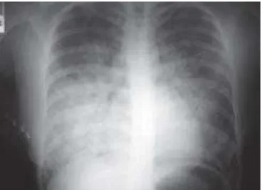
Figure 1 – Chest X-Ray Immediately after the Beginning of Symptoms. It shows diffuse and confluent alveolar opacities in the lower two thirds of the lung fields, predominantly on the right.
DISCUSSION
Transfusion-related acute lung injury is the most common transfusion-related cause of morbidity and mortality in the United States and England, which has increased the interest on its precise diagnosis and research of its pathophysiology and prevention 1,2. Although the first reports go back to 1951 3,
only in 1983 Popovski and Moore described the presentation and pathophysiology of this disorder, calling it “transfusion-related acute lung injury” – TRALI 4.
In general, TRALI presents with symptoms of respiratory distress, which begin during or up to six hours after trans-fusion. However, some cases might develop up to 48 hours after transfusion 5. Since most transfusions are
surgery-related 6, if transfusion is done during the surgery, this
di-sorder may develop early, hindering the diagnosis, which might not even be made. Thus, it is considered as a transi-tory hypoxemia because anesthesia restricts the spectrum of symptoms and since it is a self-limited process it might, depending on the severity of the case, have resolved by the end of the surgery 5. In the case presented here, transfusion
was done in the PARR with the patient awake and oriented allowing proper diagnosis despite the very early development of symptoms.
Dyspnea is another characteristic symptom of this reaction, which usually develop over a few minutes due to acute pulmo-nary edema leading to reduction in arterial oxygen saturation and in many cases cyanosis. On the chest X-ray, edema be-gins on dependent lung regions and perihilar region, simi-lar to cardiogenic pulmonary edema 5. The radiologic aspect
progresses rapidly to a generalized form (white-out) affecting the entire lung. Auscultation has discrete changes that do not explain the reduction in oxygen saturation and radiographic images 5. The interstitial nature of the fluid, in contrast with
the intra-alveolar fluid of cardiogenic pulmonary edema, des-pite the lack of scientific evidence to support this affirmation
could possibly explain the paucity of auscultation 5. During
general anesthesia the elimination through the trachea tube of frothy secretion can be seen especially in cases with rapid progression 5. In the present case, since the patient was
awake, she developed cough and eliminated frothy material through the mouth, which indicated the diagnosis of pulmo-nary edema. If the patient is intubated and under mechanical ventilation it might present with difficulty to ventilate, increa-sed endotracheal pressure, and abrupt reduction in blood pressure. However, the clinical presentation is characterized by a constellation of non-specific signs and symptoms com-mon to several disorders, making for a difficult differential diagnosis. Even with the presentation of this patient, TRALI can be mistaken with ALI (Acute Lung Injury) caused by sepsis, trauma, aspiration of gastric contents, disseminated intra-vascular coagulation, or ventilation-related pulmonary lesion. Consensus on its pathophysiology is lacking, although HLA class I (Human Leukocyte Antigen) and HLA class II antigens in the donor blood have been implicated. Less than 10% of the cases are due to leukocyte antigens in the blood of the receptor6. This explains the need to contact the blood bank
to take proper measures regarding other blood products involving the donor that might be still in storage. Along with the high prevalence of HLA antigens in blood products im-plicated in TRALI, the high incidence of multiparous donors, sensitized during pregnancy by fetal antigens, is also asso-ciated with this disorder. Despite the apparent association between prior sensitization during pregnancy and TRALI, blood with high HLA sensitization of up to 26.7% in 332 multiparous donors has not been implicated in the develop-ment of TRALI 3. Recent studies indicated the need of
an-tigen-antibody concordance, as well as some specificity of the antibodies for combinations. In the present case, the donor was male, healthy, the blood was compatible (ABO and RH), and other blood products with his blood had been previously transfused without intercurrences (one unit of fresh frozen plasma and one unit of platelets). Data regarding the patient, as well as the complication resulting from the trans-fusion, were recently included in the data bank of a health agency.
Acute pulmonary edema, despite most of the time the radio-logic characteristics suggest non-cardiogenic pulmonary edema, as in the case presented here, is the main differen-tial diagnosis and the exclusion of a cardiac cause is para-mount. Treatment of TRALI consists of supportive measures and up to 72% of the patients require intubation and mecha-nical ventilation5. When intubation is not necessary,
suppor-tive measures include oxygen administration. Hypotension can be treated with the administration of fluids or, in refractory cases, vasopressors. Fluid administration presupposes the exclusion of fluid overload and cardiogenic pulmonary ede-ma. Diuretics are not recommended when volume overload is not present. The use of corticosteroids is empirical, and data supporting or contraindicating their administration do not exist.
When compared to ALI, TRALI has a low mortality rate (6 to 10%), and it does not lead to late pulmonary dysfunction 5.
In the case presented here, despite the early development and rapid progression of symptoms, the patient did not require tracheal intubation, allowing fast and complete recovery of pulmonary function.
The present case report emphasized the importance of judicious care when transfusing blood, since the inherent risks go beyond the transmission of viral diseases, the most feared complication although rare in countries with elevated human development index. On the other hand, TRALI is common and notoriously underestimated due to the diversity of diagnostic hypotheses, justifying the dissemination of the knowledge of this disorder, especially in our country, where the anesthesiologist is involved directly in blood transfusions.
REFERÊNCIAS — REFERENCES
01. United States Government Food and Drug Administration, 2004 http://www.fda.org/
02. SHOT, 2003. http://www.shotuk.org
03. Barrett NA, Kam PC — Transfusion-related acute lung injury: a literature review. Anaesthesia, 2006;61:777-785.
04. Popovski MA, Abel MD, Moore SB — Transfusion-related acute lung injury associated with passive transfer of anti-leukocyte antibodies. Am Rev Respir Dis, 1983;128:185-189.
05. Moore SB — Transfusion-related acute lung injury (TRALI): clinical presentation, treatment, and prognosis. Crit Care Med, 2006;34(5/Suppl):s114-s117.
06. Curtis BR, McFarland JG — Mechanisms of transfusion-related acute lung injury (TRALI): anti-leukocyte antibodies. Crit Care Med, 2006;34(5/Suppl):s118-s123.
07. Densmore TL, Goodnough LT, Ali S et al — Prevalence of HLA sensitization in female apheresis donors. Transfusion, 1999;39: 103-106.
RESUMEN
Sluminsky BG, Silva RC — Aparecimiento de Lesión Pulmonar Agu-da RelacionaAgu-da con la Transfusión (TRALI - Transfusion Related Acute LungInjury) en Postoperatorio de Mastectomía con Recons-trucción Micro quirúrgica de Mama.
JUSTIFICATIVAS Y OBJETIVOS: Después de su descripción hace más de 20 años, la TRALI - Transfusion Related Acute Lung Injury
se convirtió, en los Estados Unidos de América y en Inglaterra, en la principal causa de morbidez y mortalidad relacionada con la transfusión sanguínea. Por el hecho de no haber datos confiables con relación a su epidemiología en Brasil, su difícil diagnóstico, al cuadro clínico variado y la ausencia de datos de laboratorio espe-cíficos, los relatos de casos son importantes. Es el primer relato de esta reacción transfusional indexado a la base de datos del LILACS.
RELATO DEL CASO: Paciente femenino, de 36 años, sometida a la mastectomía con reconstrucción micro quirúrgica de mama bajo anestesia general. Inmediatamente después del término de la transfusión de un concentrado de hematíes, en la sala de recupe-ración postanestésica, evolucionó con insuficiencia respiratoria no necesitando re-intubación traqueal. Fue realizado el tratamiento de soporte en la unidad de cuidados intensivos después de descar-tar otras hipótesis diagnósticas. Evolucionó bien y recibió el alta al cuarto día del postoperatorio, sin secuelas.