2009; 59: 1: 46-55 SCIENTIFIC ARTICLE
RESUMO
Cunha Jr RJ, Barrucand L,Verçosa N — Estudo das Alterações Eletrocardiográficas com o Uso de Antidepressivos Tricíclicos em Pacientes com Dor Crônica.
JUSTIFICATIVA E OBJETIVOS: Os antidepressivos tricíclicos (ADT) são amplamente utilizados como analgésicos para lombal-gias crônicas e dores neuropáticas. O objetivo deste estudo foi avaliar as alterações eletrocardiográficas dos pacientes com dor crônica em uso de amitriptilina ou imipramina.
MÉTODO: Foram estudados 40 pacientes com idade entre 26 e 81 anos (57,27 ± 13,65 anos), de ambos os gêneros (feminino 19, masculino 21), com síndromes neuropáticas (lombociatalgias, síndromes pós-laminectomia, neurites pós-herpética, entre outras); 60% com doenças cardiovasculares; 30% tinham ECG alterado (BRD, BRE, BAV 1o grau, HBAE ou extra-sístoles). Foram
realiza-dos e analisarealiza-dos três ECGs: antes do início realiza-dos ADT, 30 e 60 dias após o início do tratamento, avaliando os parâmetros PR, QRS, QT, QTc, DQT, DQTc e FC. Trinta e dois pacientes fizeram uso de amitriptilina e oito de imipramina. A dose média ao final do estudo foi de 54,29 mg de amitriptilina e de 46,87 mg de imipramina.
RESULTADOS: A análise das variáveis eletrocardiográficas após o uso dos ADT apresentou a amitriptilina com aumento na frequên-cia cardíaca transitoriamente no gênero feminino (p = 0,049) e a duração do QRS nos pacientes com idade igual ou maior que 60 anos e nos cardiopatas na segunda avaliação (p = 0,01). Nos pa-cientes que receberam amitriptilina, doses de 75 mg, o intervalo QTc
foi maior quando comparado com doses de 25 mg (p = 0,0044). O aumento desses parâmetros evidenciou o efeito da amitriptilina so-bre a condução cardíaca; no entanto, não houve comprometimen-to clínico, pois os valores permaneceram dentro dos limites de normalidade (QRS < 110ms e QTc < 470ms).
CONCLUSÕES: O uso clínico dos ADT em dores crônicas mostrou-se mostrou-seguro e eficaz, não apremostrou-sentando distúrbio da condução car-díaca com repercussão clínica.
Unitermos: DOR, Crônica: neuropática; DROGAS, Antidepressivos tricíclicos; EXAMES COMPLEMENTARES: eletrocardiograma.
SUMMARY
Cunha Jr RJ, Barrucand L,Verçosa N — A Studyon Electrocardio-graphic Changes Secondary to the Use of Tricyclic Antidepressants in Patients with Chronic Pain.
BACKGROUND AND OBJECTIVES: Tricyclic antidepressants (TCAs) are widely used as analgesics in chronic lumbar pain and neuropathic pain. The objective of this study was to evaluate the electrocardiographic changes in patients with chronic pain treated with amitriptyline or imipramine.
METHODS: Forty patients, ages 26 to 81 years (57.27 ± 13.65 years) of both genders (female 19, male 21), with neuropathic syn-dromes (lumbosciatalgia, postlaminectomy synsyn-dromes, and post-herpetic neuritis, among others) participated in this study; 60% had cardiovascular diseases; 30% had changes in the ECG (RBBB, LBBB, first-degree AVB, LAHB, or PVCs). Three ECGs were done in each patient: one ECG was done before beginning treatment, and 30 and 60 days after beginning treatment evaluating PR, QRS, QT, QTc, DQT, DQTc, and HR. Thirty-two patients were on amitriptyline and eight on imipramine. The mean dose at the end of the study was 54.29 mg of amitriptyline and 46.87 mg of imipramine.
RESULTS: Analysis of electrocardiographic parameters after the use of TCAs showed that amitriptyline caused a transitory increase in heart rate in females (p = 0.049), and the duration of the QRS in patients 60 years or older and patients with cardiopathies (p = 0.01). In patients who received 75 mg of amitriptyline, the QTc interval was greater when compared to that of patients who received 25 mg of the drug (p = 0.0044). The increase in those parameters demonstra-ted the effects of amitriptyline on cardiac conduction; however, clinical compromise was not seen, since they remained within nor-mal limits (QRS < 110 msec and QTc < 470 msec).
CONCLUSIONS: The chronic use of TACs proved to be safe and effective, and it did not show changes in cardiac conduction with clinical repercussion.
Key Words: COMPLEMENTARY EXAMS: electrocardiogram; DRUGS: tricyclic antidepressants; PAIN, chronic: neuropathic.
Estudo das Alterações Eletrocardiográficas com o Uso de
Antidepressivos Tricíclicos em Pacientes com Dor Crônica*
A Study on Electrocardiographic Changes Secondary to the Use
of Tricyclic Antidepressants in Patients with Chronic Pain
Ricardo Joaquim da Cunha Jr.1, Louis Barrucand2, Nubia Verçosa3
*Recebido do (Received from) Programa de Pós-Graduação em Cirurgia Ge-ral — Setor Anestesiologia — do Departamento de Cirurgia da Faculdade de Medicina da Universidade Federal do Rio de Janeiro (FM/UFRJ), RJ
1. Mestre em Medicina do Curso de Pós-Graduação em Cirurgia Geral — Se-tor Anestesiologia — FM/UFRJ; Anestesiologista e Coordenador do Progra-ma de Tratamento da Dor e Cuidados Paliativos do Hospital Universitário Clementino Fraga Filho (HUCFF)
2. Professor Titular de Patologia do Departamento de Anatomia Patológica da FM/UFRJ
3. Professora-Associada, Mestre e Doutora em Medicina do Departamento de Cirurgia da FM/UFRJ; Coordenadora da Graduação e Pós-Graduação em Anestesiologia da FM/UFRJ; Responsável pelo Ambulatório de Avaliação Pré-Anestésica do HUCFF/FM/UFRJ; Certificado de Área de Atuação em Dor SBA-AMB
Apresentado (Submitted) em 24 de julho de 2007
Aceito (Accepted) para publicação em 12 de setembro de 2008
Endereço para correspondência (Correspondence to): Dr. Ricardo Joaquim da Cunha Jr.
Rua Dr. Satamini, 183/803 — Tijuca 20270-233 Rio de Janeiro, RJ E-mail: [email protected]
ções quanto à segurança em baixas doses, refere-se à metanálise com seis estudos comparando os ADT em
bai-xas doses (< 100 mg), com doses-padrão para depressão
(> 100 mg), mostrando menor incidência de efeitos
colate-rais nas doses baixas 1.
Neste estudo prospectivo longitudinal, os antidepressivos foram utilizados em doses analgésicas máximas de 75 mg por dia, em uma amostra representativa da população-fon-te no que diz respeito a distribuição por gênero, quadro clí-nico doloroso e dose média de ADT empregada.
A interferência na frequência cardíaca, com discreto aumen-to dos valores, foi observada após 30 dias de uso dos
fár-macos (M1), mas com significância estatística (Tabela II). O
aumento da frequência cardíaca surge com o início da tera-pêutica, tendendo a adaptação em torno de quatro sema-nas. Essa variação ocorreu apenas com o gênero feminino.
Ao final do acompanhamento (M2) houve um retorno aos
va-lores basais (p = 0,01). Esse efeito transitório não teve im-portância clínica, sendo, porém, relatado na literatura 18.
A duração do complexo QRS evidenciou discreto aumento
em M1, com pequena significância estatística (p = 0,049 e IC
= 0,014-6,58). Em M2, porém, os valores se elevaram um
pouco mais em relação ao basal (M0), com p-valor de 0,003
(IC 2,02-8,90), sem repercussão clínica (Tabela II). Essa pe-quena variação do QRS foi observada em idosos e cardiopa-tas (Tabelas IV e V). Nenhum paciente registrou QRS maior ou igual a 120 ms ao final do acompanhamento, valor que in-dicaria maior tendência à arritmia, registrada na literatura 19.
Neste trabalho, não foi evidenciada nenhuma arritmia no eletrocardiograma.
No que diz respeito ao ECG prévio, ocorreu variação no QRS tanto nos pacientes com ECG iniciais alterados quanto nos normais (Tabela VI). Nesta pesquisa os pacientes com dis-túrbio de condução prévia não apresentaram maior variação do QRS diferindo da literatura consultada, quando os ADT
são usados em doses antidepressivas 20.
Ficou constatado que a variação do QRS ocorreu no mo-mento em que era utilizada a dose mais elevada. Isso cor-robora a idéia de que a influência desse grupo de fármacos na eletrofisiologia cardíaca é sobretudo dose-dependente. Na avaliação dos efeitos dos ADT nas diversas doses utili-zadas foi comprovado, neste trabalho, aumento significati-vo do QTc durante o uso da amitriptilina na dose de 75 mg (p = 0,004). Quanto ao uso da imipramina, foi observada re-dução do QTc com a dose de 50 mg, porém sem importân-cia estatística significativa.
O aumento do QT pode levar à arritmia ventricular grave
con-forme citação de Antzelevitch 21. A dispersão do QT (DQT)
re-flete as diferenças regionais do tempo de despolarização ventricular. Quando a heterogeneidade intrínseca do miocárdio ventricular é ampliada, ela se reflete em um maior DQT. Nos pacientes estudados a dispersão do QT não au-mentou de forma significativa durante o tratamento (Tabela III). Nesta pesquisa, não foi detectada nenhuma disritmia apesar de haver relatos na literatura comprovando a
presen-ça de complicações cardiovasculares, inclusive bloqueios
cardíacos, com doses antidepressivas de ADT 22,23.
Quando havia uma diferença em relação à dose utilizada, pode-se constatar que houve influência sobre o QTc em vi-gência apenas de amitriptilina (Tabela III). Contudo, o número reduzido de pacientes em uso de imipramina (oito pacientes) não permitiu concluir que esse fármaco não interfira nesse intervalo. Idosos e mulheres são mais propensos ao aumento
do intervalo QT induzido pelos ADT 24, o que não foi
demons-trado nas doses aqui empregadas.
O estudo concluiu que os antidepressivos tricíclicos mos-traram atuação no sistema de condução, porém sem com-prometimento clínico nos pacientes cardiopatas e idosos, porque as doses utilizadas para analgesia das dores neu-ropáticas foram menores que 100 mg.
A Study on Electrocardiographic
Changes Secondary to the Use of
Tricyclic Antidepressants in Patients
with Chronic Pain
Ricardo Joaquim da Cunha Jr., M.D.; Louis Barrucand, M.D.; Nubia Verçosa, M.D.
INTRODUCTION
Tricyclic antidepressants (TACs) are used worldwide in psychiatric patients and in the treatment of chronic pain syn-dromes. In the United Kingdom, a 40% increase in the pres-cription of those drugs was seen from 1991 to 1996. In the USA, data from 2000 and 2001 demonstrated that those drugs are
prescribed more often than serotoninergic drugs 1. They are
indicated in the treatment of neuropathic pain 2,
fibromyal-gia, irritable bowel syndrome, migraines (prophylaxis) and headaches of cervical origin, and in chronic myofascial pain. Those syndromes are common in patients treated in pain cli-nics and are difficult to treat, even in state-of-the art centers requiring, frequently, a pharmacological and non-pharmaco-logical approach by the multidisciplinary team.
The main side effects of TACs include: dry mouth, constipa-tion, somnolence, weight gain, mental confusion, urinary re-tention, postural hypotension, and cardiac arrhythmias. Some authors, based on studies during psychiatric treatment in which they are used in higher doses than the doses used in the treatment of chronic pain syndromes (25 to 100 mg) re-commend that TACs should be avoided in the elderly and patients with cardiomyopathies.
It is known that, in Brazil 4 and in the rest of the world 5, the
IN PATIENTS WITH CHRONIC PAIN
and on the other hand, the concern about possible delete-rious cardiovascular effects. However, the literature lacks evidence proving the incidence of those effects in the doses used for pain relief.
The objective of this study was to evaluate the use of TACs in chronic pain syndromes and their electrocardiographic repercussions.
METHODS
After approval by the Ethics on Research Committee of the Hospital Universitário Clementino Fraga Filho da Universi-dade Federal do Rio de Janeiro (HUCFF/UFRJ) and signing of the informed consent, 40 patients of the Pain Treatment and Palliative Care Program (PTDCP, from the Portuguese), ages 18 years or older, with neuropathic pain were included in this study. Exclusion criteria were as follows: patients on class Ia anti-arrhythmic drugs (pro-arrhythmic action); consumptive syndromes, liver disease, unstable cardiomyopathies, ma-jor mood disturbances (depression, bipolar) disorders, and altered cognition. Fifty-two patients were enrolled in the study; however, 12 were excluded for the following reasons: the dose was reduced in four patients due to a non-cardiac colla-teral effect (somnolence); one presented persistent palpi-tation and the drug was discontinued; four did not attend follow-up appointments, and three started TACs before the baseline ECG. Twenty one patients were males and 19 fema-les. Patients on amitriptyline or imipramine on doses equal or greater than 25 mg/day for relief of neuropathic pain par-ticipated in this study. Patients underwent three electrocar-diograms (ECG), which were analyzed posteriorly: before beginning TACs, and 30 and 60 days after its onset. Standard
12-lead ECG, at a speed of 25 mm.sec-1, was done with the
patient at rest. The following parameters were measured and analyzed: HR (heart rate), PR, QRS, QT, QTc, QT dispersion, and QTc dispersion. As for the PR interval, measured from the beginning of the P wave to the beginning of the QRS, the highest value among the bipolar derivations in the frontal pla-ne (D1, D2, D3) was considered, and 0.21 sec was the upper
normal limit 6. A QRS of up to 0.10 sec was considered normal.
For the QT interval, it was established a value of up to 0.46 sec, in V2 or V3, by the Lepeschkin method 7. For the QT
disper-sion (normal up to 60 msec), it was calculated the difference between the lower and higher QT of the 12 leads. The Bazett
formula (QTc = QT/RRsec1/2) was used to calculate the QTc
(QT interval corrected for the mean heart rate) 8. The QTc
dispersion (DQTc – QTcmax – QTcmin) was also calculated. For the statistical analysis, the results obtained for each pa-rameter were evaluated by the Kolmogorov-Smirnov test, which did not reject the hypothesis of normalcy. Mean stan-dard deviation and stanstan-dard error were calculated. Analysis of Variance (ANOVA) for repeated measures was used to compare measurements along time (pre-, and 30 and 60 days of treatment). Paired Student t test was used to evaluate the progression of the doses of the drugs along the study. A
level of significance of 5% was established for all statistical tests. The SPSS statistical program version 13.0 was used.
RESULTS
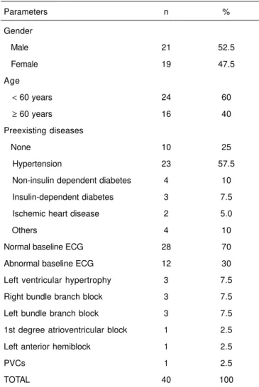
Table I shows the characteristics of all 40 patients regarding gender, age, use of imipramine or amitriptyline, preexisting diseases, and pattern of prior ECGs. Amitriptyline was used at a mean dose of 54.3 mg, and imipramine, 46.8 mg. Painful syndromes included: herniated intervertebral disk (17.5%), post-traumatic neuritis (17.5%), lumbosciatalgia (10%), herpetic and leprosy neuritis (7.5% each), and post-laminectomy syndrome, central pain and postoperative neu-roma (5.0% each).
Heart rate, PR, QRS, QT, QTc, DQT, and DQTc of all 40 pa-tients during the eight weeks of treatment with TACs were analyzed and the results only showed statistically significant differences in the variation of the HR and duration of the QRS (Table II).
Table I – Patient Characteristics
Parameters n %
Gender
Male 21 52.5
Female 19 47.5
Age
< 60 years 24 60
≥ 60 years 16 40
Preexisting diseases
None 10 25
Hypertension 23 57.5
Non-insulin dependent diabetes 4 10 Insulin-dependent diabetes 3 7.5
Ischemic heart disease 2 5.0
Others 4 10
Normal baseline ECG 28 70
Abnormal baseline ECG 12 30
Left ventricular hypertrophy 3 7.5
Right bundle branch block 3 7.5
Left bundle branch block 3 7.5
1st degree atrioventricular block 1 2.5
Left anterior hemiblock 1 2.5
PVCs 1 2.5
TOTAL 40 100
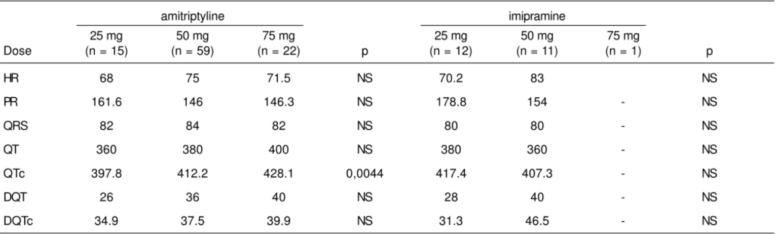
When compared with the dose of the TACs, the QTc de-monstrated statistically significant differences with the use of amitriptyline (Student t test – Table III). Due to the reduced number of patients on imipramine, confirmation of this
infor-mation was not possible. Stratified analysis was done for: gender, age, baseline cardiomyopathy, baseline ECG, and type of TCA used (Tables IV, V, and VI).
Table II – Antidepressant Actions of Tricyclic Antidepressants versus Time
Amitriptyline + Imipramine (n = 40)
Parameters M0 M1 M2 p
HR (bpm) 72.76 ± 13.21 76.79 ± 15.48 76.48 ± 14.52 < 0.05
PR (ms) 150.15 ± 27.04 155 ± 26.27 151.90 ± 31.17 NS
QRS (ms) 82.30 ± 10.61 85.60 ± 10.30 87.77 ± 9.49 < 0.05
QT (ms) 377.78 ± 28.82 371.05 ± 34.82 370.33 ± 28.98 NS
QTc (ms) 412.71 ± 27.06 414.80 ± 21.80 413.50 ± 26.53 NS
DQT (ms) 34.70 ± 14.05 36.10 ± 14.02 35.23 ± 14.80 NS
DQTc (ms) 37.74 ± 14.75 40.77 ± 16.11 39.30 ± 16.18 NS
ANOVA
M0 – pre-medication ECG; M1 – ECG 30 after beginning TACs; M2 – ECG 60 days after beginning TACs.
Table III – Effects of the Different Doses of Tricyclic Antidepressants
amitriptyline imipramine
25 mg 50 mg 75 mg 25 mg 50 mg 75 mg
Dose (n = 15) (n = 59) (n = 22) p (n = 12) (n = 11) (n = 1) p
HR 68 75 71.5 NS 70.2 83 NS
PR 161.6 146 146.3 NS 178.8 154 - NS
QRS 82 84 82 NS 80 80 - NS
QT 360 380 400 NS 380 360 - NS
QTc 397.8 412.2 428.1 0,0044 417.4 407.3 - NS
DQT 26 36 40 NS 28 40 - NS
DQTc 34.9 37.5 39.9 NS 31.3 46.5 - NS
Student t test.
p < 0,05 – significant; NS – non-significant.
Table IV – Effects of Tricyclic Antidepressants on Mean QRS Duration versus Age
Parameter Age < 60 years (n=24) Age ≥ 60 years (n=16)
QRS (ms) ∆ mean p CI 95% ∆ mean p CI 95%
M1 – M0 1.75 0.30 (NS) -1.69; 5.19 5.62 0.09 (NS) -1.17; 12.42
M2 – M0 3.70 0.07 (NS) -0.45; 7.86 8.12 0.01 1.79; 14.45
M2 - M1 1.95 0.25 (NS) -1.47; 5.39 2.50 0.18 (NS) -1.37; 6.37
ANOVA; NS – non-significant.
IN PATIENTS WITH CHRONIC PAIN
DISCUSSION
The Pain Clinic of the Hospital Universitário Clementino Fra-ga Filho da Universidade Federal do Rio de Janeiro was opened in 1983 and it has a multidisciplinary team that in-cludes anesthesiologists, psychiatrists, clinicians, physical therapists, and social workers. It treats oncology patients undergoing palliative care and patients with non-oncologic chronic pain. Tricyclic antidepressants are prescribed for approximately 40% of the patients with chronic pain followed-up by the Program, which justifies the importance of the present study.
Tricyclic antidepressants have been used for more than 40 years in the treatment of depression, and more than 20 years in the treatment of neuropathic pain, starting with the stu-dies of Watson 9, Max 10, and Leijon 11. In the decade of 1960,
when their use became more common, heart block or arrhyth-mias were the main cause of death in cases of overdose. As for their cardiac effects, they interfere with conduction, which can be seen in the electrocardiogram and electrophy-siological studies, with increases in PR, QRS, and QTc in-tervals. However, those increases are seldom symptomatic
in patients without heart disease 12. More recently, it has
been speculated that low doses of those drugs are not as-sociated with additional risks, even in the elderly and patients
with heart disease 13. The results of the present study
cor-roborate those in the literature. However, it should be em-phasized that the doses used in psychiatry are higher than
those used for analgesia 14,15, what can affect patients with
heart disease 16 and the elderly 17. One of the rare
publica-tions on their safety in low doses is a meta-analysis of six
stu-dies comparing low doses of TACs (< 100 mg) with standard
doses used in the treatment of depression (> 100 mg),
sho-wing the lower incidence of side effects in low doses 1.
In this longitudinal, prospective study, antidepressants were used in maximal analgesic doses of 75 mg/day in a repre-sentative sample of the population as far as gender, pain syndrome, and mean dose of TACs are concerned.
Interferences with the HR, with a mild increase, were obser-ved after 30 days of treatment (M1), and this was statistically significant (Table II). The increase in heart rate begins at the onset of treatment, with a tendency for adaptation in appro-ximately four weeks. This variation only affected females. At the end of the follow-up (M2), a return to baseline levels was observed (p = 0.01). This transitory effect was not clinically significant, but it has been reported in the literature 18.
The duration of the QRS complex was discretely increased in M1, with a small statistical significance (p = 0.049 and CI
= 0.014-6.58). However, values in M2 increased further when
compared to baseline levels (M0), with a p = 0.03 (CI
2.02-8.90) and without clinical repercussion (Table II). This small variation of the QRS was seen in elderly patients and patients with cardiomyopathies (Tables IV and V). QRSs complexes equal or greater than 120 msec were not obser-ved until the end of the follow-up; this level according to the literature would indicate greater tendency for arrhythmia 19. In
the present study arrhythmias were not observed on the electrocardiogram.
Table V – Effects of Tricyclic Antidepressants on Mean QRS versus Heart Disease
Parameter Absence of heart disease (n = 16) Heart disease (n = 24)
QRS (ms) ∆ mean p CI 95% ∆ mean P CI 95%
M1 – M0 4.00 0.08(NS) -0.57; 8.57 2.83 0.23 (NS) -1.98; 7.65
M2 - M0 4.56 0.07(NS) -0.47; 9.59 6.08 0.01 1.12; 11.04
M2 - M1 0.56 0.66(NS) -2.14; 3.27 3.25 0.09 (NS) -0.54; 7.04
ANOVA; NS – non-significant.
∆ mean – mean difference; M0 – before TACs; M1 - 30 days after beginning TACs; M2 - 60 days after beginning TACs; CI – confidence interval.
Table VI – Effects of Tricyclic Antidepressants on the Mean Duration of the QRS versus Baseline ECG
Parameter Normal baseline ECG (n = 28) Abnormal baseline ECG (n = 12)
QRS (ms) ∆ mean P CVC 95% ∆ mean p CI 95%
M1 – M0 4.00 0.02 0.55; 7.44 1.66 0.67 (NS) -6.77; 10.11
M2 – M0 4.28 0.02 0.51; 8.09 8.25 0.01 0.01; 16.48
M2 - M1 0.28 0.82 (NS) -2.40; 2.97 6.58 0.01 1.57; 11.59
ANOVA; NS - non significant.
As for the baseline ECG, QRS changes were observed in pa-tients whose baseline ECG was altered as well as in those with normal ECGs (Table IV). In this study, patients with pri-or conduction changes did not have greater variation of the QRS, which is not supported by the literature when TACs are
used in antidepressant doses 20.
It was demonstrated that change in the QRS occurred at the moment the highest dose was used. This supports the no-tion that the influence of this group of drugs on cardiac elec-trophysiology is mainly dose-dependent.
Evaluating the effects of different doses of TACS, this study demonstrated a significant increase in QTc during the use of 75 mg of amitriptyline (p = 0.04). On the other hand, a reduc-tion in the QTc was observed with the use of 50 mg of imi-pramine, but this was not statistically important.
An increase in QT can lead to severe ventricular arrhythmias,
according to Antzelevitch 21. QT dispersion (DQT) reflects
regional differences in ventricular depolarization time. When the intrinsic heterogeneity of the ventricular myocardium is widened, it is reflected on a greater DQT. In the present study, QT dispersion did not increase significantly during treatment (Table III). Despite reports in the literature, which demons-trate the presence of cardiovascular complications, including heart blocks, with antidepressant doses of TACS, arrhyth-mias were not detected in this study 22,23.
In cases of differences in the dose used, one could de-monstrate an influence on the QTc, but only in cases treated with amitriptyline (Table III). But the reduced number of pa-tients treated with imipramine (eight) did not allow the con-clusion that this drug does not interfere with the QT interval. Elderly patients and women are more prone to TAC-induced
increases of the QT interval 24, which was not demonstrated
in the present study.
To conclude, tricyclic antidepressants affect the conduction system, but without clinical significance in patients with heart disease and the elderly, because the doses used for anal-gesia of neuropathic pain were lower than 100 mg.
REFERÊNCIAS — REFERENCES
01. Furukawa AT, McGuire H, Barbui C — Meta-analysis of effects and side effects of low dosage tricyclic antidepressants in depression: systematic review. BMJ, 2002;325:991-995. 02. Moore A, Edwards J, Barden J et al. — Bandolier’s Little Book of
Pain. New York, Oxford, 2003.
03. Baldessarini RJ — Antidepressivos, em: Goodman & Gilman — As Bases Farmacológicas da Terapêutica, 10a Ed., Rio de Janei-ro, Guanabara Koogan, 2003;339-364.
04. Teixeira MJ, Teixeira WJ, Kraychete DC — Epidemiologia Geral da Dor, em: Teixeira MJ — Dor: contexto interdisciplinar. 1a Ed., Curitiba, Maio, 2003;53-66.
05. Kareholp I, Brattberg G — Pain and mortality risk among elderly persons in Sweden. Pain, 1998;77:271-278.
06. Decacche W — ECG para o Clínico. 1a Ed. Rio de Janeiro: Re-vinter, 2004.
07. Lepeschkin E, Surawicz B — The measurement of the QT inter-val of electrocardiogram. Circulation, 1952;6:378-388.
08. Funck-Brentano C, Jaillon P — Rate-corrected QT interval: Techniques and limitations. Am J Cardiol,1993;72:17b-22b. 09. Watson CP, Evans RJ, Reed K et al — Amitriptyline versus
pla-cebo in postherpetic neuralgia. Neurology, 1982;32:671-673. 10. Max MB, Culnane M, Schafer SC et al. — Amitriptyline relieves
diabetic neuropathy pain in patients with normal or depressed mood. Neurology, 1987;37:589-596.
11. Leijon G, Boive J — Central post-stroke pain: a controlled trial of amitriptyline and carbamazepine. Pain, 1989;36:27-36.
12. Glassman AH, Rodrigues AI, Shapiro PA — The use of antide-pressant drugs in patients with heart disease. J Clin Psychiatry, 1998;59(suppl 10):16-21.
13. Ray WA, Meredith S, Thapa PB et al. — Cyclic antidepressants and the risk of sudden cardiac death.Clin Pharmacol Ther, 2004; 75:234-241.
14. Berger A, Dukes E, Edelsberg J et al — Use of tricyclic antide-pressants in older patients with diabetic peripheral neuropathy. Clin J Pain, 2007;23:251-258.
15. Chen H, Lamer TJ, Rho RH et al — Contemporary management of neuropathic pain for the primary care physician. Mayo Clin Proc, 2004;79:1533-1545.
16. Cohen HW, Gibson G, Alderman MH — Excess risk of myocardial infarction in patients treated with antidepressant medications: as-sociation with use of tricyclic agents. Am J Med, 2000; 108:2-8. 17. Bingefors K, Isacson D, Knorring LV et al — Antidepressant-treated patients in ambulatory care mortality during a nine-year period after first treatment. Br J Psychiatry, 1996;169:647-654. 18. Glasmann AH — Cardiovascular effects of antidepressant drugs
update. J Clin Psychiatry, 1998;59(suppl 15):13-18.
19. Buckley NA, Chevalier S, Leditschke IA et al. — The limited utility of electrocardiography variables used to predict arrhythmia in psychotropic drug overdose. Critical Care, 2003;7:101-107 20. Roose SP, Glassman AH, Dalack GW — Depression, heat
di-sease and tricyclic antidepressants. J Clin Psychiatry, 1989; 50: 12-18.
21. Antzelevitch C — Arrhythmogenic mechanisms pf QT prolonging drugs: is QT prolongation really the problem? J Electrocardiol, 2004;37(suppl):15-24.
22. Sadanaga T — Abnormal QT prolongation and psychotropic drug therapy in psychiatric patients: significance of bradycardia-dependent QT prolongation. J Electrocardiol, 2004;37:267-273. 23. Chow BJ, Gollob M, Birnie D — Brugada syndrome precipitated
by a tricyclic antidepressant. Heart, 2005;91:651.
24. Morganroth J — A definitive or thorough phase 1 QT ECG trial as a requirement for drug safety assessment. J Electrocardiol, 2004;37:25-29.
RESUMEN
Cunha Jr RJ, Barrucand L,Verçosa N — Estudio de las Alteracio-nes Electrocardiográficas con el Uso de Antidepresivos Tricíclicos en Pacientes con Dolor crónico.
JUSTIFICATIVA Y OBJETIVOS: Los antidepresivos tricíclicos (ADT) son muy utilizados como analgésicos para lumbalgias cró-nicas y dolores neuropáticos. El objetivo de este estudio fue evaluar las alteraciones electrocardiográficas de los pacientes con dolor crónico que usan amitriptilina o imipramina.
BRE, BAV 1°G, HBAE o extra-sístoles). Se realizaron y se analiza-ron tres ECGs: antes del inicio de los ADT, 30 y 60 días después del inicio del tratamiento, evaluando los parámetros PR, QRS, QT, QTc, DQT, DQTc y FC. Treinta y dos pacientes usaron amitriptilina y ocho imipramina. La dosis promedio al final del estudio fue de 54,29 mg de amitriptilina y de 46,87 mg de imipramina.
RESULTADOS: El análisis de las variables electrocardiográficas después del uso de los ADT arrojó lo siguiente: la amitriptilina aumentó la frecuencia cardíaca transitoriamente en el sexo feme-nino (p = 0,049) y la duración del QRS en los pacientes con edad igual o superior a los 60 años y en los cardiópatas en la segunda
evaluación (p = 0,01). En los pacientes que recibieron amitriptilina, dosis de 75 mg, el intervalo QTc fue mayor cuando se le comparó a las dosis de 25 mg (p = 0,0044). El aumento de esos parámetros mostró el efecto de la amitriptilina sobre la conducción cardíaca, sin embargo, no se registró comprometimiento clínico, pues los valores permanecieron dentro de los límites de la normalidad (QRS
< 110ms y QTc < 470ms).
CONCLUSIONES: El uso clínico de los ADT en dolores crónicos, arrojó resultados seguros y eficaces, y no presentó disturbio de la conducción cardíaca con repercusión clínica.