Oliveira KF et al. / Massive thymoma of the mid-posterior mediastinum
Radiol Bras. 2016 Nov/Dez;49(6):403–405 403
0100-3984 © Colégio Brasileiro de Radiologia e Diagnóstico por Imagem
Case Report
Massive thymoma of the mid-posterior mediastinum:
an unprecedented case in a young adult
*
Timoma volumoso de mediastino medioposterior em adulto jovem: relato de caso inédito
Oliveira KF, Rodrigues MM, Lopes GP, Almeida RS, Lusvarghi JL, Santos JPV. Massive thymoma of the mid-posterior mediastinum: an unprecedented case in a young adult. Radiol Bras. 2016 Nov/Dez;49(6):403–405.
Abstract
R e s u m o
We report an unprecedented case of ectopic thymoma in a young adult. A 33-year-old male presented with a 10-day history of non-productive cough and fever. Investigation revealed mediastinal widening without pulmonary involvement. Computed tomography showed a large mass—14.8 × 10.8 × 8.4 cm—in the mid-posterior mediastinum, and a biopsy obtained by video-assisted thoracoscopy indicated that the mass was a tumor. Immunohistochemistry showed combined thymoma type AB1. Because of the considerable proportions of the tumor and its close proximity to major structures, the patient was treated with chemotherapy.
Keywords: Thymoma; Middle mediastinum; Posterior mediastinum; Ectopic tumor.
Apresentamos caso inédito de volumoso timoma ectópico em adulto jovem. Homem de 33 anos de idade, encaminhado com tosse seca e febre diária havia 10 dias. Durante investigação observou-se alargamento mediastinal sem comprometimento pulmonar. Um tumor mediastinal medioposterior, medindo 14,8 × 10,8 × 8,4 cm, foi diagnosticado após tomografia computadorizada e biópsia videotoracoscópica. A imuno-histoquímica revelou timoma misto AB1. Devido à íntima relação com estruturas nobres e grandes propor-ções, optou-se pela quimioterapia.
Unitermos: Timoma; Mediastino médio; Mediastino posterior; Tumor ectópico.
* Study conducted in the Department of Radiology and Diagnostic Imaging at the Universidade Federal do Triângulo Mineiro (UFTM), Uberaba, MG, Brazil.
1. MD, Resident in Dermatology at the Hospital Universitário de Brasília (HUB), Brasília, DF, Brazil.
2. Fellow in Oncological Radiology at the Instituto do Câncer do Estado de São Paulo Octavio Frias de Oliveira (Icesp), São Paulo, SP, Brazil.
3. Head of the Department of Radiology and Diagnostic Imaging at the Universi-dade Federal do Triângulo Mineiro (UFTM), Uberaba, MG, Brazil.
4. MD, Resident in Clinical Medicine at the Universidade Federal de Uberlândia (UFU), Uberlândia, MG, Brazil.
5. MD, Resident in Plastic Surgery at the Hospital Heliópolis, São Paulo, SP, Brazil.
6. Thoracic Surgeon at the Hospital de Clínicas da Universidade Federal do Triân-gulo Mineiro (UFTM), Uberaba, MG, Brazil.
Mailing address: Dra. Karen Fernandes de Oliveira. SGAN 911, Bloco H, conjunto G, ap. 02, Edifício Garden Park, Asa Norte. Brasília, DF, Brazil, 70790-110. E-mail: [email protected].
Received March 29, 2014. Accepted after revision June 20, 2014.
sion, the physical examination showed no alterations. Labo-ratory tests showed normocytic, normochromic anemia (Hb = 9.7 g/dL); inversion of the albumin/globulin ratio (A/G ratio = 0.65); a C-reactive protein level of 33.1 mg/dL; thrombocytosis (platelet count = 592,000/mm3
); and leu-kocytosis without a shift (leukocyte count = 14,290/mm3
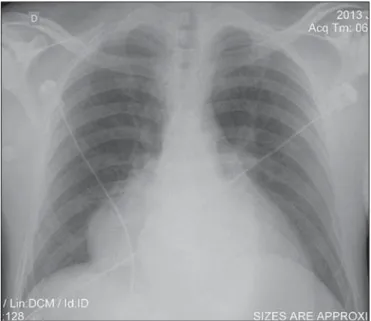
). The patient evolved to daily fever spikes and tachycardia. A chest X-ray revealed mediastinal widening (Figure 1), and
Karen Fernandes de Oliveira1, Marcio Maciel Rodrigues2, Gesner Pereira Lopes3, Renan Sandoval de Almeida4,
Juliana Lopes Lusvarghi5, João Paulo Vieira dos Santos6
http://dx.doi.org/10.1590/0100-3984.2014.0025 INTRODUCTION
Although thymoma is the most common primary tumor of the anterior mediastinum, it accounts for less than 1% of all neoplasms in adults(1,2). The involvement of middle and
posterior mediastinum is rare, only 16 cases of thymoma in the middle mediastinum having been reported(2–10). The
incidence of thymoma peaks between 50 and 60 years of age. Here, we describe the challenge of diagnosing this rare neo-plasm in a mildly symptomatic young adult.
CASE REPORT
A 33-year-old man was hospitalized in July of 2013 to investigate a 10-day history of dry cough and fever. At
Oliveira KF et al. / Massive thymoma of the mid-posterior mediastinum
Radiol Bras. 2016 Nov/Dez;49(6):403–405 404
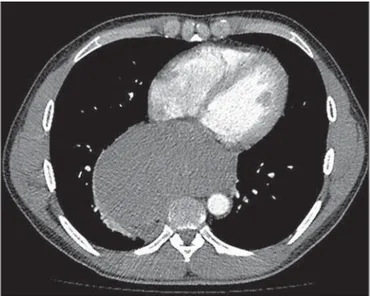
echocardiography was therefore required. The echocardiog-raphy showed extrinsic compression of the left atrium by a mediastinal mass, with an ejection fraction of 56%. Com-puted tomography (CT) of the chest (Figures 2 and 3) showed that the mass measured 14.8 cm at its greatest di-ameter and was located in the mid-posterior mediastinum. Upper gastrointestinal endoscopy identified extrinsic com-pression of the distal esophagus and gastric cardia. Barium swallow allowed us to visualize a significant delay in empty-ing, and protein electrophoresis showed a polyclonal increase in gamma globulins. The main diagnostic hypotheses were lymphoma, giant leiomyoma of the esophagus, neurogenic tumor, and plasmacytoma. Empirical antibiotic therapy was started and resulted in clinical improvement.
For diagnostic clarification, a video-assisted thoraco-scopic biopsy was performed. Frozen section analysis of the biopsy material was suggestive of lymphoma. However, im-munohistochemistry revealed thymoma type AB1.
The patient was discharged with a referral for outpatient chemotherapy. A follow-up CT scan showed partial regres-sion of the tumor. When the CT scan was reevaluated by the thoracic surgery team in February of 2014, the tumor was still considered unresectable.
DISCUSSION
Half of all mediastinal tumors—including thymoma, germ cell tumors, thyroid diseases, and lymphoma—have an anterior origin. In the middle mediastinum, congenital cysts prevail, whereas neurogenic tumors prevail in the posterior mediastinum(1,2,11,12).
The thymus is a lymphoid organ that plays a critical role in the maturation of lymphocytes and in cellular immunity. Embryologically, it originates from the third and fourth pharyngeal pouches. During their migration, thymic tissue fragments or accessory lobes can erroneously locate to the cervical region (in 4% of cases) or to the middle mediasti-num(1,7,11,12). Including the case presented here, only 17 cases
of mediastinal thymoma have been reported. Among those cases, ours is the only one in which the tumor was unre-sectable, and the individual affected in our cases was younger than those affected in the other cases reported, underscor-ing the rarity of the case presented here.
The annual incidence of thymoma is 0.15 cases/100,000 population, with no difference between genders, and its inci-dence increases in adulthood, peaking at 50–60 years of age(2).
Most patients are asymptomatic, being diagnosed on the basis of incidental findings on imaging studies. Approximately Figure 2. Contrast-enhanced CT of the chest, with coronal (A) and sagittal (B) reconstructions, showing a mass with a heterogeneous texture and discrete hetero-geneous contrast enhancement, measuring 10.8 × 8.4 × 14.8 cm, in the mid-posterior mediastinum. The mass presents an intimate relationship with the great mediastinal vessels, right main bronchus, vertebral bodies, right atrium, diaphragm, and esophagus, albeit without direct signs of invasion of any of those structures.
A B
Oliveira KF et al. / Massive thymoma of the mid-posterior mediastinum
Radiol Bras. 2016 Nov/Dez;49(6):403–405 405
40% of symptomatic patients present with myasthenia gravis, the paraneoplastic syndrome most often associated with thy-moma(8,9). Our patient reported having a normal diet and
showing no signs of dysphagia.
Thymomas originate from the epithelial cells of the thy-mus. Histologically, the thymus has two regions: the cor-tex, which is rich in lymphocytes, and the medulla, which is composed of epithelial cells. The histological classification is based on the morphology of the epithelial cells and the proportional relationship between those cells and lympho-cytes. Most of the thymomas that are determined to be type A, AB, or B1 have a benign course. Thymoma types B2 and B3 are considered to be malignant, with metastatic poten-tial(13).The most widely used staging system is that devised by Masaoka et al.(14), which involves postoperative
patho-logical evaluation of capsular invasion of the thymus. Because the tumor could not be resected, radiology played a key role in the case presented here. The initial di-agnosis and staging were based on imaging studies, with an emphasis on the detection of locoregional or distant inva-sion. Approximately 45–80% of thymomas are visible on conventional chest X-rays, which initiated the diagnostic in-vestigation in our patient. For the evaluation of mediastinal masses, CT is the tool of choice(2,15).
Complete surgical resection is the main therapy for in-vasive and noninin-vasive thymomas, being the most important predictor of long-term survival. However, radiation therapy and chemotherapy, alone or in combination, produce favor-able results, increasing survival and improving the progno-sis(12,16).
Thymoma can mimic a variety of diseases, including those with compressive symptoms and paraneoplastic dis-eases, as well as mediastinal widening. Hence the importance of this case—to expand diagnostic reasoning for a tumor in the middle mediastinum, because it is a rare differential di-agnosis to be considered, especially in young patients.
REFERENCES
1. Shikada Y, Katsura M, Takenaka T, et al. A case of middle medias-tinal thymoma. Gen Thorac Cardiovasc Surg. 2012;60:664–7. 2. Juanpere S, Cañete N, Ortuño P, et al. A diagnostic approach to
the mediastinal masses. Insights Imaging. 2013;4:29–52. 3. Bradford R, Cohen SL, McLlelland J, et al. Thymoma presenting
as a middle mediastinal mass. Postgrad Med J. 1984;60:611–3. 4. Wu Z, Wang Z, Li S. Thymoma of the middle mediastinum: case
report. Chinese J Med Imaging Technol. 2010;26:777.
5. Venayaga K, Ooi JSM, Shabir B. A rare case of middle mediastinal thymoma mimicking left lower lobe lung tumor. Med J Malaysia. 2005;60:508–10.
6. Adebonojo SA, Grillo IA, Falase AO, et al. Middle mediastinal thy-moma simulating pericardial cyst. Int Sur. 1977;62:343–5. 7. Koezuka S, Sato F, Hata Y, et al. Video-assisted thoracoscopic
sur-gery for ectopic mediastinal thymoma in a patient with myasthenia gravis. Ann Thorac Surg. 2013;95:e67–8.
8. Chung SR, Kim IS, Kim J. Thymoma of the middle mediastinum. Korean J Thorac Cardiovasc Surg. 2012;45:267–8.
9. Takizawa M, Oda M, Matsumoto I, et al. Myasthenia gravis compli-cated with lung cancer and middle mediastinal thymoma. Asian Cardiovasc Thorac Ann. 2012;20:486–8.
10. Shiryazdi SM, Ayatollahi S, Moghimi M. Cystic thymoma in middle mediastinum – a rare case report. Pol Przegl Chir. 2013;82:35–8. 11. Minniti S, Valentini M, Pinali L, et al. Thymic masses of the middle mediastinum: report of 2 cases and review of the literature. J Thorac Imaging. 2004;19:192–5.
12. Kim JY, Hofstetter WL. Tumors of the mediastinum and chest wall. Surg Clin North Am. 2010;90:1019–40.
13. Rosai J. Histological typing of tumours of the thymus. In: WHO International histological classification of tumours. 2nd ed. New York, NY: Springer-Verlag; 1999. p. 5–15.
14. Masaoka A, Monden Y, Nakahara K, et al. Follow-up study of thy-momas with special reference to their clinical stages. Cancer. 1981; 48:2485–92.
15. Takahashi K, Al-Janabi NJ. Computed tomography and magnetic resonance imaging of mediastinal tumors. J Magn Reson Imaging. 2010;32:1325–39.