Patients with chronic high spinal cord injury can be
safely treated with neuromuscular electrical stimulation:
cardiovascular function is unafected
Letícia Vargas AlmeidaI, Carolina LinsI, Janaina TancredoI, Renato VarotoI, Wilson Nadruz JuniorI, Alberto Cliquet
JuniorI,II
DOI: 10.5935/MedicalExpress.2017.05.03
I Universidade de Campinas - UNICAMP, Faculdade de Ciências Médicas, Departamento de Ortopedia e Traumatologia, Laboratório de Biomecânica e Reabilitação
do Sistema Locomotor, Campinas, SP, Brazil.
II Universidade de São Paulo – USP, Escola de Engenharia de São Carlos, Departamento de Engenharia Elétrica e de Computação, Laboratório de Biocibernética
e Engenharia de Reabilitação, São Carlos, SP, Brazil.
OBJECTIVE: To identify changes in blood pressure and heart rate in individuals with chronic paraplegia undergone neuromuscular electrical stimulation treatment.
METHOD: Design: Observational prospective. Participants: Twenty individuals with chronic paraplegia (neurological level above T6) belonging to two diferent groups (G1 and G2) were submitted to an upper limb exercise test. G1 patients (n=13) had been treated with neuromuscular electrical stimulation (25Hz, pulses of 300μs, 100V) for 2 years or more, at least once a week; G2 patients (n=7) did not receive neuromuscular electrical stimulation treatment; G3 individuals (n=6) were healthy volunteers. Procedures: Arterial blood pressure and heart rate were measured during four phases of the exercise test: at initial rest, during warmup, during the exercise itself, and at rest after the exercise.
RESULTS: Systolic and diastolic blood pressures showed no statistical diference between groups. In the comparison between exercise phases, regardless of the group, systolic pressure was signiicantly higher and diastolic pressure signiicantly lower at the end of the exercise itself, when compared to all other phases. Resting heart rate was signiicantly lower in healthy controls vs. G1 and G2, which were not signiicantly diferent between themselves. Exercise increased heart rate in all groups.
CONCLUSION: This study showed that the groups are normotensive and homogeneous in their results; heart rate was higher in both paraplegic groups compared to healthy controls, but no diference was found between treated vs. untreated groups. Thus, neuromuscular electrical stimulation is a safe and efective way to treat individuals with chronic paraplegia.
KEYWORDS: Spinal Cord Injury, Paraplegia, Blood Pressure, Heart Rate, Neuromuscular Electrical Stimulation, Autonomic Dysrelexia.
Almeida LV, Lins C, Tancredo J, Varoto R, Nadruz-Junior W, Cliquet-Junior A. Patients with chronic high spinal cord injury can be safely treated with neuromuscular electrical stimulation: cardiovascular function is unafected. MedicalExpress (São Paulo, online). 2017 October;4(5):M170503.
Received for Publication on June 23, 2017; First review on July 27, 2017; Accepted for publication on September 10, 2017; Online on September 26, 2017
E-mail: [email protected]
■
INTRODUCTIONSpinal Cord Injury (SCI) is one of the most serious and complex pathologies in the neurological domain and is, in turn, responsible for a series of deleterious compli-cations in the human body. In addition to all the physical, psychic and social damages that affect these individuals,
who, in Brazil, are usually young men, aged 15-35 years, they will still have to deal with Autonomic Dysreflexia, the silent and potentially life -threatening complication.1
The population of individuals with SCI is increas-ing in Brazil and in the world, because of car accidents, firearms, cold weapons, diving, falls, among others, or because of natural disasters, such as earthquakes.2-5
The American Spinal Injury Association (ASIA) Im-pairment Scale (AIS) was applied by a single professional trained and qualified to do so. Briefly, AIS classifies the severity of spinal cord injury, identifying sensory levels (scored for 28 locations, maximum 112 points) and motor levels (scored to a maximum of 100 points).9,10
G1 patients had been treated with NMES for more than 2 years, at least once a week. (treatment initiated at least 1 year after injury). For this, a 4-channel electrical stimulator was used, with a monophasic signal of 25 Hz, rectangular pulses of 300 μs and maximum intensity of 100 V (1 kΩ load). NMES was applied on the quadriceps and on the fibular nerve for 20 and 15 minutes, respectively.
G1 and G2 subjects performed the upper limb exer -tion test in their daily use wheelchairs during the year 2015; G3 subjects carried out the test in an ordinary chair. A custom-made table with adjustable height was used to accommodate individuals of different stature and various models of wheelchair.
The following materials were used for upper limbs exertion test: static cycle ergometer (Hand Cycle Endorphin Professional Series, Endorphin Corporation, Pinellas Park, FL, USA), automatic blood pressure monitor (Omron Healthcare, Inc., São Paulo, SP, Brazil), heart rate monitor (Polar S810- T31 cod, Polar Electro Inc., New York, NY, USA) and a 1.2x0.8 m table with adjustable height.
During the upper limbs exertion test all subjects were monitored for systolic and diastolic blood pressures, and for heart rate data, which were acquired simultaneously.
The exertion test used was the Conconi model, which was adapted for the wheelchair users.11,12 This test was
ap-plied three times to all participants on consecutive weeks. The entire activity was divided into four phases: initial rest (5 minutes), warming up, test itself (until fatigue) and final rest (5 minutes). After the initial rest period, the warming up phase consisted of cyclic movements with the upper limbs at a predetermined speed of 5 km/h for 2 minutes. Next came the test itself, which was started with a speed of 7 km/h during the first minute. Verbal commands were given to the subjects to increase speed by 1 km/h at every minute until fatigue set in, at which point the test was concluded. The tests were performed without load, with the test proper lasting in average 11.34 minutes throughout the groups.
Data analysis was done through measures of mean, median, standard deviation, minimum and maximum va-lues. Phases and groups were compared through ANOVA technique for repetitive measures with response variables transformed to segmented factors. The significance level adopted was 5%.
■
RESULTSConsequently, health complications have become more evident on account of the increase of the SCI popula-tion and of their life expectancy. Autonomic Dysreflexia is a complication due to the breakdown of the Autonomic Nervous System function resulting from SCI; 90% of the cases are observed in spinal cord injuries at high neurologi-cal levels, i.e. above the sixth thoracic nerves. The clinineurologi-cal signs of autonomic dysreflexia were first observed in 1830 in a patient presenting intense sweating and cutaneous ery-thema in the head and neck during a vesical catheterization. In 1917, autonomic reflexes were associated to the vesical and cutaneous stimuli below the level of SCI. The relation-ship between paroxysmal hypertension and the increased number of individuals with SCI was only confirmed in 1947.7
It is a well-established fact that negative cardiologic changes are events caused by reduced sympathetic and increased parasympathetic control. The disordered rela-tionships between these two systems results in variations of heart rate and blood pressure and may cause arrhyth-mias, namely bradycardias with a risk of cardiac arrest in individuals with high cervical and thoracic injuries.8
In view of the relationship between harmful skin and vesical stimuli below the lesion level and signs and symptoms generated by the lack of harmony within the autonomic nervous system, the objective of this study was to describe the blood pressure and heart rate changes observed in a group of individuals with high paraplegia who underwent treatment with Neuromuscular Electrical Stimulation (NMES).
■
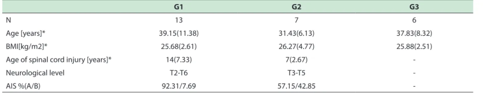
MATERIALS AND METHODSTable 1 - Demographic and clinical data of subjects (n=26).
G1 G2 G3
N 13 7 6
Age [years]* 39.15(11.38) 31.43(6.13) 37.83(8.32)
BMI[kg/m2]* 25.68(2.61) 26.27(4.77) 25.88(2.51)
Age of spinal cord injury [years]* 14(7.33) 7(2.67)
-Neurological level T2-T6 T3-T5
-AIS %(A/B) 92.31/7.69 57.15/42.85
-* Signiicant diference between G1 vs. G2 (p<0.05)
blood pressures, and heart rate throughout the test, ac-cording to phases and the groups are displayed in Figures 1-3, respectively.
Figure 1 shows that systolic blood pressure exhibi-ted no significant differences between groups (p = 0.147), regardless of phase. However, at the end of the fatigue phase systolic pressure was significantly higher (p < 0.001), compared to the values of the other three phases.
Diastolic blood pressure (Figure 2) shows that simi-larly to systolic pressure, no significant differences occurred between groups (p = 0.311). However, and independently of groups, diastolic pressure was significantly lower (p < 0.001) at the end of the fatigue phase, compared to the other three phases.
Figure 3 shows that significant differences (p < 0.001) were observed between the groups for heart rate. Regardless of the phase, G3 presented significantly lower values than the G2 (p = 0.037) and G1 (p-value = 0.009). There were no significant differences between G1 and G2 (p = 0.9538). Significant differences were also observed between phases independently of the group: the initial rest phase presented the lowest, whereas the fatigue phase presented the highest values of the series.
■
DISCUSSIONIn this work, the aim was to analyze heart rate and blood pressure during the physical exertion test of upper limbs and NMES, identifying possible changes caused by the treatment in individuals with chronic paraplegia. It is known that NMES provides benefits to individuals with SCI such as recruitment of muscle fibers, improve-ment of muscle tissue oxygenation and venous return, orthostatism, gait and adequacy of upper limb tasks, as well as increased cardiac blood flow.13 Faced with
so many benefits and considering its important role in the rehabilitation of these individuals, it was necessary to investigate the cardiologic aspects and its possible changes in this treatment strategy.
Figure 1. Systolic arterial pressure for G3 (controls), G2 (no previous NMES) and G1 (previous NMES). * indicates that systolic pressure was signiicantly higher for all groups at the end of the fatigue phase, for all groups.
Figure 2. Diastolic arterial pressure for G3 (controls), G2 (no previous NMES) and G1 (previous NMES). * indicates that diastolic pressure was signiicantly lower for all groups at the inal rest period.
Gallo et al14 have shown that physical activity
ge-nerates important metabolic alterations and autonomic modifications that influence in the cardiovascular system.
The hypothesis regarding the lack of active muscula-ture and the accumulation of adipose tissue is a significant indicator in which the increase of carotid intima-media thickness was investigated in a comparative study betwe-en spinal cord injured individuals with neurological level between C4-T12 and healthy individuals.15
In a recent study, heart rate variability was analyzed in 60 patients with spinal cord injury divided into 2 groups (above and below the T6 level) in relation to the control group. The evaluation was done from the waveforms and spectral analysis of the electrocardiogram. Statistical data showed that both groups of patients had some degree of autonomic dysfunction. However, the group of patients with spinal cord injury above the T6 level presented an increased risk of comorbidity and morbidity.16
Manogue et al,17 in a recent review have noted that
both acute and chronic SCIs are often followed by cardiac abnormalities, characterized by impairment of the Sympa-thetic Nervous System, with dominance of the Parasympa -thetic counterpart. Episodes of bradycardia, sinus arrest or total heart block are observed in the acute phase. In the chronic phase, there are episodes of autonomic dysreflexia and paroxysmal hypertension, which are frequently initia-ted by vesical distensions or other stimuli below the lesion, and may be followed by bradycardia or tachycardia. They also note that the prognosis of these individuals tends to improve due to new technological advances and improved professional care.17 We believe our contribution here shows
that cardiovascular function is essentially normal in chronic paraplegia.
Bar-On et al18 evaluated, in paraplegic patients, the
relationship between heart rate and O2 consumption at
maximal physical effort: they report that, in individuals with SCI below T3, the interaction within the Sympathetic Nervous System is intact, because a linear correlation be -tween heart rate and O2 consumption occurs due to
incre-ased exercise load. However, they do suggest the presence of some other compensatory mechanism in the regulation of cardiac control.
It is known that individuals with acute or chronic SCI exhibit cardiac impairment due to the lack of harmo-nization within the autonomic nervous system, especially when patients with a neurological level above T6 are con-sidered,14,16,18 suggesting the presence of a compensated
sympathetic impairment.16,18 In our study neither systolic
nor diastolic pressure were significantly different between the three groups in response to exercise, with or without NMES treatment. However, in the comparison between exercise phases, significant differences for systolic and
speed imposed by the test and consequently the increase of the force of ventricular contraction and cardiac output during the exercise.19 On the other hand, diastolic pressure
was lower at the end of the exercise itself. The values were significant in relation to the phase and were consistent with a drastic drop in resistance to peripheral blood flow during exercise.19
Petrofsk20 investigated the systolic and diastolic
blood pressure and heart rate responses in paraplegics having complete and incomplete injuries and healthy vo-lunteers during fatiguing isometric contraction of the grasp and quadriceps muscles. All subjects performed voluntary grasp and presented pressure and heart rate increasing from rest to fatigue. Quadriceps contraction was genera-ted by functional electrical stimulation in patients, and their systolic and diastolic responses were similar to the healthy controls. However, the heart rate of healthy con-trols reached 127 beats/min at the end of the contraction, while the paraplegic group presented an average increase of 6 beats/min. According to these findings, the control of blood pressure is peripheral in origin and heart rate seems to be centrally mediated.
This study also shows that NMES treatment did not generate sympathetic hyperexcitability and a possible hypertensive peak, nor did it cause cutaneous stimuli be-low the lesion level. Therefore, NMES is a safe and effective treatment for this group of individuals.
Healthy controls presented a lower heart rate when compared with the paraplegic patients, with no difference between treated and untreated groups. This difference is very likely a consequence of the severe deficit of active musculature in paraplegic individuals, placing them at the disadvantage in relation to the control group, a fact already expected by the study. Nonetheless all three groups exhibi -ted the physiologically expectable tachycardia during exer-cise, further confirmation of the harmless action of NMES. Study Limitations. The sample is small because of the smallness of the group of patients treated with NMES in our Institution. Recruitment of paraplegic patients (levels T2 – T6) who had not undergone NMES treatment was even more difficult. Although this study has demonstrated that NMES is an effective and safe strategy of treatment, future studies with more volunteers will be necessary to confirm these results. However, to the best of our knowledge, no published report covers this specific aspect of the mana -gement of paraplegia.
■
CONCLUSION-nos uma vez por semana; os pacientes do G2 (n = 7) não
receberam o tratamento com estimulação elétrica neuro
-muscular; os indivíduos do G3 (n = 6) eram voluntários saudáveis. Procedimentos: A pressão sanguínea arterial e a frequência cardíaca foram medidas durante quatro fases do teste de exercício: no repouso inicial, durante o aqueci -mento, durante o exercício e no repouso após o exercício.
RESULTADOS: As pressões arteriais sistólica e
diastólica não apresentaram diferença estatística entre os grupos. Na comparação entre as fases do exercício, inde -pendentemente do grupo, a pressão sistólica foi significa -tivamente maior e a pressão diastólica significa-tivamente menor no final do exercício, em comparação com todas as outras fases. A frequência cardíaca em repouso foi signi -ficativamente menor em controles saudáveis versus G1 e G2, que não foram significativamente diferentes entre eles mesmos. O exercício aumentou a frequência cardíaca em todos os grupos.
CONCLUSÃO: Este estudo mostrou que os grupos são
normotensos e homogêneos em seus resultados; a frequ
-ência cardíaca foi maior em ambos os grupos paraplégicos em comparação com controles saudáveis, mas nenhuma diferença foi encontrada entre os grupos tratados versus os não tratados. Assim, a estimulação elétrica neuromuscular é uma maneira segura e eficaz de tratar indivíduos com paraplegia crônica.
PALAVRAS-CHAVE: Lesão Medular, Paraplegia, Pres
-são Sanguínea, Frequência Cardíaca, Estimulação Elétrica Neuromuscular, Disreflexia Autonômica.
■
REFERENCES1. Ahuja CS, Wilson JR, Nori S, Kotter MRN, Druschel C, Curt A, et al . Traumatic spinal cord injury. Nat Rev Dis Primers. 2017;3:17018. DOI:10.1038/nrdp.2017.18.
2. Ferro FP, González HJ, Ferreira DM, Cliquet A Jr: Electrical stimu -lation and treadmill gait in tetraplegic patients: assessment of its effects on the knee with magnetic resonance imaging. Spinal Cord
2008;46(2):124-8. DOI:10.1038/sj.sc.3102078
3. Kim D, Tan CO. Alterations in autonomic cerebrovascular control after spinal cord injury. Auton Neurosci. 2017. pii: S1566-0702(17)30084-X. DOI:10.1016/j.autneu.2017.04.001.
4. Grove, C C, Poudel, MK, Baniya M, Rana C, House DR, Descriptive study of earthquake-related spinal cord injury in Nepal. Spinal Cord. 2017;55(7):705-10. DOI:10.1038/sc.2017.25.
5. Kriz J, Kulakovska M, Davidova H, Silov M and Kobesova A. Inciden -ce of acute spinal cord injury in the Czech Republic: a prospective
epidemiological study 2006–2015. Spinal Cord. 2017;55(9):870-4. DOI:10.1038/sc.2017.20.
6. Savic G, DeVivo MJ, Frankel HL, Jamous MA, Soni BM and Charlifue S. Long-term survival after traumatic spinal cord injury: a 70-year British study. Spinal Cord. 2017;55(7):651-8. DOI:10.1038/sc.2017.23. 7. Partida E, Mironets E, Hou S,Tom VJ. Cardiovascular dysfunction
following spinal cord injury. Neural Regen Res. 2016;11(2):189-94. DOI:10.4103/1673-5374.177707.
8. Biering-Sørensen F, Biering-Sørensen T, Liu N, Malmqvist L, Wecht JM, Krassioukov A. Alterations in cardiac autonomic control in spinal cord injury. Auton Neurosci. 2017. pii: S1566-0702(17)30044-9. DOI:10.1016/j.autneu.2017.02.004.
higher basal heart rates vs healthy controls. Overall, this study showed that NMES is a safe and effective form of tre -atment for individuals with high level paraplegia, although more studies should be done towards a better understanding of the role of NMES in autonomic dysfunctions.
■
ACKNOWLEDGEMENTSWe thank the support by grants from São Paulo Re -search Foundation (FAPESP), Coordination of Improvement of Higher Level Personnel (CAPES) and National Council for Scientific and Technological Development (CNPq), and Fernanda Furtado Camargo for contributing with her kno-wledge to this study.
■
CONFLICTS OF INTERESTThe authors declare no conflict of interest.
■
AUTHOR PARTICIPATIONLetícia Vargas de Almeida: main researcher of the described work; elaborated the proposed methodology, performed data collection and analyzed the results. Carolina Lins: auxiliary researcher, assisted in the elaboration of the proposed method and in the statistical analyses. Janaina Roland Tancredo: assisted in data collection, including AIS assessment. Renato Varoto: co-author, provided assistance on data interpretation and discussion, text elaboration and revision. Wilson Nadruz Junior: co-author, provided assis -tance on text elaboration and data interpretation and dis-cussion, based on his expertise on the human cardiovascular system. Alberto Cliquet: academic Advisor, proposed the initial concept for the presented work. Provided guidance and assistance on research development and execution.
PACIENTES COM LESÃO CRÔNICA ALTA DA ME-DULA ESPINHAL PODEM SER TRATADOS DE FOR-MA SEGURA COM ESTIMULAÇÃO ELÉTRICA NEU-ROMUSCULAR: A FUNÇÃO CARDIOVASCULAR NÃO É AFETADA
OBJETIVO: Identificar mudanças na pressão arterial
e frequência cardíaca em indivíduos com paraplegia crônica tratados com estimulação elétrica neuromuscular.
MÉTODO: Estudo prospectivo observacional.
Par-ticipantes: vinte indivíduos com paraplegia crônica (nível
9. Maynard FM, Jr, Bracken MB, Creasey G, Ditunno JF, Jr, Donovan WH, Du
-cker TB, et al. International Standards for Neurological and Functional Classification of Spinal Cord Injury. Spinal Cord. 1997;35(5):266-74. 10. Kirshblum SC, Burns SP, Biering-Sorensen F, Donovan W, Graves DE, Jha
A, et al. International standards for neurological classification of spinal cord injury (Revised 2011). J Spinal Cord Med. 2011;34(6):535-46. DO I:10.1179/204577211X13207446293695.
11. Conconi, F.; Ferrari, M.; Ziglio P. G.; Droghetti, P.; Codeca, L. Determina
-tion of the anaerobic threshold by a noninvasive field test in runners. J Appl Physiol Respir Environ Exerc Physiol. 1982;52(4):869-73. 12. Conconi F, Grazzi G, Guglielmini C, Borsetto C, Ballarin E, Mazzoni G,
et al. The Conconi Test: Methodology after 12 years of application. Int J Sports Med. 1996;17(7):509-19. DOI:10.1055/s-2007-972887 13. Carvalho DCL, Zanchetta MC, Sereni JM and Cliquet Jr A. Metabolic
and cardiorespiratory responses of tetraplegic subjects during trea-dmill walking using neuromuscular electrical stimulation and partial
body weight support. Spinal Cord. 2005;43(7):400-5. DOI:10.1038/ sj.sc.3101730
14. Gallo-Junior L, Maciel BC, Marin-Neto JA, Martins LE. Sympatheric
and parasympathetic changes in heart rate control during dynamic
exercise induced by endurance training in man. Braz J Med Biol Res 1989;22(5);631-43
15. Matos-Souza JR, Pithon KR, Ozahata TM, Gemignani T, Cliquet A Jr, Nadruz W Jr. Carotid intima-media thickness is increased in patients
with spinal cord injury independent of traditional cardiovascular risk
factors. Atherosclerosis. 2009;202(1):29-31. DOI:10.1016/j.atheros
-clerosis.2008
16. Carvalho-Abreu EM, Dias LP, Lima FP, Paula-Júnior AR, Lima MO. Car -diovascular autonomic control in paraplegic and quadriplegic. Clin
Auton Res. 2016;26(2):117-26. DOI:10.1007/s10286-015-0339-1. 17. Manogue M, Hirsh DS, Lloyd M. Cardiac electrophysiology of pa
-tients with spinal cord injury. Heart Rhythm 2017;14(6):920-7. DOI:10.1016/j.hrthm.2017.02.015
18. Bar-On ZH, Nene AV. Relationship Between Heart Rate and Oxygen Uptake in Thoracic Level Paraplegics. Paraplegia. 1990;28(2):87-95. DOI:10.1038/sc.1990.11
19. Currie KD, West CR and Andrei V. Krassioukov AV. Differences in Left Ventricular Global Function and Mechanics in Paralympic Athletes with Cervical and Thoracic Spinal Cord Injuries. Front Physiol. 2016;7:110. DOI:10.3389/fphys.2016.00110.