O
ri
gi
na
l
a
rt
ic
le
s
Analysis of the complementary measurement of nasogenian wrinkles
using Radiocef 2.0
®software in the evaluation of facial chronoaging
among women of different age groups
Rodrigo Marcel Valentim da Silva1
Gabriela Paiva de Melo2
Silvia Maria Lambert da Costa2
Jackelline Savana Vieira Estrela3
Veruschka Ramalho Araruna4
Amanda Caroline Muñoz Costa3
Janaina Maria Dantas Pinto1
Hanieri Gustavo de Oliveira5
Patrícia Froes Meyer3
1 Universidade Federal do Rio Grande do Norte, Centro de Ciências da Saúde, Programa de Pós-graduação
em Fisioterapia. Natal, RN, Brasil.
2 Universidade Potiguar, Curso de Fisioterapia. Natal, RN, Brasil.
3 Universidade Potiguar, Programa de Pós-graduação em Fisioterapia Dermato-funcional. Natal, RN,
Brasil.
4 Universidade Católica Nostra Señora de Assuncion, Programa de Pós-graduação em Fisioterapia. Assunção,
Paraguai.
5 Universidade do Estado do Rio Grande do Norte, Departamento de Odontologia. Caicó, RN, Brasil.
Correspondence
Rodrigo Marcel Valentim da Silva E-mail: marcelvalentim@hotmail.com
Abstract
Objective: To evaluate the facial aging of women of different ages using a software program to assist in the classification of wrinkles and sagging in the nasogenian region. Method: A descriptive observational study of 100 female volunteers was performed. The women were aged between 20 and 70 years old and were sorted by age group into five groups of 20 volunteers each. The instruments used were the Facial Assessment Protocol, a cephalostat for the standardization of photos, a 14.1 megapixel Sony digital camera, and the Radiocef 2.0® software program. The Kolmogorov-Smirnov (KS) test was used
for confirmation of normality and all data was statistically analyzed using ANOVA and Tukey’s post hoc analysis. The Chi-squared and Pearson’s correlation tests were also
performed. A significance level of 5% and a p value of ≤0.05 were adopted. Results: It was observed that all age groups had wrinkles in the nasolabial fold region. There was an association between age and the Goglau, Lapiere and Pierard scale. This incidence increased progressively with aging. A moderate correlation (r=0.67) was observed between age and distance from the nasolabial folds, while angle represented only a weak correlation (r=0.3), with the most significant age group that with the shortest distance and the widest angle. Conclusion: The present study demonstrated the importance of the Radiocef 2.0® software program in providing a more detailed analysis of the nasolabial
folds. It is therefore a complementary assessment to the Facial Assessment Protocol, representing a research protocol for identifying the effectiveness of treatments and improving the evaluative procedure.
INTRODUCTION
During the aging process, several changes rapidly occur in the cutaneous system, including the degeneration of collagen and elastin fiber function. The deterioration of connective tissue functioning means the fat pads under the skin are no longer uniform and the degeneration of elastic fibers, combined with a slower rate of tissue oxygen exchange, causes dehydration of the skin, resulting in wrinkles and facial sagging.1,2
The face is prematurely affected by signs of aging, such as the formation of wrinkles. A complex system of muscles, fascia and skin interconnect to form what is almost a single tissue, with similar movements in different facial expression positions.3
This process is the result of the reduction of deeper fat tissue and the loss of hydration of the skin, together with changes to the connective tissue.4
Among the signs of the facial aging process, accentuation of the nasolabial folds (NLF), associated with facial sagging, is one characteristic of aging skin.5 Evaluation of this process is
often complex, imprecise, and characterized by subjectivity. Facial evaluation provides the therapist with information that helps to determine changes in the face of the patient during the aging process, offering greater support for therapeutic measures, and facilitating more appropriate and effective treatment.3,6-8
However, medical professionals require a precise method for measuring facial aging. As a result the NLF has been adopted as a reference, as by measuring the length and angulation of the fold the degree of facial aging can be inferred.1,4,5
Cephalometrics have also emerged as a method of increasing the accuracy of measuring facial aging. This is a technique that geometrically analyzes the anatomical complexity of the human head. The captured image is analyzed by a program that measures distances and angles. The Radiocef 2.0® software program is responsible for this
analysis process. The technique is used in dentistry for diagnosis, planning and monitoring of the
dimensions of skull and facial structures to assess craniofacial growth and development, and is an essential part of orthodontic treatment, as it allows assessment of the movement and adequacy of bone and dental structures.9
Due to the scarcity of methods to evaluate facial symmetry, muscle and skin sagging and measurement of folds and wrinkles in the nasogenian region, a new proposal for analyzing facial aging through cephalometry and the Radiocef 2.0® software program, adapted to assess facial
chronoaging in women of different ages, would be of great interest to medical professionals. It would potentially represent an alternative method for evaluating the results of the treatment of wrinkles and sagging in the nasogenian region.
The aim of this study was therefore to analyze the accuracy of the objective measurement of the nasogenian wrinkles of women of different age groups using the Radiocef 2.0® software as a
complementary tool for the evaluation of facial chronoaging.
METHODS
An observational and descriptive study of individuals, including employees, attending the Universidade Potiguar in Natal in the state of Rio Grande do Norte was undertaken between February to June 2011. As the equipment used for this analysis was located at the university, individuals who were present at the location were used in the sample, to facilitate recruitment.
selection of individuals was based on the technical conditions and factors related to the use of the equipment at the university, which was limited to 100 trials. The separation into groups based on 20 year age brackets was the result of skin aging patterns observed in clinical practice.4
The data collection instruments used were the validated Protocolo de Avaliação Facial (“Facial Evaluation Protocol”) (PAF),10 which covers the
following topics: identification, age, whether or not individual has used botox, use of sunscreen, is or is not a smoker, skin color, skin type (Fitzpatrick Scale), the Glogau, Tsuji and Lapiere and Pierrard classifications, type and location of wrinkles and muscle tone. A Sony Cyber-shot DSC-W35014.1 megapixel camera with a tripod was placed 1.44 m from the face of the volunteers, at a height adjusted according to each volunteer. A cephalostat was
used to position the head and standardize the photographs, with the volunteers seated with their heads positioned level with the horizontal Frankfurt Plane, in the anteroposterior direction, with the equipment supported at the glabella and the ear tips of the apparatus inserted into the external auditory pathways. Slight upward pressure was exerted, making head movement impossible. The Radiocef Studio 2 software program was used to perform the digital cephalometric reading. This method allows the marking of anatomical points on the digital image of a photo or radiography on a computer screen, and then performs automatic measurement.8 It was used to assess facial aging
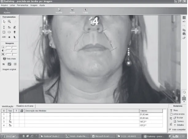
through the dimensions of the NLF. In this software it was measured the angle of both right and left NFL and the distance between nasal basis and labial comissure (NBLC), as shown in Figure 1.
Descriptive and inferential analysis of the statistical data was performed using the Statistical Package for the Social Sciences, version 19.0 (SPSS). Data normality was evaluated with the Kolmogorov-Smirnov test. To compare the parametric data of the groups the ANOVA test with Tukey post hoc
was used. The association between categorical variables was analyzed with the Chi-squared test and Pearson’s correlation test was used to compare age groups and photographic analysis values. A significance level of 5% (p<0.05) was adopted.
The study was approved by the Ethics Research Committee of the Universidade Potiguar, under protocol n° 228/2010. After screening, the objectives, methodology and procedures of the research were explained to the volunteers. All participants signed a Free and Informed Consent Form.
RESULTS
Among the population studied the skin of most of the subjects was white or Asian-Brazilian (56%), mixed-race (32%), or Afro-Brazilian (12%). Regarding type of skin, 43% had mixed skin; 32% normal skin; 12% oily skin; and 6% dry skin. On the Fitzpatrick skin phototype scale, there was a prevalence of 24% of volunteers with type II skin and 33% with type III skin.
According to the Lapiere and Pierard classification, which assesses the degree of wrinkles
24% of the sample were grade I (expression wrinkles), 35% grade II (dermal-epidermal thinning); 16% grade III (gravitational changes with dermal-epidermal and muscle changes) and 25% had no wrinkles. The prevalence of grade II, comprising 35% of the sample, is noteworthy. In terms of the location of wrinkles, only 5% of volunteers in the 21-30 years age group had wrinkles that were characteristic of the accentuation of the NLF. In the 31-40 years age group there was an increase in the number of wrinkles, predominantly of the NLF (60%). From 41-50 years onward, 100% of the volunteers had wrinkles, and all had an accentuated NLF.
Regarding the use of sunscreen, it was found that the vast majority of participants (71%) used sunscreen every day, while 29% did not. A total of 60% of participants used other types of protection, while 40% were unprotected. A total of 74% participants did not smoke, while 26% were smokers.
The average age in each age group was 23.5 ± 2.5 years in the 21-30 years group; 35.2 ± 2.99 years in the 31-40 years group; 45.2 ± 2.84 years in the 41-50 years group; 55.5 ± 3.01 years in the 51-60 years group; and 65.3 ± 2.69 years in the 61-70 years group.
Table 1 shows the association between the age groups of volunteers with the facial wrinkles evaluation scales.
Table 1. Association between age groups and wrinkle evaluation scales. Natal, RN, 2011.
X2 p
Age group x Goglau -0.35 0.001*
Age group x Tsuji 0.65 0.480
Age group x Lapiere and Pierard 0.67 0.001*
It was found that there was an association between increased age and increased Goglau (p=0.001) and Lapierre and Pierord scale (p=0.001) results. This accentuation mainly occurred in the age groups above 40 years of age.
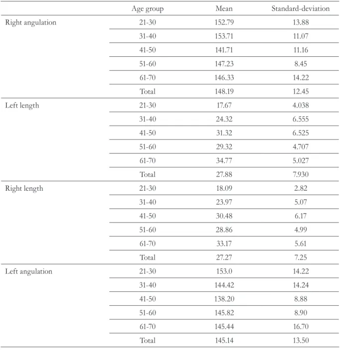
In terms of facial symmetry, the results showed that in the 21-30 years and 31-40 years age group the left and right NLF angulation was greater, respectively, especially when compared with the
61-70 year age group. Additionally, the right and left NBLC lengths were lower in the 21-30 years and 31-40 years age groups than in the 61-70 years age group, as shown in Table 2.
The comparison of the right and left NLF angulation and the right and left NBLC length for the groups and their level of significance are shown in Table 3.
Table 2. Descriptive analysis of facial symmetry measurements. Natal, RN, 2011.
Age group Mean Standard-deviation
Right angulation 21-30 152.79 13.88
31-40 153.71 11.07
41-50 141.71 11.16
51-60 147.23 8.45
61-70 146.33 14.22
Total 148.19 12.45
Left length 21-30 17.67 4.038
31-40 24.32 6.555
41-50 31.32 6.525
51-60 29.32 4.707
61-70 34.77 5.027
Total 27.88 7.930
Right length 21-30 18.09 2.82
31-40 23.97 5.07
41-50 30.48 6.17
51-60 28.86 4.99
61-70 33.17 5.61
Total 27.27 7.25
Left angulation 21-30 153.0 14.22
31-40 144.42 14.24
41-50 138.20 8.88
51-60 145.82 8.90
61-70 145.44 16.70
Table 3. Analysis of facial symmetry variables in relation to angles and distances. Natal, RN, 2011.
Variable Age group Age group p
Right angulation
Left length
21-30 31-40 0,999
41-50 0,056
51-60 0,645
61-70 0,490
31-40 41-50 0,021*
51-60 0,454
61-70 0,307
41-50 51-60 0,610
61-70 0,745
51-60
21-30
61-70 0,999
31-40 0,005*
41-50 0,000*
51-60 0,000*
31-40
61-70 0,000*
41-50 0,002*
51-60 0,048*
61-70 0,000*
41-50
51-60
51-60 0,793
61-70 0,296
61-70 0,022*
31-40 0,009*
41-50 0,002*
Right length
Left angulation
21-30 51-60 0,033
61-70 0,000*
41-50 0,002
51-60 0,867
31-40
41-50
61-70 0,478
51-50 0,867
61-70 0,075
51-60 21-30
61-70 0,407
31-40 0,579
41-50 0,375
51-60 0,375
31-40
41-50
51-60
61-70 0,416
41-50 0,375
51-60 0,997
61-70 1,000
51-60 1,000
61-70 0,416
61-70 1,000
In Table 3, it can be seen that while there were statistically significant differences among all age groups and analyzed measures, these were most significant for the right and left length (p=0.0001). When the Tukey post hoc test was applied it could be seen that the biggest difference in right angulation was between the 21-30 years age group and the 41-50 years age group (p=0.056).
For the right and left lengths all the age groups had similarly significant differences (p=0.0001). For the 21-30 years age group the biggest difference was with the 41-50, 51-60 and 61-70 years age groups. For the 31-40 years age
group the biggest difference was with the 61-70 years age group. For the 41-50 and 51-60 years age group the biggest difference was with the 21-30 years age group. For the 61-70 years age group the biggest difference was with the 21-30 and 31-40 years age groups. These comparisons applied to both the left and the right lengths. For the left angle, the biggest difference for the 21-30 years age group was with the 41-50 years age group, and vice versa (p=0.009).
Table 4 shows the correlation between the variables of age and measurements of left and right NLF angles and lengths.
Table 4. Correlation between age and measurements of angle and distance of nasolabial fold. Natal,
RN, 2011.
r p
Age x right angle -0,31 0,002*
Age x left angle -0,14 0,170
Age x right distance 0,67 0,001*
Age x left distance 0,67 0,001*
r= Pearson Correlation Coefficient; p= level of significance; *correlation between variables studied (p<0.05).
There was a moderate positive correlation between age and NLF length (r = 0.67), indicating that the NLF increased in length with age. In terms of NLF angle, there was a weak negative correlation (r=-0.31) in relation to age. With advancing age NLF length increases, while angulation decreases, with the age group that had the shortest length and the greatest angle being the most significant.
DISCUSSION
ultraviolet radiation from the sun, culminating in photoaging of the skin.11-13
Evaluation of facial aging is a complex process due to the different factors that influence facial symmetry. However, the chronological aging process is reflected in the accentuation of the NLF.14
Therefore the NLF can be an important parameter for the evaluation of facial symmetry and, indirectly, facial aging. In clinical practice, various methods have been attempted to achieve a more quantitative facial evaluation, including the cephalometric technique, which allows quantitative analysis of the NLF, serving as a parameter for facial symmetry evaluation.13,14
Some facial assessment studies have already made use of this type of facial symmetry analysis. In these studies, the technique has proven to be an effective parameter in the evaluation process, identifying a reduction in the angle and an increase in the length of the fold, findings that are associated with the chronological aging process.10,15 Combined with other evaluative
procedures such as the Goglau, Tsuji and Lapiere and Pierord scales and the Facial Assessment Protocol (PAF),10 the complementary measurement
of nasogenian wrinkles by Radiocef 2.0® software in the evaluation of the facial chronoaging of women of different age groups has been found to be an alternative method of evaluation. From the knowledge of angles and lengths common to specific age groups, a standard range, consistent for the evaluation of different groups of patients of different age groups, can be established.16,17
The NLF often becomes deeper and characteristic of facial aging early, at around 22 to 25 years of age. This region is affected by gravity, with sagging and ptosis of tissues occurring between 32 and 35 years of age.4,5 The data in the
present study shows that as age increased, the angle of the fold was reduced.
It was observed that accentuation of the NLF becomes more common with age, which is associated with changes in dental positioning that occur during the aging process, as well as an increase in skin sagging. These results were better quantified following cephalostat analysis, thereby more reliably complementing the result obtained with the PAF. Therefore, a new way of quantifying facial skin aging has been identified, namely the measurement of NLF angles and lengths.16,17
The use of this new assessment technique can therefore contribute to improved monitoring of chronological changes in the face, directly supporting the possible quantification of clinical progression in treatment of the facial region. As a result, this method can complement the PAF, facilitating the confirmation of visual findings.
It has been seen that there is a sharp increase in the characteristics of the facial aging process in older age groups. An association was found between age and the degree of wrinkles found using the Goglau, Tsuji and Lapiere and Pierord scales. The reduction of fibroblast activity, decreased skin elasticity by gradual loss of elastic and collagen fibers contribute to the appearance of wrinkles.18,19
Other methods of evaluation based on facial photographs have been tested by analyzing wrinkles in different regions of the face.19,20
However, these methods require more equipment and more complex analysis for characterizing the aging process. The assessment carried out in the present study is more streamlined and aimed at inferring results from the NLF.
Limitations of this study include its sample size, the lack of analysis of different genders, and the need to perform a more specific correlation with other factors that promote aging and the appearance of wrinkles such as solar photo-exposure, living habits, consumption of alcohol, and insomnia.
CONCLUSION
This study demonstrates the effectiveness of cephalometric measurement using Radiocef 2.0® software in providing a more detailed analysis of the nasolabial folds, representing, therefore a complement for Facial Assessment Protocol evaluation. It may, as a result, be considered a method for identifying the effectiveness of treatment by optimizing the evaluation method. Given the importance of the nasolabial fold as a surface marker for skin and muscle aging, measuring its angulation and length can be a way to evaluate this process.
REFERENCES
1. Montagner S, Costa A. Bases biomoleculares do fotoenvelhecimento. An Bras Dermatol 2009;84(3):263-9.
2. Servetto N, Cremonezzi D, Simes JC, Moya M, Soriano F, Palma JA, et al. Evaluation of inflammatory biomarkers associated with oxidative stress and histological assessment of low-level laser therapy in experimental myopathy. Lasers Surg Med [Internet] 2010 [acesso em 02 fev 2011];42(6):577-83.
3. Borghetti RL. Avaliação in vitro da citotoxicidade, genotoxicidade e mutagenicidade de materiais estéticos de preenchimento facial [dissertação]. Porto Alegre:PUCRS; 2015.
4. Pontes JK. Estimativa hierárquica da idade baseada em características globais e locais de imagens faciais [dissertação]. Curitiba: Universidade Federal do Paraná; 2015.
5. Pitanguy I, De Amorim NFG . Tratamento do sulco nasogeniano. Rev Bras Cir 1997;87(5):231-42.
6. Pascali M, Botti C, Cervelli V, Botti G. Midface rejuvenation: a critical evaluation of a 7-year experience. Plast Reconstr Surg 2015;135(5):1305-16.
7. Yeilding RH, Fezza JP. A Prospective, split-face, randomized, double-blind study comparing onabotulinumtoxinA to incobotulinumtoxinA for upper face wrinkles. Plast Reconstr Surg 2015;135(5):1328-35.
8. Duscher D, Barrera J, Wong VW, Maan ZN, Whittam AJ, Januszyk M, et al. Stem cells in wound healing: the future of regenerative medicine?: a mini-review. Gerontology 2015:1-10.
9. Vedovello MF, Berzin F, Gomes MF, Madeira MC, Siqueira JTT. Cefalometria: técnicas de diagnóstico e procedimentos. Nova Odessa: Napoleão; 2007. p. 109-14.
10. Silva RMV, Daams EFCC, Delgado AM, Silva EM, Oliveira HG, Meyer PF. Efeitos da terapia manual no rejuvenescimento facial. Rev Ter Man 2013;11(1):534-39.
11. Tsukahara K, Osanai O, Kitahara T, Takema Y. Seasonal and annual variation in the intensity of facial wrinkles. Skin Res Technol 2013;19(3):279-87.
13. Battie C, Jitsukawa S, Bernerd F, Del Bino S, Marionnet C, Verschoore M. New insights in photoaging, UVA induced damage and skin types. Exp Dermatol 2014;23(Suppl 1):7-12.
14. Tsukahara K, Sugata K, Osanai O, Ohuchi A, Miyauchi Y, Takizawa M, et al. Comparison of age-related changes in facial wrinkles and sagging in the skin of Japanese, Chinese and Thai women. J Dermatol Sci 2007;47(1):19-28.
15. Estrela JV, Duarte CCF, Araujo DN, Araruna VR, Silva RMV, Cavalcanti RL, et al. Efeito do led na flacidez tissular facial. Catussaba 2014;3(1):29-36.
16. Vasconcelos MHF, Janson G, Freitas MR, Henriques JFC. Avaliação de um programa de traçado
cefalométrico. Rev Dent Press Ortodon Ortopedi Facial 2006;11(2):44-54.
17. Reis SAB, Capelozza FLC, Cardoso MA, Scanavini MA. Características cefalométricas dos indivíduos Padrão I. Rev Dent Press Ortodon Ortopedi Facial 2005;10(1):67-78.
18. Choi JW, Kwon SH, Huh CH, Park KC, Youn SW. The influences of skin visco-elasticity, hydration level and aging on the formation of wrinkles: a comprehensive and objective approach. Skin Res Technol 2013;19(1):349-55.
19. Cula GO, Bargo PR, Nkengne A, Kollias N. Assessing facial wrinkles: automatic detection and quantification. Skin Res Technol 2013;19(1): 243-51.
20. Hatzis J. The wrinkle and its measurement: a skin surface profilometric method. Micron 2004;35(3):201-19.


