O
ri
gi
na
l
a
rt
ic
le
s
1 Universidade Estadual de Feira de Santana, Departamento de Ciências Biológicas, Laboratório de Genética
Toxicológica. Feira de Santana, BA, Brasil.
Correspondence
José Roberto Cardoso Meireles E-mail: [email protected]
Abstract
The effects of aging, gender and lifestyle factors on inducing chromosomal damage (micronuclei) and nuclear degenerative changes were assessed using the micronucleus test on exfoliated cells of the oral mucosa. The sample included 80 healthy subjects divided into four groups according to age and gender: men and women aged 19-29 years (M19, W19) and men and women aged over sixty years (M60, W60). An interview questionnaire was used to characterize the sample and to determine an index reflecting lifestyle (HLI). The frequency of micronuclei and nuclear degenerative changes was significantly higher among the elderly (p<0.001) and did not differ by gender among young people (p>0.05). The occurrence of micronuclei was similar among elderly men and women (p>0.10), but karyorrhexis and karyolysis were more frequent among men (p<0.005 and p<0.025, respectively), who also had a lower HLI than the other groups (p<0.0004). The results of the study indicate that age is the main factor associated with the induction of genetic material damage.
Keywords: Age Groups; Gender; Micronucleus; Apoptosis
Effects of age on the frequency of micronuclei and degenerative
nuclear abnormalities
Gregory Alves Ferraz1
Antônio de Oliveira Costa Neto1
Eneida de Moraes Marcílio Cerqueira1
INTRODUCTION
Advancing age is characterized by a general reduction in physiological efficiency, resulting in homeostatic imbalance and the subsequent onset of illnesses inherent to the aging process. Studies suggest that this process is associated with an increase in genomic instability due to the reduced capacity to repair damaged DNA.1,2 It has been
reported that, in old age, biomarkers of genomic instability, such as micronuclei (MN), are more common in the peripheral lymphocytes3,4 and
exfoliated epithelial cells.5,6
The accumulation of DNA damage is a significant event in the aging of cells. During this process, there is a progressive decrease in metabolic enzymes and DNA-repair enzymes, which increases the predisposition and susceptibility of the cells to exogenous and/or endogenous genotoxic agents. These factors contribute to an increase in age-related spontaneous DNA damage.7,8
Therefore, modifications to genetic material do not only occur as a result of exposure to mutagens. They can also be caused by chemical reactions related to physiological processes.9 In addition to
age, studies have shown that differences in the occurrence of genetic damage can also be related to gender.10-12 Trzeciak et al.11 reported that the
removal of active carcinogens through tobacco is lower in women, thereby indicating biochemical differences between the genders. Larmarcovai
et al.10 and Kažimírová et al.12 reported a greater
occurrence of MN as a result of age and gender.
The prevalence of DNA damage is also influenced by factors related to lifestyle. Smoking, drinking alcohol, working long hours, not sleeping enough, physical inactivity, type of diet and psychological stress contribute to an increased prevalence and the consequent development of illnesses, including cancer.13,14,15
Cancer leads to alterations in the genes involved in controlling cellular proliferation, cellular differentiation, DNA repair and apoptosis. Thus, the quantification of genetic damage is important when assessing the risks of developing cancer.16,17
The micronucleus test for exfoliated cells from the oral mucosa is considered an effective method of identifying genetic damage through chromosomal losses and breaks, particularly when conducted in accordance with the protocols described by Tolbert et al.18.19 and Thomas et al.17
These protocols calculate MN and degenerative nuclear alterations that are indicative of apoptosis and necrosis.
Therefore, the aim of the present study was to assess the effects of age and gender on the prevalence of micronuclei and degenerative nuclear alterations, while also considering lifestyle factors.
METHODS
Ethical aspects
The present study was conducted in accordance with the legislation of the Brazilian National Health Council (CNS 196/96), which is based on the Declarations of Helsinki/Hong Kong. The research received approval from the Research Ethics Committee of the Universidade Estadual de Feira de Santana (Feira de Santana State University) (Protocol 063/2009). All of the participants signed a free and informed consent form.
Study period
The present study was conducted between March and November of 2010.
Sample
The sample contained 80 individuals, who were divided into four groups of twenty:
• M19: men aged between 19 and 29 years;
• W19: women aged between 19 and 29 years;
• M60: men aged 60 years or more;
Characterization of the sample
The sample was characterized using an interview questionnaire (adapted from Caires20)
that contained questions about gender, age, diet, smoking, alcohol consumption, hours of sleep, professional occupation, physical exercise, stress levels, the use of oral antiseptics, chronic illnesses and exposure to genotoxic agents and/or toxic products. All of the variables except age and gender were used to calculate the Healthy Living Index.
Healthy living index
Lifestyle was assessed using the Healthy Living Index (HLI), based on the parameters adopted by Morimoto et al.,21 which are: don’t
smoke; don’t consume alcoholic beverages on a daily basis; eat breakfast every day; sleep between seven and eight hours per night; work less than ten hours per day; exercise at least once a week; maintain a nutritionally-balanced diet; and suffer a moderate level of mental stress (self-reported by the interviewee). The following extra variables were also considered: the use of oral antiseptics; the absence of chronic illnesses and the non-exposure to genotoxic agents and toxic products. One (1) point was attributed for each of the variables classified as healthy. Answers that differed from these specifications were attributed a score of zero (0). The HLI of each participant was calculated by summing the scores obtained in the abovementioned parameters. Three HLI categories were applied: good (11-12 points); moderate (9-10 points) and poor (0-8 points).
Micronucleus test
Exfoliated cells of the oral mucosa of each individual were collected using an endocervical brush and transferred by smear to microscopic slides containing two drops of physiological saline (0.9% NaCl). After drying to room temperature, the slides were submerged in methanol / acetic acid (3:1) for fixation. After 24 hours, the material
was stained using the reactive shift method and counter-stained using fast green (1%).
Cytological analysis of the data obtained via the questionnaires was conducted under an optical microscope (blind test). In total, 2000 cells were analyzed for each individual. The MN identification criteria described by Sarto et al.22 were
adopted. Thus, MN structures were considered to be morphologically similar to a nucleus when they exhibited up to 1/3 of its size and were visible on the same plane. Degenerative nuclear alterations (NA) indicative of apoptosis (karyorrhexis, condensed chromatin, pyknosis) and necrosis (karyolysis) were also calculated.
Statistical analysis
The mean age and HLI values for the groups were compared using the Kruskal-Wallis test. The occurrence of MN and NA in the groups was compared using a conditional test for the comparison of proportions in situations of rare events.23 The Kruskal-Wallis test was also used
to assess the (mean) occurrence of MN and NA in relation to HLI and age group.
RESULTS
Table 1 displays the mean ± standard deviation values for the age and HLI of the participants.
The Kruskal-Wallis test showed that the mean age of the individuals in the younger groups was significantly lower than that of the older groups (p<0.00001). In a single age group, men and women did not differ in terms of age. Individuals in the M19, W19 and W60 groups exhibited a significantly higher HLI value (p<0.0004).
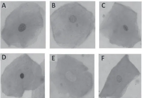
Figure 1. Photomicrographs of cells exfoliated from the oral mucosa (stained using the Felgen/Fast green method) exhibiting normal nuclear morphology: (A) a micronucleus (B); condensed chromatin ©; pyknosis (D); karyolysis (E); karyorrhexis.
Table 1. Mean age and HLI of the sample. Feira de Santana, Bahia, 2010.
Group Mean age ± standard deviation Mean HLI ± standard deviation
M60 69.00 ± 9.30 7.65 ± 1.31
W60 81.25 ± 9.11 8.65 ± 0.81
M19 22.20 ± 1.79 9.15 ± 0.99
W19 21.90 ± 1.71 9.20 ± 1.11
HLI = Healthy Life Index.
Table 2. Occurrence of micronuclei and nuclear alterations in individuals from two different age groups. Feira de Santana, Bahia, 2010.
Endpoints Observed Expected χ2 p
19-29 ≥ 60 19-29 ≥ 60
Micronuclei 5 38 21.50 21.50 25.3256 <0.001
Karyolysis 2 56 29.00 29.00 50.2758 <0.001
Karyorrhexis 12 209 110.50 110.50 175.6064 <0.0001
Condensed chromatin 192 533 362.50 362.50 160.3876 <0.0001
Pyknosis 151 740 445.50 445.50 389.3614 <0.00001
No differences were found between men and women in the younger group for the occurrence of any of the endpoints. In the elderly group, the frequency of karyolysis and karyorrhexis was higher among men (Table 3).
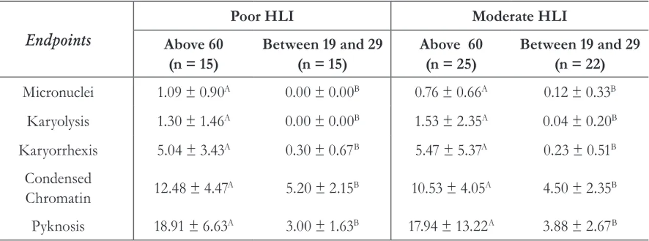
The assessment of the occurrence of MN and NA in relation to HLI and age was conducted by dividing the sample into two groups (poor HLI and moderate HLI), since only three individuals
(one man and two women from the younger group) were classified with a good HLI. These groups were divided into two subgroups based on age group. The analysis revealed that there were no significant differences in the frequency of MN and NA in the same age group, regardless of HLI score. Conversely, older individuals exhibited a greater occurrence of the endpoints analyzed, irrespective of their HLI score (Table 4).
Table 3. Occurrence of micronuclei and nuclear alterations in men and women aged ≥60. Feira de Santana, Bahia, 2010.
Endpoints Observed Expected χ2 p
Men Women Men Women
Micronuclei 23 15 19.00 19.00 1.6842 > 0.10
Karyolysis 37 19 28.00 28.00 5.7857 < 0.025
Karyorrhexis 130 79 104.50 104.50 12.4450 < 0.005
Condensed chromatin 264 269 266.59 266.59 0.0469 > 0.10
Pyknosis 370 370 370.00 370.00 0.0000 = 1.000
Endpoints = markers of chromosomal damage (micronuclei) and nuclear alterations (karyolysis, karyorrhexis, condensed chromatin and pyknosis).
Table 4. Mean quantity of micronuclei and nuclear alterations caused by the Healthy Living Index (HLI) and age. Feira de Santana/BA. 2010.
Endpoints
Poor HLI Moderate HLI
Above 60
(n = 15)
Between 19 and 29 (n = 15)
Above 60
(n = 25)
Between 19 and 29 (n = 22)
Micronuclei 1.09 ± 0.90A 0.00 ± 0.00B 0.76 ± 0.66A 0.12 ± 0.33B
Karyolysis 1.30 ± 1.46A 0.00 ± 0.00B 1.53 ± 2.35A 0.04 ± 0.20B
Karyorrhexis 5.04 ± 3.43A 0.30 ± 0.67B 5.47 ± 5.37A 0.23 ± 0.51B
Condensed
Chromatin 12.48 ± 4.47
A 5.20 ± 2.15B 10.53 ± 4.05A 4.50 ± 2.35B
Pyknosis 18.91 ± 6.63A 3.00 ± 1.63B 17.94 ± 13.22A 3.88 ± 2.67B
DISCUSSION
The micronucleus test of exfoliated cells is an effective method of detecting genetic damage and has been widely used in population biomonitoring.24
Micronuclei are structures that cause chromosomal breaks or complete chromosomes that fail to bond with the spindle during cellular division and are not included in the nucleus of daughter cells. Therefore, the micronucleus is an endpoint of chromosomal damage caused by a clastogenic or aneugenic event.
As well as chromosomal damage, other endpoints that are indicative of apoptosis and necrosis can be identified by this test. Alterations in the level of apoptosis, inferred by the occurrence of karyorrhexis, condensed chromatin and pyknosis, demonstrate genotoxic effects and have been correlated with the onset of cancer.17-19 Necrosis,
in turn, is inferred by the occurrence of karyolysis, which demonstrates the cytotoxic effect associated with cancer promotion.17 In this context, the
calculation of these endpoints is significant in order to increase the sensitivity of the micronucleus test.
One of the many possible applications of this test includes the assessment of the mutagenic effects of smoking and/or consuming alcoholic beverages,25,26 as well as the effects of the depletion
of nutrients in the diet.27 The effects of age and
gender on the prevalence of micronuclei have been more commonly assessed in lymphocytes than in isolation (as was the case in the present study). This may have affected the results of studies related to other variables.28,29
According to Bonassi et al.,27 the effects of gender
on the frequency of MN in exfoliated cells are not significant, unlike those observed with lymphocytes (where women tend to exhibit higher frequencies). In the present study, gender did not affect the frequency of MN, although karyolysis and karyorrhexis were more common among men aged ≥60 years, which may be associated with the lower HLI scores exhibited by this group. The assessments of associations between the occurrence of micronuclei, nuclear alterations and lifestyle (inferred by the HLI) recorded no significant differences in the present study. Further investigations of the influence of gender and lifestyle on the occurrence of nuclear
alterations are required, given that a greater occurrence of MN has previously been associated with an unhealthy lifestlye.15
The influence of age on the promotion of damage to genetic material has been widely discussed in literature. In terms of this association, the results of the present study corroborate the findings of several other authors who have reported a higher occurrence of genetic damage among more elderly individuals.12,28,29 According
to Huang et al.,15 this is due to the fact that aging
is linked with genetic instability. However, further studies with larger samples are required, given that the quantity of individuals analyzed could be considered a limitation of the present study.
According to Fenech and Bonassi,30 the increase
of MN over time is probably due to a combination of factors, including: (a) the cumulative effect of mutations in genes involved in DNA repair, chromosomal segregation and checkpoints of the cellular cycle; and (b) numerical and structural alterations in chromosomes that are induced by endogenous and/or exogenous genotoxins, as well as a wide range of unhealthy lifestyle factors.
Thus, the effects of aging seem to be a combination of genetically programmed processes and genetic alterations caused by exogenous and endogenous factors. During the aging process, the enzymes involved in DNA repair become less and less common, which increases the susceptibility of cells to genotoxic agents.31,32 Kirsch-Volders et al.33 suggested that flaws in the cellular defense
systems that protect against DNA damage, as well as the reduced efficiency of DNA repair, can lead to an accumulation of mutations. These mutations, either in isolation or in combination with other age-related alterations, can contribute to aging and the development of age-related illnesses.
CONCLUSION
REFERENCES
1. Garm C, Moreno-Villanueva M, Bürkle A, Petersen I, Bohr VA, Christensen K, et al. Age and gender effects on DNA strand break repair in peripheral blood mononuclear cells. Aging Cell 2013;12(1):58-66.
2. Trzeciak AR, Barnes J, Ejiogu N, Foster K, Brant LJ, Zonderman AB, et al. Age, sex, and race influence single-strand break repair capacity in a human population. Free Radic Biol Med 2008;45(12):1631-41.
3. Kazimírováa A, Barancokováa M, Dzupinkováa
Z, Wsólová L, Dusinskáa M. Micronuclei and
chromosomal aberrations, important markers of ageing: Possible association with XPC and XPD polymorphisms. Mutat Res 2009;661(1-2):35-40.
4. Joseph LJ, Patwardhan UN, Samuel AM. Frequency of micronuclei in peripheral blood lymphocytes from subjects occupationally exposed to low levels of ionizing radiation. Mutat Res 2004;564(1):83-8.
5. Pinto D, Ceballos JM, García G, Guzmán P, Del Razo LM, Vera E, et al. Increased cytogenetic damage in outdoor painters. Mutat Res 2000;467(2):105-11.
6. Wu PA, Loh CH, Hsieh LL, Liu TY, Chen CJ, Liou SH. Clastogenic effect for cigarette smoking but not areca quid chewing as measured by micronuclei in exfoliated buccal mucosal cells. Mutat Res 2004;562(1-2):27-38.
7. Milosevic-Djordjevic O, Grujicic D, Novakovic T, Arsenijevic S, Marinkovic D. Micronuclei and ageing in a sample of yugoslavian population. Genetika 2002;38(2):264-7.
8. Picerno I, Chirico C, Condello S, Visalli G, Ferlazzo N, Gorgone G, et al. Homocysteine induces DNA damage and alterations in proliferative capacity of T-lymphocytes: a model for immunosenescence? Biogerontology 2007;8(2):111-9.
9. Heuser VD, Andrade VM, Peres A, Braga LMGM, Chies JAB. Influence of age and sex on the
spontaneous DNA damage detected by micronucleus test and comet assay in mice peripheral blood cells. Cell Biol Int 2008;32(10):1223-9.
10. Iarmarcovai G, Bonassi S, Botta A, Baan RA, Orsière T. Genetic polymorphisms and micronucleus formation: a review of the literature. Mutat Res 2008;658(3):215-33.
11. Trzeciak AR, Barnes J, Ejiogu N, Foster K, Brant LJ, Zonderman AB, et al. Age, sex, and race influence single-strand break repair capacity in a human population. Free Radic Biol Med 2008;45(12):1631-41.
12. Kazimírová A, Barancoková M, Dzupinková
Z, Wsólová L, Dusinská M. Micronuclei and
chromosomal aberrations, important markers of ageing: possible association with XPC and XPD polymorphisms. Mutat Res 2009;661(1-2):35-40.
13. Johnson JV, Lipscomb J. Long working hours, occupational health and the changing nature of work organization. Am J Ind Med 2006;49(11):921-9.
14. Metcalfe C, Smith GD, Macleod J, Hart C. The role of self-reported stress in the development of breast cancer and prostate cancer: a prospective cohort study of employed males and females with 30 years of follow-up. Eur J Cancer 2007;43(6):1060-5.
15. Huang P, Huang B, Weng H, Nakayama K, Morimoto K. Effects of lifestyle on micronuclei frequency in human lymphocytes in Japanese hard-metal workers. Prev Med 2009;48(4):383-8.
16. Holland N, Bolognesi C, Kirsch-Volders M, Bonassi S, Zeiger E, Knasmueller S, et al. The micronucleus assay in human buccal cells as a tool for biomonitoring DNA damage: the HUMN project perspective on current status and knowledge gaps. Mutat Res 2008;659(1-2):93-108.
17. Thomas P, Holland N, Bolognesi C, Kirsch-Volders M, Bonassi S, Zeiger E, et al. Buccal micronucleus cytome assay. Nat Protoc 2009;4(6):825-37.
18. Tolbert PE, Shy CM, Allen JW. Micronuclei and other nuclear anomalies in buccal smears: a field test in snuff users. Am J Epidemiol 1991;134(8):840-50.
19. Tolbert PE, Shy CM, Allen JW. Micronuclei and other nuclear anomalies in buccal smears: methods development. Mutat Res 1992;271(1):69-77.
20. Caires NFR. Sobrepeso e obesidade entre os
funcionários da Universidade Estadual de Feira
de Santana-BA [dissertação]. Feira de Santana: Universidade Estadual de Feira de Santana; 2004.
21. Morimoto K, Takeshita T, Inoue-Sakurai C, Maruyama S. Lifestyles and mental health status are associated with natural killer cell and lymphokine-activated killer cell activities. Sci Total Environ 2001;270(1-3):3-11.
23. Bragança-Pereira CA. Teste estatístico para comparar proporções em problemas de citogenética. In: Rabello-Gay MN, Rodrigues MAR, Monteleone Neto R, organizadores. Mutagênese, carcinogênese e teratogênese: métodos e critérios de avaliação. São Paulo: Sociedade Brasileira de genética; 1991. p.113-21.
24. Thomas P, Fenech M. Buccal micronucleus cytome assay. In: Didenko VV, organizator. DNA damage detection in situ, ex vivo, and in vivo: methods and protocols. New York: Springer Science+Business Media; 2011. p. 235-48.
25. Pellicioli AC, Visioli F, Ferreira LA, Danilevicz CK, Carrard VC, Rados PV. Cytogenetic abnormalities in exfoliated oral mucosal cells and their association with oral cancer. Anal Quant Cytol Histol 2011;33(5):271-6.
26. Pradeep MR, Yadavalli G, Maji J, Kartikay S, Deepa K, Vishnudas P. Comparative study of genotoxicity in different tobacco related habits using micronucleus assay in exfoliated buccal epithelial cells. J Clin Diagn Res 2014;8(5):21-4.
27. Bonassi S, Coskun E, Ceppi M, Lando C, Bolognesi C, Burgaz S, et al. The HUman MicroNucleus project on eXfoLiated buccal cells (HUMN(XL)): the role of life-style, host factors, occupational exposures, health status, and assay protocol. Mutat Res 2011;728(3):88-97.
28. Wojda A, Zietkiewicz E, Witt M. Effects of age and gender on micronucleus and chromosome nondisjunction frequencies in centenarians and younger subjects. Mutagenesis 2007;22(3):195-200.
29. Nefic H, Handzic I. The effect of age, sex, and lifestyle factors on micronucleus frequency in peripheral blood lymphocytes of the Bosnian population. Mutat Res 2013;753(1):1-11.
30. Fenech M, Bonassi S. The effect of age, gender, diet and lifestyle on DNA damage measured using micronucleus frequency in human peripheral blood lymphocytes. Mutagenesis 2011;26(1):43-9.
31. Chen SK, Hsieh WA, Tsai MH, Chen CC, Hong AI, Wei YH, et al. Age-associated decrease of oxidative repair enzymes, human 8-oxoguanine DNA glycosylases (hOgg1), in human aging. J Radiat Res 2003;44(1):31-5.
32. Nassour J, Martien S, Martin N, Deruy E, Tomellini E, Malaquin N, et al. Defective DNA single-strand break repair is responsible for senescence and neoplastic escape of epithelial cells. Nat Commun 2016;29(7):10399.
33. Kirsch-Volders M, Mateuca RA, Roelants M, Tremp A, Zeiger E, Bonassi S, et al. The effects of GSTM1 and GSTT1 polymorphisms on micronucleus frequencies in human lymphocytes in vivo. Cancer Epidemiol Biomark Prev 2006;15(5):1038-42.