O
ri
gi
na
l
a
rt
ic
le
s
Influence of body mass index and age on the lung function of
obese women
Dayla Sgariboldi1 Fernanda Aparecida Faria2 Jéssica Cristina Carbinatto2 Eli Maria Pazzianotto-Forti1,2,3
1 Universidade Metodista de Piracicaba - UNIMEP, Programa de Pós-Graduação em Fisioterapia. Piracicaba,
SP, Brasil
2 Universidade Metodista de Piracicaba - UNIMEP, Curso de Graduação em Fisioterapia. Piracicaba, SP,
Brasil.
3 Universidade Metodista de Piracicaba – UNIMEP, Programa de Pós-Graduação em Ciências do Movimento
Humano. Piracicaba, SP, Brasil.
Correspondence Eli Maria Pazzianotto Forti E-mail: [email protected]
Abstract
Introduction: Obesity and aging may cause changes in lung function. Objective: to assess whether body mass, body mass index (BMI) and age influences vital capacity (VC) and
forced expiratory volume in the first second (FEV1) in women. Methods: 81 women aged
between 30 and 75 years participated in the study. The sample included obese and morbidly obese, non-smoking, sedentary individuals without chronic lung disorders. Anamnesis, anthropometric and spirometric evaluations were performed. Statistical analysis was carried out using the Pearson correlation and Spearman tests, adopting a significance
level of 5%. Results: It was observed that age had significant and negative correlations
with VC and its components: inspiratory reserve volume (IRV), expiratory reserve volume
(ERV) and tidal volume (TV), and with FEV1. There was also a significant positive
correlation between body mass and VC and IRV and a significant negative correlation
between BMI and ERV. Conclusion: Pulmonary function declines over time. Body mass
appears to exert a greater influence on IRV, whereas a greater BMI is associated with a decline in ERV.
INTRODUCTION
Obesity is a chronic disease that has grown rapidly in recent years and is related to several factors such as hereditariness, poor diet, physical inactivity and hormonal factors1. In Brazil, 56.9% of the adult population is overweight, representing 82 million people, with 13% classified as obese2. The condition is associated with several chronic and/or degenerative co-morbidities such as type II diabetes, systemic arterial hypertension, coronary heart disease and strokes3.
It can also promote changes in lung function and reductions in functional residual capacity (FRC), tidal volume (TV), lung compliance and thoracic expandibility4 as well as decreased expiratory reserve volume (ERV). This has been described as the key finding related to alterations in lung function due to obesity5, as abdominal fat exerts a mechanical effect on the chest and diaphragm, reducing lung volumes even in individuals with no changes in lung function6.
Aging can also affect lung function due to physical changes associated with increased age such as loss of height, weight and the replacement of muscles with adipose tissue, triggering muscle weakness, and leading to changes in lung mechanics.7-9
According to Sekhri et al,10 obesity may have a greater impact on lung function in older individuals due to the deposition of body fat which varies with increasing age. As the body ages, there is reduced secretion of the growth hormone and a reduction in the basal metabolic rate, reducing lean body mass and increasing body fat, especially in women.11
Therefore, the hypothesis of the present study is that excess body fat and age can be causative factors of alterations in lung function behavior in women.
The aim of the study was to evaluate if body mass, BMI and age influence the vital capacity (VC) and forced expiratory volume in the first second (FEV1) in women.
METODOLOGY
Subjects
An observational cross-sectional study based on a convenience sample was carried out at the Cardiorespiratory and Physiotherapy Evaluation and Intervention Laboratory of the Universidade Metodista de Piracicaba (the Piracicaba Methodist University) (UNIMEP). The study evaluated 81 sedentary women aged between 30 and 75 years with a body mass index (BMI) between 30 and 55 kg/m2, from August 2012 to July 2013.
The study was approved by the Ethics Research Committee of the Universidade Metodista de Piracicaba (UNIMEP), under protocol 48/12, in accordance with the guidelines of Resolution 196/96 of the National Health Council. The objectives of the study were explained to all the participants, all of whom signed a Free and Informed Consent Form.
Sample Calculation
The calculation of sample size was based on a pilot study and a significant correlation between BMI and ERV. An r value of -0.60 and the linear correlation test were used for the calculation, with a statistical power of 95% and an alpha of 0.05 applied. A minimum of 31 volunteers in each group was determined to analyze BMI. The sample calculation for each group according to BMI and age was also carried out, and a minimum of eight volunteers per group was established. The sample size calculation was performed using the BioEstat software version 5.3 (Belém, Brazil).
The women evaluated and included in the study were from obesity treatment centers in the city of Piracicaba in the state of Sao Paulo and presented recent and normal clinical, laboratory and spirometry test results, in accordance with medical evaluations, before being included in the groups.
understanding how to carry out the evaluations and with an absence of systemic arterial hypertension, diabetes, cardiovascular and pulmonary diseases and respiratory infections in the previous two weeks were included in the study.
The women studied were divided into four groups according to BMI and age: 38 obese women (BMI between 30.0 and 39.9 kg/m²), of whom 27 were aged between 30 and 55 years and 11 were aged between 56 and 75 years, and 43 morbidly obese women (BMI>40 kg/m²), of whom 28 were aged between 30 and 55 years and 15 were aged between 56 and 75 years.
Experimental Procedure
The volunteers underwent anamnesis for the collection of clinical data, the evaluation of anthropometric measurements and subsequently the evaluation of lung volume and capacity by spirometry. Each volunteer attended the lab once and evaluations were carried out in a single day by researchers previously trained for the experimental procedure. To carry out the experimental procedure, the laboratory was properly prepared and air conditioned with the room temperature controlled by Split air conditioning equipment (Trane; Curitiba, Paraná, Brazil) to between 22 and 24° C and the relative humidity maintained by a humidifier at between 40 and 60%.
During the anthropometric evaluation, the volunteers remained in the orthostatic position without shoes or heavy clothing. Body mass was measured by a digital scale (Welmy; Santa Bárbara D’Oeste, São Paulo, Brazil), with a maximum capacity of 300 Kg. Height was measured by the stadiometer of the scale and BMI was calculated using the equation: body mass (kg)/height² (m).13
A computerized spirometer with a flow sensor, calibrated daily, was used to measure lung function (Microquark; Cosmed, Roma, Italy). The volunteers remained seated and used a nasal clip during the realization of slow vital capacity (SVC) and forced vital capacity (FVC) maneuvers, in accordance with the guidelines of the American Thoracic Society14 and norms for the tests of lung function.15
The maneuvers were performed until three acceptable and two reproducible curves were achieved, without exceeding eight attempts. The values are expressed in liters and percentage of predicted, according to the equations established for the Brazilian population.16
Statistical Analysis
The BioEstat version 5.3 program was used for statistical analysis. To verify the normality of the data the Kolgomorov-Smirnov normality test was used. For intragroup and intergroup comparison of age, anthropometric characteristics and the spirometric variable values, the Student t and Mann-Whitney tests were used. For analysis of the correlation of body mass, BMI and age with spirometric variables, the Pearson and the Spearman correlations were used. A significance level of 5% was adopted for all analyses.
RESULTS
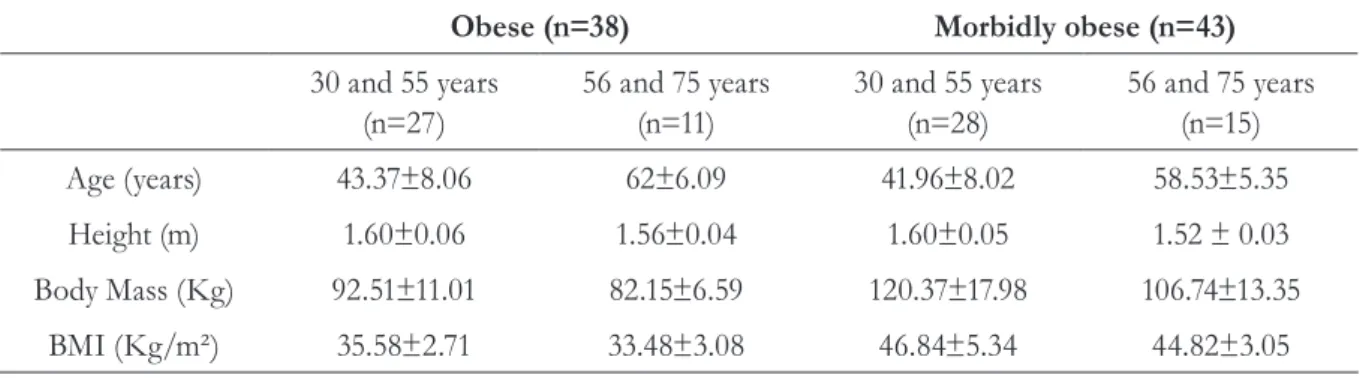
Table 1 shows the data of the age and anthropometric characteristics of the study volunteers divided into groups.
Table 1. Age and anthropometric characteristics of the volunteers studied. Piracicaba, São Paulo, 2012-2013.
Obese (n=38) Morbidly obese (n=43)
30 and 55 years (n=27)
56 and 75 years (n=11)
30 and 55 years (n=28)
56 and 75 years (n=15)
Age (years) 43.37±8.06 62±6.09 41.96±8.02 58.53±5.35
Height (m) 1.60±0.06 1.56±0.04 1.60±0.05 1.52 ± 0.03
Body Mass (Kg) 92.51±11.01 82.15±6.59 120.37±17.98 106.74±13.35
BMI (Kg/m²) 35.58±2.71 33.48±3.08 46.84±5.34 44.82±3.05
BMI: body mass index.
Table 2. spirometric variable values in absolute and percentage of predicted form of the studied volunteers.
Piracicaba, São Paulo, 2012-2013.
Obese (n=38) Morbidly obese (n=43)
30 to 55 years (n=27)
p
intragroup 56 to 75 years (n=11)
30 to 55 years (n=28)
p
intragroup 56 to 75 years (n=15)
p
intergroup
value value value
VC (L) 2.99 ±0.65 0.02* 2.43 ±0.66 2.85 ±0.39 0.0056* 2.49 ±0.36 0.42
%Pred 90.40±15.22 0.56 86.79±22.27 87.26±9.38 0.22 91.21±11.03 0.82
ERV (L) 0.44±0.27 0.05 0.27±0.08 0.39+ 0.11 0.29 0.23±0.13 0.04*
%Pred 41.21±22.73 0.25 36.51±11.45 37.36+14.44 0.05 21.64±12.51 0.02*
IRV (L) 1.52±0.54 0.42 1.35±0.64 1.78±0.37 0.02* 1.43±0.56 0.02*
TV (L) 0.98±0.42 0.27 0.83±0.24 0.81±0.32 0.1 0.65±0.26 0.04*
FEV1 (L) 2.63±0.61 0.09 2.25 ±0.64 2.50 ±0.43 0.01* 2.16 ±0.32 0.27
%Pred 94.94±16.70 0.37 101.34±26.43 90.99±13.19 0.07 98.69±13.42 0.41
FEV1: forced expiratory volume in one second; VC: vital capacity; ERV: expiratory reserve volume; IRV: inspiratory reserve volume; TV: tidal
volume, %Pred: percentage of predicted. *p<0.05.
In intragroup analysis, it was observed that VC suffered a decline over the years, irrespective of the level of obesity.
In terms of intergroup analysis, it was evident that irrespective of age, the morbidly obese
presented a decline in lung function when compared to the obese.
Therefore, the greater the age, the lower the spirometric variable values, while VC and IRV values increased with body weight. As BMI values increased, so ERV values declined.
DISCUSSION
The purpose of the study was to determine whether body mass, BMI and age have an influence on VC and FEV1 in women. As ERV is part of VC and is the marker volume of morbid obesity, it was decided to present and discuss not only the results of VC but also its development, represented by IRV, ERV and TV.
In this context, the results obtained in this study showed a significant negative correlation between age and VC, IRV, ERV, TV and FEV1, with the higher the age, the lower the respective variable values.
Fabron et al.17 analyzed the spirometric measurements of the participants of groups of elderly persons and identified a significant correlation between age and VC, suggesting a decrease in VC values with increasing age, corroborating the results of the present study. The decrease in SVC due to aging was also observed by Matsudo et al.18 and Ruivo et al.8 who described the influence of age on both VC and FEV1 in healthy subjects, suggesting that aging causes a genuine impact on lung function, leading to a decrease in VC and FEV1.
FEV1 is a variable of interest when studying lung function and is considered to be a reproducible measure as it is effort-independent. According to the ATS,14 the severity of ventilator disorders is characterized by changes in FEV1 percentage, and this value is commonly used to stratify severity in patients with an obstructive, restrictive or mixed component. The reduction in FEV1 in the elderly may signify obstructive changes, which can be explained by a decrease in elastic lung retractility, a reduction in chest wall compliance and a reduction in the strength of the respiratory muscles, leading to the progressive decline in lung function over time.19,20 With respect to the decrease in respiratory muscle strength, Santos
et al.21 suggested the application of respiratory muscle training among this population, as this can potentiate the respiratory muscles of the elderly and thus constitute a preventive strategy against the decline of respiratory muscle strength and pulmonary function.
In a study aimed at evaluating the harm obesity can cause to lung function, Melo et al.22 found significant differences in FVC and FEV1 between the obese and normal weight groups. The authors demonstrated pulmonary damage through the concept of lung age and found that this measure increases in patients with morbid obesity, suggesting early damage and accelerated lung aging in these individuals.
In the present study, there was also a significant and positive correlation between body mass and VC and IRV, demonstrating that
Table 3. Correlation between age and anthropometric characteristics and spirometric variables of the
volunteers studied (n=81). Piracicaba, São Paulo, 2012-2013.
Age Body Mass BMI
VC r=-0.55 p<0.0001* r=0.29 p=0.0078* r=0.07 p=0.4802
ERV r=-0.25 p=0.020* r=-0.04 p=0.7223 r=-0.30 p=0.029*
IRV r=-0.37 p=0.0006* r=0.29 p=0.0076* r=0.19 p=0.0785
TV r=-0.15 p=0.15 r=0.13 p=0.2316 r=-0.03 p=0.7566
FEV1 r=-0.50 p<0.0001* r=0.16 p=0.1382 r=-0.06 p=0.5627
VC and IRV increase with body mass and that there is a negative correlation between BMI and ERV, where a higher BMI results in a lower ERV. Similar results were described by Melo et al.23 who observed that the higher the BMI, the greater the degree of lung function impairment, corroborating the present study.
In the study by Rasslan et al.,24 the aim of which was to correlate BMI and waist circumference with spirometric values in obese individuals, ERV was also significantly lower in men with increased abdominal fat deposition. As in the study of Jones and Nzekwu,25 it was observed that ERV decreased exponentially with an increase in BMI.
Guimarães, Martins and Santos,26 evaluated the lung function of morbidly obese individuals and concluded that obesity influenced ERV and FRC. The authors explained that the reduction of ERV and FRC in obesity occurs due to mechanical changes in the chest wall, a decrease in total respiratory compliance, and reduced lung flow and volume.
Clinically, this change is important as morbidly obese individuals are more prone to the appearance of atelectasis, which can be explained by the reduction of ERV and consequently FRC.27
Rasslan et al.24 reported that VC and FEV 1 were significantly lower in obese women than in
normal-weight women, arguing that obesity can reduce VC as it can interfere with the movement of the diaphragm and excursion of the chest wall.
However, according to Benício et al.28 there were no differences between obtained and predicted VC and FEV1 values in subjects with varying degrees of obesity. According to the authors there is controversy regarding pulmonary function and obesity, as some studies have found decreased lung volumes and capacities, while others report that these volumes remain constant.
Among the limitations of this study were the absence of men, since the results are limited to a population of obese women and cannot be extrapolated to other BMI classifications nor to the male gender.
CONCLUSION
Based on these results, it can be concluded that as obese and morbidly obese women age there is a decline in VC, ERV, IRV and FEV1. Body mass in isolation can increase VC and IRV, however, as the BMI increases, there is a decline in ERV.
Therefore, obesity combined with aging can increase the deterioration of pulmonary function, especially with regard to a reduction in ERV. Thus, preventive strategies should be proposed for the population studied.
REFERENCES
1. Chen Y, Rennie D, Cormier YF, Dosman J. Waist circumference is associated with pulmonary function in normal-weight, overweight, and obese subjects. Am J Clin Nutr 2007;85(1):35-9.
2. Portal Brasil. Mais da metade dos adultos está acima do peso [Internet]. Brasília, DF: Ministério da Saúde; 2015 [acesso em 26/02/2016]. Disponível em: http://www.brasil.gov.br/saude/2015/08/ mais-da-metade-dos-adultos-estao-acima-do-peso#acontent
3. Yurcisin BM, Gaddor MM, DeMaria EJ. Obesity and Bariatric Surgery. Clin Chest Med 2009;30(3):539-53.
4. Costa TR, Lima TP, Gontijo PL, De Carvalho HA, Cardoso FPF, Faria OP, et al. Correlação da força muscular respiratória com variáveis antropométricas de mulheres eutróficas e obesas. Rev Assoc Med Bras 2010;56(4):403-8.
5. Littleton SW. Impact of obesity on respiratory function. Respirology 2012; 17(1):43-9.
7. Nóbrega ACL, Freitas EV, Oliveira MAB, Leitão, Lazzoli JK, Nahas RM, et al. Posicionamento Oficial da Sociedade Brasileira de Medicina do Esporte e da Sociedade Brasileira de Geriatrifa e Gerontologia: Atividade Física e Saúde no Idoso. Rev Bras Med Esporte1999;5(6):207-11.
8. Ruivo S, Viana P, Martins C, Baeta C. Efeito do envelhecimento cronológico na função pulmonar. Comparação da função respiratória entre adultos e idosos saudáveis. Rev Port Pneumol 2009;15(4)629-53.
9. Tramont CVV, Faria ACD, Lopes AJ, Jansen JM, Melo PL. Influence of the ageing process on the resistive and reactive properties of the respiratory system. Clinics 2009;64(11):1065-73.
10. Sekhri V, Abbasi F, Ahn CW, Delorenzo LJ, Aronow WS, Chandy D. Impact of morbid obesity on pulmonary function. Arch Med Sci 2008;4(1):66-70.
11. Rudman D, Feller AG, Nagraj HS, Gergans GA, Lalitha PY, Goldeberg AF, et al. Effects of human growth hormone in men over 60 years old. N Engl J Med 1990;323:1-6.
12. Baecke JA, Burema J, Frijters JE. A short questionnaire for the measurement of habitual physical activity in epidemiological studies. Am J Clin Nutr 1982;36(5):936-42.
13. Keys A, Fidanza R, Karvonen MJ, Kimura N, Taylor HL. Indices of relative weight and obesity. J Chronic Dis 1972;25(6-7):329-43.
14. Miller MR, Hankinson J, Brusasco V, Burgos F, Casaburi R, Coates A, et al. Standardization of Spirometry. Eur Respir J 2005;26:319-38. (Series ATS\ERS Task Force: Standardisation of lung function testing).
15. Pereira CAC. Directives for pulmonary function tests. J Pneumol 2002; 28(3):1-82.
16. Pereira CAC, Barreto SP, Simões JG, Pereira FWL, Gerstler JG, Nakatami J. Valores de referência para espirometria em uma amostra da população brasileira adulta. J Pneumol 1992;18(1):10-22.
17. Fabron EMG, Sebastião LT, Oliveira GAG, Motonaga SM. Medidas da dinâmica respiratória em idosos participantes de grupos de terceira idade. Rev CEFAC 2011;13(5):895-901.
18. Matsudo SMM. Envelhecimento, atividade física e saúde. Rev Min Educ Fís 2002;10(1):195-209.
19. Krumpe PE, Knudson RJ, Parsons G, Reiser K. The aging respiratory system. Clin Geriatr Med 1985;1(1):143-75.
20. Janssens J P, Pache JC, Nicod LP. Physiological changes in respiratory function associated with ageing. Eur Respir J 1999;13(1):195-205.
21. Santos LA, Borgi JR, Daister JLN, Pazzianotto-Forti EM. Efeitos da estimulação diafragmática elétrica transcutânea na função pulmonar em idosos. Rev Bras Geriatr Gerontol 2013;16(3):495-502.
22. Melo SMD, Melo VA, Melo EV, Menezes RS Filho, Castro VL, Barreto MSP. Envelhecimento pulmonar acelerado em pacientes com obesidade mórbida. J Bras Pneumol 2010;36(6):746-52.
23. Melo SMD, Melo VA, Menezes RS Filho, Santos FA. Efeitos do aumento progressivo do peso corporal na função pulmonar em seis grupos de índice de massa corpórea. Rev Assoc Med Bras 2011;57(5):509-15.
24. Rasslan Z, Junior RS, Stirbulov R, Fabbri RMA, Lima CAC. Evaluation of pulmonary function in class I and II obesity. J Bras Pneumol. 2004;30(6):508-14.
25. Jones RL, Nzekwu MMU. The effects of body mass index on lung volumes. Chest 2006;130(3):827-33.
26. Guimarães C, Martins MV, Santos JM. Função pulmonar em doentes obesos submetidos a cirurgia bariátrica. Rev Port Pneumol 2012;18(3):115-19.
27. Delgado PM, Lunardi AC. Complicações respiratórias pós-operatórias em cirurgia bariátrica: revisão da literatura. Fisioter Pesqui 2011;18(4): 388-92.
28. Domingos-Benício NC, Gastaldi AD, Perecin JC, Avena KM, Guimarães RC, Sologuren MJJ, et al. Medidas espirométricas em pessoas eutróficas e obesas nas posições ortostática, sentada e deitada. Rev Assoc Med Bras 2004;50(2):142-47.