Cop
yright
© ABE&M t
odos os dir
eit
os r
eser
vados
.
1 Divisão de Endocrinologia e Diabetes, Hospital Agamenon Magalhães, Sistema Único de Saúde (SUS), Universidade do Estado de Pernambuco (UPE), Recife, PE, Brasil 2 Divisão de Cirurgia Geral, Hospital Agamenon Magalhães, SUS/UPE, Recife, PE, Brasil
Correspondence to: Francisco Bandeira Av. Rui Barbosa, 1435 52050-450 − Recife, PE, Brasil [email protected]
Received on Nov/15/2009 Accepted on Feb/9/2010
Factors associated with
secondary hyperparathyroidism
in premenopausal women
undergoing Roux-en-Y gastric
bypass for the treatment of obesity
Fatores associados com hiperparatireoidismo secundário
em mulheres na pré-menopausa submetidas a bypass
gástrico em Y de Roux para tratamento de obesidade
Renata Simões de Vasconcelos1, Maíra Viégas1, Thyciara Fontenele Marques1, Erik Trovão Diniz1, Cynthia Salgado Lucena1, José Bezerra Câmara Neto2, Francisco Bandeira1
ABSTRACT
Objective: To evaluate bone metabolism, bone density (BMD) and vertebral fractures in morbi-dly obese individuals. Subjects and methods: Case series of 29 premenopausal obese patients, 15 of whom had been submitted to bariatric surgery. Serum calcium, albumin, PTH and 25-hydroxy vitamin D (25OHD) were measured as well as bone densitometry of the lumbar spine and proxi-mal femur, and lateral spine x-rays. Results: High parathyroid hormone (PTH) levels were recor-ded in 46.6% of the surgical patients and in 21.4% of the non-surgical patients (p = 0.245); 26.7% of the former and 28.6% of the latter revealed levels 25OHD ≤ 30 ng/mL (p = 1.000). Calcium in-take was higher in the surgical group (p = 0.004) along with lower serum calcium concentrations (p = 0.019). There were no signiicant differences in bone mineral density (BMD) or in the preva-lence of vertebral fractures between groups. Conclusion: In premenopausal women undergoing Roux-en-Y gastric bypass there is important intestinal calcium mal absorption which seems to be the main factor causing secondary hyperparathyroidism. Arq Bras Endocrinol Metab. 2010;54(2):233-8
Keywords
Bone mass; fracture; bone disease; bone remodeling; bariatric surgery
RESUMO
Objetivo: Avaliar o metabolismo ósseo, a densidade mineral óssea (DMO) e as fraturas vertebrais em obesos mórbidos. Sujeitos e métodos: Série de casos de 29 mulheres obesas e na pré-me-nopausa das quais 15 foram submetidas à cirurgia bariátrica. Realizadas dosagem sérica de cál-cio, albumina, hormônio da paratireoide (PTH) e 25-hidróxi-vitamina D (25OHD), densitometria óssea de coluna lombar e fêmur e radiograia de coluna vertebral. Resultados: Os níveis de PTH estavam elevados em 46,6% das pacientes operadas e em 21,4% das pacientes não operadas (p = 0,245); 26,7% das operadas e 28,6% das não operadas apresentavam 25OHD ≤ 30 ng/mL (p = 1,000). A ingestão de cálcio foi maior no grupo operado (p = 0,004) e o cálcio sérico foi menor neste grupo (p = 0,019). Não houve diferenças signiicativas na densidade mineral óssea (DMO) e na prevalência de fraturas vertebrais entre os grupos. Conclusão: Em mulheres na pré-menopausa, submetidas ao bypass gástrico em Y de Roux, há considerável má absorção intestinal de cálcio, que parece ser o principal fator causador do hiperparatireoidismo secundá-rio. Arq Bras Endocrinol Metab. 2010;54(2):233-8
Descritores
Cop
yright
© ABE&M t
odos os dir
eit
os r
eser
vados
.
INTRODUCTION
O
besity is currently considered a global epidemic and is regarded as one of the most important risk factors for a number of diseases such as type 2 diabetesmellitus, dyslipidemia, sleep apnea, gall bladder diseases,
coronary diseases, hypertension, musculoskeletal disor-ders, cancer and psychosocial disturbances (1,2). Recent studies have demonstrated that body mass index (BMI) is in itself a strong predictor of overall mortality (3).
Despite the tendency to associate morbid obesity with the treatment of metabolic bone diseases, vitamin D and calcium deiciencies and high levels of parathyroid hormone (PTH) are common indings among the obese population thereby increasing the risk of bone loss (4-9). The mechanism responsible for these abnormalities in the morbidly obese remains uncertain, although evi-dence points to the storage of vitamin D in fatty tissue, resulting in a reduction in its bioavailability (4,9,10).
In patients submitted to bariatric surgery, a num-ber of studies have demonstrated an increased risk of secondary hyperparathyroidism (SHPT) and reduced bone mineral density (BMD) (2,4,11-13). Metabolic alterations have been reported just eight weeks after bariatric surgery and the lack of an eficient supplemen-tation of calcium, vitamin D, magnesium, vitamin B12 and proteins may cause bone loss, resulting in fractures and other adverse effects (4,11,13-17).
There appears to be a strong correlation between the percentage of bone loss and the rate of weight loss. After bariatric surgery many patients lose weight very quickly. This abrupt loss of weight combined with a restricted oral intake, reduced calcium absorption and a vitamin D deiciency are factors that put patients at high risk for the rapid development of metabolic bone diseases (2,8,18-21).
The aim of the present study was to evaluate altera-tions in bone turnover markers and bone mass as well as the presence of vertebral spinal fractures in morbidly obese patients divided into two cohorts, one of which had undergone bariatric surgery.
SUBJECTS AND METHODS
A series of 29 premenopausal women over the age of 18 years, 15 of whom had undergone bariatric surgery and 14 who were obese, with a BMI ≥ 40 kg/m2 or
≥ 35 kg/m2 associated with comorbidities, and who had
not undergone surgery. The patients were recruited from the Endocrine and General Surgery Departments
at Agamenon Magalhães Hospital. All patients agreed to participate in the study after providing the informed consent. The study was approved by the Agamenon Magalhães Hospital Ethics Committee.
Fifteen patients were submitted to a Roux-en-Y gastric bypass (Capella technique), carried out with a gastric camera with a capacity of approximately 30 mL and a feed cable of 150 cm. All the operations were per-formed between September 2007 and October 2008. Information regarding weight, height and BMI prior to bariatric surgery was analyzed from the patients’ medi-cal records.
Serum alkaline phosphatase (AP), calcium, magne-sium, phosphorus, albumin and 24-hour urinary cal-cium excretion were determined using the VITROS 950 system (Johnson-Johnson Company). Serum PTH was measured by the immunometric two-site chemi-luminescence method, with intra-assay and inter-assay coeficients of variation of 4.2%-5.7% and 6.3%-6.8%, respectively. Serum 25-hydroxy vitamin D (25OHD) was measured by high performance chromatographic liquid method, with intra-assay and inter-assay coefi-cients of variation of 8.4% and 12.9%, respectively.
Bone mineral density was determined by Dual En-ergy X-ray Absorptiometry (Lunar Corporation Madi-son, Wisconsin, USA), at the following skeletal sites: antero-posterior lumbar spine (LS) L1 to L4, proximal femur, including the femoral neck and total femur, the results being expressed in g/cm2 and T-score. The in
vivo precision error of the equipment used in the study
is expressed as a percentage coeficient of variation (%CV = standard deviation, mean BMD of repeated determinations) and was 0.9% for the lumbar spine and 1.2% for the femoral neck.
Cop
yright
© ABE&M t
odos os dir
eit
os r
eser
vados
.
Data analysis provided the absolute and percent-age distributions and statistical measurements: mean and standard deviation (techniques of descriptive sta-tistics) and statistical tests: the Pearson chi-square test, or the Fisher Exact test when conditions to apply the chi-square test were not met, and the Student t-test with equal or unequal variations. The Levene f-test was applied to verify the equality of variance hypothesis.
Statistical calculations were carried out with SPSS (Statistical Package for the Social Sciences) version 15. The margin of error employed in the analysis of the statistical tests was 5.0%.
RESULTS
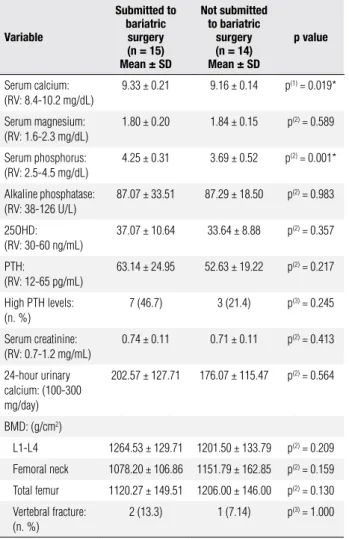
Table 1 illustrates the clinical characteristics and mean intake of calcium, vitamin D and vitamin B12 in the two groups.
Table 1. Clinical characteristics of study patients
Variable
Submitted to bariatric
surgery (n = 15) Mean ± SD
Not submitted to bariatric
surgery (n = 14) Mean ± SD
p value
Age: (years) 35.07 ± 7.36 34.71 ± 8.09 p(1) = 0.903
Height: (cm) 163.60 ± 4.73 160.14 ± 6.24 p(1) = 0.103
Preoperative weight: (kg) 123.07 ± 10.94 114.35 ± 15.94 p(1) = 0.096
Postoperative weight: (kg) 82.31 ± 13.06 ** **
Weight loss: (kg) 40.49 ± 6.61 ** **
Preoperative BMI: (kg/m2) 46.08 ± 5.17 44.59 ± 5.93 p(1) = 0.477
Postoperative BMI: (kg/m2) 30.81 ± 5.30 ** **
Time elapsed since surgery: (months)
12.53 ± 5.32 ** **
Calcium intake – total: (mg/day)
824.20 ± 513.11 328.86 ± 301.03 p(1) = 0.004*
Calcium intake – diet: (mg/day)
623.33 ± 440.09 328.86 ± 301.03 p(1) = 0.046*
Calcium intake – supplement: (mg/day)
200.87 ± 142.25 0.00 ± 0.00 p(2) < 0.001*
Vitamin D supplement: (U/day)
533.33 ± 445.08 0.00 ± 0.00 p(2) < 0.001*
Vitamin B12 supplement: (mcg/day)
7.00 ± 5.57 0.00 ± 0.00 p(2) < 0.001*
DM: n(%) 1 (6.7) 3 (21.4) p(3) = 0.330
SAH: n(%) 7 (46.7) 11 (78.6) p(4) = 0.077
Regular menstrual cycle: n(%)
15 (100.0) 11 (78.6) p(3) = 0.100
(*): significant difference of 5.0%. Obs.: Mean ± SD signifies mean and standard deviation. (**): Not possible to calculate due to the existence of information on only one group. (1): Using the Student t-test with equal variances. (2): Using the Student t-test with unequal variances. (3): Using the Fisher exact test. (4): Using the Pearson chi-square test. BMI: body mineral index. DM:
diabetes mellitus. SAH: systemic arterial hypertension.
Serum calcium was within normal limits in all pa-tients in both groups.
Serum magnesium was below normal in 13.3% of the patients submitted to surgery and normal in those not submitted to surgery. Serum alkaline phosphatase was high in 6.7% of the patients submitted to surgery as opposed to the normal levels of those who had not undergone surgery.
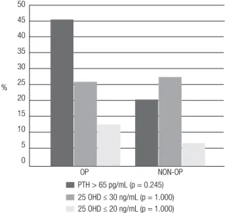
Serum PTH levels were higher than normal in 46.6% of the surgical patients and in 21.4% of the non-surgical patients (p = 0.245). Serum 25OHD was 30 ng/mL or less in 26.7% of the former and 28.6% of the latter (p = 1.000), while 13.3% of the surgery group and 7.1%, of the non-surgery group had levels of 20 ng/mL or less and these differences were not statistically signiicant (p = 1.000)(Table 2, Figure 1).
Table 2. Laboratory and bone mineral density data from study patients
Variable
Submitted to bariatric
surgery (n = 15) Mean ± SD
Not submitted to bariatric
surgery (n = 14) Mean ± SD
p value
Serum calcium: (RV: 8.4-10.2 mg/dL)
9.33 ± 0.21 9.16 ± 0.14 p(1) = 0.019*
Serum magnesium: (RV: 1.6-2.3 mg/dL)
1.80 ± 0.20 1.84 ± 0.15 p(2) = 0.589
Serum phosphorus: (RV: 2.5-4.5 mg/dL)
4.25 ± 0.31 3.69 ± 0.52 p(2) = 0.001*
Alkaline phosphatase: (RV: 38-126 U/L)
87.07 ± 33.51 87.29 ± 18.50 p(2) = 0.983
25OHD: (RV: 30-60 ng/mL)
37.07 ± 10.64 33.64 ± 8.88 p(2) = 0.357
PTH:
(RV: 12-65 pg/mL)
63.14 ± 24.95 52.63 ± 19.22 p(2) = 0.217
High PTH levels: (n. %)
7 (46.7) 3 (21.4) p(3) = 0.245
Serum creatinine: (RV: 0.7-1.2 mg/mL)
0.74 ± 0.11 0.71 ± 0.11 p(2) = 0.413
24-hour urinary calcium: (100-300 mg/day)
202.57 ± 127.71 176.07 ± 115.47 p(2) = 0.564
BMD: (g/cm2)
L1-L4 1264.53 ± 129.71 1201.50 ± 133.79 p(2) = 0.209
Femoral neck 1078.20 ± 106.86 1151.79 ± 162.85 p(2) = 0.159
Total femur 1120.27 ± 149.51 1206.00 ± 146.00 p(2) = 0.130
Vertebral fracture: (n. %)
2 (13.3) 1 (7.14) p(3) = 1.000
Cop
yright
© ABE&M t
odos os dir
eit
os r
eser
vados
.
In relation to vertebral fractures, two patients from the surgery group (13.3%) were found to have grade I morphometric fractures in the thoracic spine, and one pa-tient from the non-surgery group (7.14%) had a grade I fracture, also in the thoracic spine. The patient from the non-surgery group presented low LS BMD and normal serum 25OHD and PTH. The patients with fractures from the surgery group did not present any alterations in the serum PTH, 25OHD or BMD values.
DISCUSSION
Vitamin D deiciency, an inadequate intake of calcium and SHPT are common in obese individuals and have been described in previous studies (4,6,7,8). The pres-ent study found a prevalence of 21.4% of SHPT and 28.6% of vitamin D deiciency in the morbidly obese patients with normal renal function and who had not been submitted to surgery which is similar to the data reported by a number of other authors (6,7,9). The mean intake of calcium in the non-surgery group was less than 50% of the recommended levels for premeno-pausal women. The abovementioned metabolic altera-tions may intensify during the postoperative period and careful preoperative screening for alterations in bone metabolism and an adequate supplementation of vita-mins and minerals are therefore essential to prevent or minimize osteometabolic complications that may occur after gastric bypass.
The mean calcium intake in the surgery group de-spite being signiicantly greater than in the non-surgery group was still lower than the recommended levels for such a speciic group of patients, which should be be-tween 1,000 and 1,800 mg/day (4). The intake of vita-mins D and B12 was also lower than the recommended levels. It may be inferred from these data that postop-erative vitamin and mineral supplementation should not consist only of multivitamins, most of which do not contain the recommended daily requirements of calcium and vitamins D and B12.
The main absorption sites of calcium are the duode-num and the proximal jejuduode-num, where the greatest con-centration of calcium transporters is found. After gas-tric bypass, food intake no longer passes through these portions of the intestine, which may result in calcium deiciency. Associated with this, there is intolerance to foods rich in calcium, such as milk, and an absorption deiciency of vitamin D through the poor absorption of fat. Chronic poor absorption of calcium combined with Mean serum 25OHD in patients submitted to
bar-iatric surgery who presented normal serum PTH was 41.13 ± 6.13 ng/mL, while for those who presented high levels of PTH (> 65 pg/mL) it was 32.43 ± 13.16 ng/mL (p = 0.147). Of the seven patients with high serum PTH in the surgery group, only 3 had serum 25OHD lower than 30 ng/mL. In the non-surgical patients with normal and high serum PTH, the mean serum 25OHD was 33.5 ± 9.69 and 34 ± 6.56 ng/mL, respectively (p = 0.941).
Mean higher LS BMD was observed and lower val-ues in the femoral neck and total femur in the group submitted to surgery, in comparison to the non-surgery group, but with no statistical signiicance (p = 0.209) (Table 2). Of the 15 surgical patients, 13 (86.7%) pre-sented a normal T-score, two (13.3%) revealed low bone mass and no patients had osteoporosis accord-ing to World Health Organization criteria. One patient with low bone mass had normal serum PTH, AP and 25OHD, while the other had high serum PTH and AP with low 25OHD. Spine X-rays of the two patients with low bone mass did not reveal any fractures. Of the 14 patients in the group not submitted to surgery, 12 (85.7%) presented normal T-scores, two (14.3%) pre-sented low LS BMD and no patients had osteoporosis. The two patients with low bone mass in this group had normal serum AP, PTH and 25OHD and one of these had a grade I fracture.
50
45
40
35
30
25
20
15
10
5
0
OP NON-OP
PTH > 65 pg/mL (p = 0.245) 25 OHD ≤ 30 ng/mL (p = 1.000) 25 OHD ≤ 20 ng/mL (p = 1.000)
Figure 1. Secondary hyperparathyroidism and vitamin D deficiency in study patients.
OP: operated on; NON-OP: non-operated on; 25OHD: 25-hydroxy vitamin D; PTH: parathyroid hormone.
Cop
yright
© ABE&M t
odos os dir
eit
os r
eser
vados
.
inadequate supplementation may lead to SHPT and the individuals affected will have high serum PTH associ-ated with normal or low serum calcium (4,22,23). The fact that there is no signiicant difference between the mean 25OHD values in patients submitted to bariatric surgery with normal PTH and high PTH (41.13 ± 6.13 vs. 32.43 ± 13.16), and that only three of the seven patients with abnormal PTH levels had serum 25OHD less than 30 pg/mL, suggests that vitamin D deiciency is not the main cause of SHPT in this group of patients and that the poor absorption of calcium appears to be a predominant factor for the development of SHPT.
Despite the lower calcium absorption, it is not com-mon to ind hypocalcemia in patients submitted to gas-tric bypass, probably due to the compensatory action of SHPT. In the present study, none of the patients submitted to bariatric surgery presented low serum cal-cium, as conirmed by data from other authors (24,25), demonstrating that hypocalcemia is a low-sensitivity marker for evaluating bone disease in this population.
Youssef and cols. (24) conducted a prospective study with 193 morbidly obese women submitted to bariatric surgery and detected high serum PTH in 53.3%, and vitamin D deiciency using a cutpoint of < 20 ng/ml in 20.2% (24). These indings are similar to those of the present study, with 46.6% of high PTH and 28.6% vitamin D deiciency (< 30 ng/ml).
Pereira and cols. studied 39 women, 11 non-obese, 12 obese and 16 obese submitted to Roux-en-Y gastric bypass. They did not ind any signiicant differences in serum AP, magnesium, PTH or 25OHD between the surgical, non-surgical and non-obese groups, which is in agreement with the results of the present study (26).
Several studies have negatively associated obesity with 25OHD status (27-31), including data that illus-trate insuficient levels of up to 80% in obese individuals (32). One hypothesis is that vitamin D is stored by the fatty tissue of obese patients and that it may be grad-ually released into the circulation during weight loss (11). However, contrary to common belief, there is no signiicant improvement in 25OHD status after bariat-ric surgery, as illustrated by several longitudinal stud-ies (16,17,33-36). This was conirmed by the present study, in which no signiicant differences in the serum 25OHD levels were recorded in patients submitted to bariatric surgery. If vitamin D is released postoperative-ly from fatty tissue, the lack of high blood levels during this period appears to suggest that the steroid is being absorbed at even lower proportions.
In the present study, the time elapsed between sur-gery and the evaluation ranged from 7 to 22 months, and there were no signiicant differences between mean BMD and the prevalence of vertebral fractures in either group. It is possible that the relatively short duration of the study and the selection of eugonadic premenopausal women may have contributed to the bone sparing ef-fect. Little data is available concerning the incidence or prevalence of fractures in patients submitted to bariatric surgery. In a recent 7-year study from Mayo Clinic pre-sented at the 91st Annual Meeting of the Endocrine
So-ciety a retrospective cohort of 97 patients submitted to bariatric surgery including men and pre- and postmeno-pausal women 90% of whom had undergone a gastric bypass, showed that the risk of a irst fracture at any site was 1.8 (1,1-2,8, 95% CI) in patients submitted to bar-iatric surgery as compared with what would be expected for the general population for the same age (21).
Some longitudinal studies have shown bone loss re-lated to the degree of weight loss, but none of them spe-ciically included premenopausal women (2,11,13,26). None of the patients with vertebral fractures sub-mitted to surgery had low bone mass or osteoporosis, suggesting that bone fragility and increased risk of frac-ture were not exclusively related to bone mineral den-sity in our group of patients.
The limitations of this study lie in the fact that it is a case series with a small sample. On the other hand, the selection of a speciic group of premenopausal, eugo-nadic women made the analysis of bone metabolism al-terations resulting from bariatric surgery more reliable since we excluded menopausal effects on bone health. More prospective studies are required to clarify the is-sue of bariatric surgery and bone loss.
In conclusion, the present study demonstrated that af-ter Roux-en-Y gastric bypass there is evidence of poor ab-sorption of calcium and vitamin D resulting in SHPT in a speciic group of premenopausal, eugonadic women. Low serum 25OHD levels were not the main cause of SHPT, suggesting that the poor absorption of calcium could play a vital role. Close observation of nutritional aspects and bone density before and after bariatric surgery is essential to prevent or minimize metabolic complications.
Disclosure: no potential conlict of interest relevant to this article was reported.
REFERENCES
Cop
yright
© ABE&M t
odos os dir
eit
os r
eser
vados
.
2. Carrasco F, Ruz M, Rojas P, Csendes A, Rebolledo A, Codoceo J, et al. Changes in bone mineral density, body composition and adi-ponectin levels in morbidly obese patients after bariatric surgery. Obes Surg. 2009;19(1):41-6.
3. Whitlock G, Lewington S, Sherliker P, Clarke R, Emberson J, Halsey J, et al. Body-mass index and cause-speciic mortality in 900,000 adults: collaborative analyses of 57 prospective studies. Lancet. 2009;373(9669):1083-96.
4. Williams SE, Cooper K, Richmond B, Schauer P. Perioperative ma-nagement of bariatric surgery patients: focus on metabolic bone disease. Cleve Clin J Med. 2008;75(5):333-49.
5. Ybarra J, Sánchez-Hernández J, Pérez A. Hypovitaminosis D and morbid obesity. Nurs Clin North Am. 2007;42(1):19-27.
6. Goldner WS, Stoner JA, Thompson J, Taylor K, Larson L, Erickson J, et al. Prevalence of vitamin D insuficiency and deiciency in morbidly obese patients: a comparison with non-obese controls. Obes Surg. 2008;18(2):145-50.
7. Hamoui N, Anthone G, Crookes PF. Calcium metabolism in the morbidly obese. Obes Surg. 2004;14(1):9-12.
8. Vilarrasa N, Maravall J, Estepa A, Sánchez R, Masdevall C, Navar-ro MA, et al. Low 25-hydNavar-roxyvitamin D concentrations in obese women: their clinical signiicance and relationship with anthro-pometric and body composition variables. J Endocrinol Invest. 2007;30(8):653-8.
9. Stein EM, Straint G, Sinha N, Ortiz D, Pomp A, MacMahon DJ, et al. Vitamin D insuficiency prior to bariatric surgery: risk factors and a pilot treatment study. Clin Endocrinol (Oxf). 2009;71:176-83. 10. Wortsman J, Matsuoka LY, Chen TC, Lu Z, Holick MF.
Decre-ased bioavailability of vitamin D in obesity. Am J Clin Nutr. 2000;72:690-3.
11. Fleischer J, Stein EM, Bessler M, Della Badia M, Restuccia N, Oli-vero-Rivera L, et al. The decline in hip bone density after gastric bypass surgery is associated with extent of weight loss. J Clin Endocrinol Metab. 2008;93(10):3735-40.
12. Viégas MA, Bandeira L, Bandeira F. Complicações metabólicas da cirurgia bariátrica. In: Bandeira F, Graf H, Griz L, Faria M, Lazaretti-Castro M, eds. Endocrinologia e Diabetes. 2nd ed. Rio de Janeiro: Medbook; 2009. p. 1038-45.
13. Coates PS, Fernstrom JD, Fernstrom MH, Schauer PR, Greens-pan SL. Gastric bypass for morbid obesity leads to an increase in bone turnover and a decrease in bone mass. J Clin Endocrinol Metab. 2004;89(3):1061-5.
14. Mahdy T, Atia S, Farid M, Adulatif A. Effect of Roux-en Y gastric bypass on bone metabolism in patients with morbid obesity: Mansoura experiences. Obes Surg. 2008;18(12):1526-31. 15. Von Mach MA, Stoeckli R, Bilz S, Kraenzlin M, Langer I, Keller U.
Changes in bone mineral content after surgical treatment of mor-bid obesity. Metabolism. 2004;53(7):918-21.
16. Abbasi AA, Amin M, Smiertka JK, Grunberger G, MacPherson B, Hares M, et al. Abnormalities of vitamin D and calcium metabo-lism after surgical treatment of morbid obesity: a study of 136 patients. Endocr Pract. 2007;13(2):131-6.
17. Ybarra J, Sánchez-Hernández J, Gich I, De Leiva A, Rius X, Ro-dríguez-Espinosa J, et al. Unchanged hypovitaminosis D and secondary hyperparathyroidism in morbid obesity after bariatric surgery. Obes Surg. 2005;15(3):330-5.
18. Compher CW, Badellino KO, Boullata JI. Vitamin D and the baria-tric surgical patient: a review. Obes Surg. 2008;18(2):220-4.
19. Clements RH, Yellumahanthi K, Wesley M, Ballem N, Bland KI. Hyperparathyroidism and vitamin D deiciency after laparoscopic gastric bypass. Am Surg. 2008;74(6):469-74.
20. Carlin AM, Rao DS, Meslemani AM, Genaw JA, Parikh NJ, Levy S, et al. Prevalence of vitamin D depletion among morbidly obe-se patients obe-seeking gastric bypass surgery. Surg Obes Relat Dis. 2006;2(2):98-103.
21. Haglind EGC, Kennel KA, Collazo-Clavell ML, Achenbach SJ, Atkinson EJ, Melton JL, et al. Fracture risk after bariatric surgery. 91st Annual Mee-ting of the Endocrine Society, Washington DC, 2009; OR07-4. 22. Shah M, Simha V, Garg Abhimanyu. Review: long-term impact of
bariatric surgery on body weight, comorbidities, and nutritional status. J Clin Endocrinol Metab. 2006;91(11):3223-4231.
23. DeMaria EJ. Bariatric surgery for morbid obesity. N Engl J Med. 2007;356(21):2176-83.
24. Youssef Y, Richards WO, Sekhar N, Kaiser J, Spagnoli A, Abu-mrad N, el al. Risk of secondary hyperparathyroidism after lapa-roscopic gastric bypass surgery in obese women. Surg Endosc. 2007;21(8):1393-6.
25. Diniz Mde F, Diniz MT, Sanches SR, Salgado PP, Valadão MM, Araújo FC, et al. Elevated serum parathormone after Roux-en-Y gastric bypass. Obes Surg. 2004;14(9):1222-6.
26. Pereira FA, de Castro JA, dos Santos JE, Foss MC, Paula FJ. Impact of marked weight loss induced by bariatric surgery on bone mineral density and remodeling. Braz J Med Biol Res. 2007;40(4):509-17.
27. Arunabh S, Pollack S, Yet J, Aloia JF. Body fat content and 25-hydroxyvitamin D levels in healthy women. J Clin Endocrinol Metab. 2003;88(1):157-61.
28. Kamycheva E, Sundsfjord J, Jorde R. Serum parathyroid hormo-ne level is associated with body mass index. The 5th Tromso Stu-dy. Eur J Endocrinol. 2004;151(2):167-72.
29. Parikh SJ, Edelman M, Uwaifo GI, Freedman RJ, Semega-Janneh M, Reynolds J, et al. The relationship between obesity and serum 1,25-dihydroxy vitamin D concentrations in healthy adults. J Clin Endocrinol Metab. 2004;89(3):1196-9.
30. Yanoff LB, Parikh SJ, Spitalnik A, Denkinger B, Sebring NG, Slau-ghter P, et al. The prevalence of hypovitaminosis D and secondary hyperparathyroidism in obese Black Americans. Clin Endocrinol. 2006;64(5):523-9.
31. Snijder MB, van Dam RM, Visser M, Deeg DJ, Dekker JM, Bouter LM, et al. Adiposity in relation to vitamin D status and parathyroid hormone levels: a population-based study in older men and wo-men. J Clin Endocrinol Metab. 2005;90(7):4119-23.
32. Bischof MG, Heinze G, Vierhapper H. Vitamin D status and its re-lation to age and body mass index. Horm Res. 2006;66(5):211-5. 33. El-Kadre LJ, Rocha PRS, Tinoco AC, Tinoco RC. Calcium
meta-bolism in pre- and post-menopausal morbidly obese women at baseline and after laparoscopic Roux-en-Y gastric bypass. Obes Surg. 2004;14(8):1062-6.
34. Johnson JM, Maher JW, DeMaria EJ, Downs RW, Wolfe LG, Kellum KM. The long-term effects of gastric bypass on vitamin D metabolism. Ann Surg. 2006;243(5):701-4.
35. Sánchez-Hernández J, Ybarra J, Gich I, De Leiva A, Rius X, Rodrí-guez-Espinosa J, et al. Effects of bariatric surgery on vitamin D status and secondary hyperparathyroidism: a prospective study. Obes Surg. 2005;15(10):1389-95.