Cop
yright
© ABE&M t
odos os dir
eit
os r
eser
vados
.
1 Departamento de Medicina
Interna, Faculdade de Medicina de Ribeirão Preto, Universidade de São Paulo (FMRP-USP), Ribeirão Preto, SP, Brasil
2 Maine Medical Center
Research Institute, Scarborough, ME, Estados Unidos
Correspondence to:
Clifford J. Rosen
Maine Medical Center Research Institute, 81
Research Drive
04074-7205 − Scarborough, ME Estados Unidos
Received on Nov/16/2009 Accepted on Jan/28/2010
Obesity,
diabetes mellitus
and
last but not least, osteoporosis
Obesidade, diabete melito e osteoporose
Francisco J. A. de Paula1,2, Clifford J. Rosen2
SUMMARY
Knowledge about the inluence of bone on intermediary metabolism corresponds to a deve-loping area that has gained prominence. The old concept of bone and adipose tissues as inert metabolic tissues, with minor contributions to metabolic adaptations has been reconsidered in light of indings that bone is involved in the development of insulin sensitivity. Similarly adipo-se tissue exerts important inluences on bone mass development and maintenance. Moreover, the use of drugs in the treatment of metabolic disorders such as diabetes mellitus can impact
bone metabolism. These networks linking osteoporosis to obesity and diabetes mellitus have
reinvigorated investigations in the pathophysiology of osteoporosis. The present review exami-nes this aspect and calls attention to health care providers and potential treatments of skeletal disorder. Arq Bras Endocrinol Metab. 2010;54(2):150-7
Keywords
Osteoporosis; obesity; diabetes mellitus; bariatric surgery; thiazolidinediones
SUMÁRIO
O estudo sobre a inluência do tecido ósseo no metabolismo intermediário corresponde a uma área em desenvolvimento que tem ganho recente destaque. O conceito prévio de que os te-cidos ósseo e adiposo seriam metabolicamente inativos foi reconsiderado à luz de estudos que mostram que metabólitos ósseos podem inluenciar a sensibilidade à insulina. Da mesma forma, o tecido adiposo exerce inluência importante no desenvolvimento e na manutenção da massa óssea. Além disso, o uso de drogas no tratamento de doenças metabólicas como o diabetes melito pode afetar o metabolismo ósseo. A rede de conexões existentes que ligam a osteoporose à obesidade e ao diabetes melito tem revigorado investigações sobre a isiopato-logia da osteoporose. A presente revisão analisa esse aspecto e destaca a necessidade de aten-ção para esses pontos por parte de serviços de saúde voltados para o atendimento de diabetes melito e da obesidade quanto ao potencial impacto sobre o tecido ósseo. Arq Bras Endocrinol Metab. 2010;54(2):150-7
Descritores
Osteoporose; obesidade; diabetes melito; cirurgia bariátrica; tiazolidinediona
INTRODUCTION
H
uman being image has changed signiicantly in the last ifty years due to the evolving tendency to increase in body weight. Obesity, the metabolic di-sorder characterized by excessive fat storage relects the imbalance between energy intake and expenditure. The epidemic characteristic of the disturbance can be veriied by the impressive 100% increase in the rate of obese pe-ople in the USA in data from the period of 1976-1980 (15% of adult population) toward 1999-2002 (31% ofadult population) (1). Data obtained in South America were similar since in Brazil the increase in obesity rate was 92% in men and 63% in women from 1975 toward 1989 (2).
Cop
yright
© ABE&M t
odos os dir
eit
os r
eser
vados
.
which was similar to other Western countries for the same period (4). Type 2 DM (T2DM) comprises 90% of all cases of DM syndrome. A major risk factor for developing T2DM is excessive adiposity.
Bone loss and degenerative changes of bone charac-teristics are a progressive process occurring after matu-rity. Therefore, the increase in life expectancy places os-teoporosis as one of the major health public problems in the world. Among US population aged more than 50 years, 10 million have osteoporosis (5). In a recent Brazilian population-based study the prevalence of fra-gility fracture in women and men aged higher than 40 years were 15.1 and 12.8%, respectively (6).
Insulin resistance, the central metabolic disturbance associated with fat accumulation, is linked to metabolic (i.e. increased free fat acids and hypertriglyceridemia), endocrine (i.e. increased glucocorticoid and andro-gens) and pro-inlammatory (i.e. cytokines and tumor necrosis factor) alterations. During the last decade, the role of adipose tissue as an exclusive site of energy stor-age has shifted to one that is an endocrine source of ac-tive modulators of insulin sensitivity such as leptin, adi-ponectin, resistin and cytokines. Furthermore, visceral adipose tissue has an increased lipolytic activity and releases FFA swiftly to the liver and pancreas, thereby provoking lipotoxicity. Insulin, as well as those adipose tissue factors, also inluences bone remodeling.
Recently, investigators have identiied polypeptides such as osteocalcin and osteopontin, which can also modulate insulin sensitivity (7). These new networks link metabolic disturbances to osteoporosis in a new bidirectional pattern. The clinical impact of this associa-tion cannot be currently predicted due to the heteroge-neity of each disorder. However, it cannot be neglected, particularly since one of the most promising groups of drugs to treat diabetes (thiazolidinediones) can induce bone fragility. In the present review we discuss several aspects of the relationship between hard and soft tissues. The clinical association between osteoporosis, obesity and DM and its mechanisms are contemplated, our principal focus is to call attention to the potential recip-rocal relationship of these common diseases.
OSTEOPOROSIS AND OBESITY
Previous studies showed that body weight correlates positively with bone mineral density. In addition, rapid weight loss by severely obese individuals can induce bone loss, as high as 10 percent in the femoral neck,
one year after bariatric surgery (8). Notwithstanding the evidence suggesting a beneicial effect of adipose tissue on bone maintenance, other data argue against this effect. A large population-based study in China did not verify positive correlation between bone mass and fat mass (9). Similar results were obtained in adoles-cents and young adults of both sexes (10).
Bone mineral density (BMD) alone, although cur-rently considered the strongest single factor correlated to risk of fracture, does not permit a comprehensive recognition of fracture risk. There are several other in-dependent factors contributing to fracture risk (age, glucocorticoid therapy, previous fracture, smoking, al-cohol abuse, and rheumatoid arthritis). Also, low body mass index (BMI) only affects the risk of fracture in-dependently of BMD in the case of hip fracture, most likely through an association with frailty and increased risk of fall (11).
Population-based studies revealed that less than a half of the people with a sustained fragility fracture have BMD values below the threshold established by the WHO report as osteoporosis (12-13). Consequently, the majority of fractures that occur in individuals is clas-siied at most as osteopenic. Also, a signiicant num-ber of individuals have osteoporotic fractures despite a normal T-Score (12-13). In addition to revealing the limitations of BMD measurements these data highlight the important contribution of independent risk factors to fracture risk.
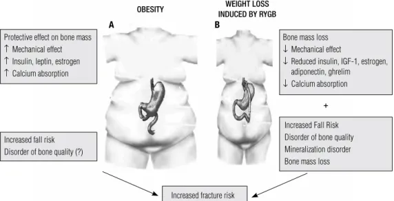
Evidence showing an association between obesity and fracture were acquired in a pediatric clinical inves-tigation. The study exhibited higher forearm fracture incidence in obese children compared with normal weight, age-matched individuals. Recently, more com-pelling data was obtained by Premaor and cols. (14), when they audited the proile of patients aged less than 75 years, attending the Liason Fracture Service, in the United Kingdom, with a fragility fracture (14). Ap-proximately, 40% of this group had normal BMD and more than 50% were obese or morbidly obese patients. Within the obese and morbid obese patient subgroups 80% and 89% had normal value of BMD T-Score of total hip. Thus, these results conirm the tenet that obesity has a protective role on BMD, but at the same time suggest the occurrence of high fracture risk in this population (Figure 1).
in-Cop
yright
© ABE&M t
odos os dir
eit
os r
eser
vados
.
Bone mass loss ↓ Mechanical effect
↓ Reduced insulin, IGF-1, estrogen, adiponectin, ghrelim
↓ Calcium absorption Protective effect on bone mass
↑ Mechanical effect ↑ Insulin, leptin, estrogen ↑ Calcium absorption
OBESITY WEIGHT LOSS
INDUCED BY RYGB
Increased Fall Risk Disorder of bone quality Mineralization disorder Bone mass loss Increased fall risk
Disorder of bone quality (?)
Increased fracture risk
+
Figure 1. (A) Pathophysiology of the increased fracture risk in obesity. (B) Pathophysiology of bone loss and of the increased fracture risk associated to weight loss after bariatric surgery.
A B
clude fat accumulation on one side and bone catabo lism on the other. The same pattern is observed in hypercor-tisolism. Another circumstance emerged with the use of activators of the PPARγ (thiazolidinediones) for the treat-ment of T2DM. At the same time patients using these drugs experience decrease in blood glucose levels and gain weight due to the increase in peripheral adipose tis-sue. Clinical investigations showed bone loss and higher rates of fracture in diabetics using rosiglitazone compared to others on metformin or glyburide (16). Coincidentally with bone loss associated with glucocorticoids and ag-ing, rosiglitazone enriches bone marrow fat while bone volume is reduced. Thus, like insulin resistance which has visceral fat as a marker; fat allocation in bone marrow is an indicator of osteoporosis. While the waist to hip rate is a surrogate identiier of obesity linked to insulin resis-tance, currently there is no clinical means to distinguish the obese patient associated with osteoporosis.
Osteoporosis and obesity treatments
Independent of its inluence on skeleton, obesity is as-sociated with several major health problems and has a strong psychological impact. Hypertension, DM and dyslipidemia are all closely associated to obesity and together increase signiicantly the risk of cardiovascu-lar disease (17). Obese patients also have increased risk to develop breast, colon, prostrate and endometrial cancer (17). Moderate weight loss is an eficient way to prevent the risk to develop DM in overweight and obese individuals, while drastic weight loss ameliorates
the expectancy of life of severely obese patients. Thus, weight loss is pursued by patients and encouraged by physicians to decrease the risk of cardiovascular disease and related disorders. However, the quality of life of a considerable number of these patients can be affected, since the consequences of weight loss on body compo-sition is not limited to adipose tissue. Bone tissue seems exquisitely sensitive to decrements in body weight.
Previous studies have shown that even 10% weight loss can induce about 2% bone loss (18,19). The impact can be higher in individuals exposed to more intense and rapid weight loss (20) compared to moderate dec-rement in weight and for longer periods (21).
Cop
yright
© ABE&M t
odos os dir
eit
os r
eser
vados
.
The Roux-en-Y gastric bypass (RYGB) procedure combines restriction and malabsorption techniques, creating both a small gastric pouch and a deviation of a segment of the small intestine. Presently, RYGB is the most frequent and eficient surgery option to treat se-vere obesity and will be the only one we will discuss in the present review.
Patients may be at risk of postgastrectomy bone disease. Metabolic bone disease is a well-documented long-term complication of obesity surgery. It is often undiagnosed, or misdiagnosed, because of lack of phy-sician and patient awareness. Abnormalities in calcium and vitamin D metabolism begin shortly after gastro-intestinal bypass operations; however, clinical and bio-chemical evidence of metabolic bone disease may not be detected until many years later. RYGB can potential-ly be associated to two metabolic bone disorders osteo-malacia and osteoporosis. Isolated cases of osteoosteo-malacia in patients submitted to gastric bypass for morbid obe-sity treatment have been published in the last few years, resembling old cases of osteomalacia after gastric re-section for management of peptic ulcer (23,24). While severe osteomalacia seems a latter and less frequent complication, bone loss appears to be an early effect on almost all patients submitted to RYGB (Figure 1).
Osteometabolic changes after Roux-en-Y gastric bypass
Vitamin D has an important role in calcium absorption. Estrogen levels, dietary intake, age and body weight also inluence calcium absorption. True fractional cal-cium absorption can be approximately 10% higher in se-verely obese patients compared to overweight individu-als, most likely as a result of higher estrogen levels and increased gastrointestinal mucosal surface (25). Thus, RYGB has signiicant impact on calcium and vitamin D metabolism. Diet restriction reduces the exogenous load of calcium and vitamin D and decreases intake of macronutrients that positively affect their absorption. In RYGB, the proximal jejunum is bypassed, excluding an important site of calcium absorption, which contributes to the decreased calcium load.Additionally, the reduc-tion in food intake leads to increased release of cortisol and decrease in IGF-I serum levels, both adaptations potentially impair calcium absorption (25) (Figure 1).
Adipose tissue shrinkage modiies the adipocyte se-cretion proile. Whereas leptin declines, adiponectin increases after weight loss, though the impact of these changes on bone remodeling is still to be understood. Leptin induces bone loss indirectly by the central
stim-ulation of the sympathetic system, but acts directly on osteoblasts, stimulating bone formation. Conlicting results have been obtained on clinical investigation about bone effects of adiponectin, suggesting neutral, positive or negative association to BMD. Estrogens, IGF-I and insulin fall after weight loss. Gastrointestinal hormones are other variables to be considered in the global evaluation of the weight loss impact on bone. Ghrelin, a stomach-derived peptide, increases during food deprivation to stimulate appetite. In contrast, weight loss due to gastric bypass produces signiicant reduction of ghrelin levels (25). Previous data have shown that ghrelin stimulates osteoblast proliferation and differentiation. Glucagon-like peptide-2 (GLP-2), an enteroendocrine L-cell derived hormone, is an in-hibitor of gastric emptying. GLP-2 also decreases bone resorption (26) and has positive effect on bone miner-alization (27). Overall, the nutritional, metabolic and hormonal changes produced by rapid weight loss favor bone resorption. Considering that obesity can be asso-ciated with fracture and that obesity treatment also can damage skeleton, it is reasonable to conclude that the primary target should be obesity prevention.
Bone mineral density evaluation in obese patients
The evaluation of BMD in obese patients by DXA is limited by the capacity of equipment to support and to measure individuals weighing more than 115-160 kg, depending on the manufacturer. New machines can ac-commodate individuals of up to 180 kg, but it is still insuficient to satisfy the current proile of obesity. The follow up of total body BMD during weight loss is con-founded by variables such as fat density and distribu-tion.
Peripheral BMD is the alternative suitable to evalu-ate bone mass in obese patients. Peripheral DXA mea-surements at phalanges and proximal/distal forearm have been shown in studies to correlate directly with BMD measurements of the spine and hip by central DXA. Quantitative ultrasound seems to be less closely correlated with hip and spine BMD by DXA.
Management after Roux-en-Y gastric bypass
Cop
yright
© ABE&M t
odos os dir
eit
os r
eser
vados
.
Figure 2. Pathophysiology of the increased fracture risk in type 1 and type 2 diabetes mellitus.
Low weight ↓ IGF-I ↓ Insulin ↓ Estrogen
Low bone mass Equilibrium
disorders
↑ Fracture risk
Microdamage accumulation
?
↑ proteolysis ↓ Muscle strength
Oculopathy neuropathy
Protein glycation
Obesity
Diabetes mellitus
Type 1 Type 1-Type 2 Type 2
Patients undergoing RYBG should be screened for osteoporosis with bone density measurement. Labora-tory evaluation includes calcium, albumin, magnesium, PTH and 25(OH)D.
Operated patients should be encouraged to perform regular weight-bearing physical exercise. Recent data suggest increased fall risk in post-operative bariatric surgery patients (28). Thus, mechanical risks for falls should be addressed and physical activity is part of the strategy to reduce fracture risk.
Calcium and vitamin D supplementation should be prescribed in all bariatric patients. One should con-sider slightly higher doses than RDI recommendations: 1,500 mg calcium and 2000 UI vitamin D. Check 25(OH)D serum levels every three months to ensure adequate levels. One should consider pharmacological treatment in patients if BMD is below -1.5. In patients with severe malabsorption parenteral administration may be more convenient.
DIABETES MELLITUS
AND OSTEOPOROSIS
The term heterogeneous is correct in deining diabetes mellitus (DM) due to the large spectrum of variation in its etiopathogenesis and clinical manifestations. Al-most certainly, the only rule valid to all diabetic pa-tients is the tendency to hyperglycemia. Osteoporosis can be added to the large list of disorders associated to DM whose mechanistic association and magnitude of clinical impact have been hard to clarify. When BMD is used as surrogate indicator of osteoporosis, most data
suggest that type 1 diabetes mellitus (T1DM) is linked to osteoporosis. Also, results from the Nord-Trondelag Health Survey from Norway showed a signiicant in-crease in hip fracture rates among female type 1 dia-betic patients (relative risk 6.9, conidence interval 2.2-21.6) compared to nondiabetic female patients (29). However, the results are quite undeined in relation to T2DM, existing indications of bone loss, bone mainte-nance and protective effects on bone.
Cop
yright
© ABE&M t
odos os dir
eit
os r
eser
vados
.
metabolism and recent studies have shown impairment of muscle function and balance in diabetic patients. Therefore, diabetic patients have a greater susceptibil-ity to fall and bone microdamage. Additionally, a wide range of pathogenic pathways have been proposed that underlie bone quality dysfunction in osteoporosis sec-ondary to DM. Protein glycation is a common post-translational modiication of proteins induced by the spontaneous condensation of glucose and metabolic in-termediates (e.g. triose phosphate, glyoxal and methy-glyoxal) with free amino groups in lysine or arginine residues. This process leads to an irreversible formation of advanced glycation end products (AGE) from an ar-ray of precursor molecules (32). Alfa-oxaloaldehydes such as glyoxal, methylglyoxal (MGO) and 3-deoxy-glucosone occur at high levels in diabetic plasma or are signiicantly elevated in cells exposed to high glucose concentrations (32). They can react directly with pro-tein to yield intracellular and extracellular AGEs, which have been shown to have signiicant pathogenic effects in cells and tissues (33,34). Collagen cross-linking, a major post-translational modiication of collagen, plays important roles in the biological and biomechanical features of bone. Collagen cross-links can be divided into lysyl hydroxylase and lysyl oxidase-mediated en-zymatic immature divalent cross-links, mature trivalent pyridinoline and pyrrole cross-links and glycation- or oxidation-induced non-enzymatic cross-links (ad-vanced glycation end products) such as glucosepane and pentosidine. These types of cross-links differ in the mechanism of their formation and function, determin-ing differences in the mineralization process as well as in the bone tendency to microdamage. Pentosidine is one of the well-known AGEs, and its concentration in cortical and trabecular bone is negatively associated with bone strength. Previous studies showed that pa-tients with femoral neck fractures had higher concen-trations of pentosidine in cortical(35) and cancellous bone (36) than in controls.In vitro, highglucose and AGEs synergistically inhibit the mineralization activity of MC3T3-E1 cells through glucose-induced increase in the receptor for AGE (RAGE) (37). Diabetic patients have signiicantly higher levels of serum pentosidine than control individuals. Furthermore, in a recent clini-cal study, Yamamoto and cols. (38) observed in a small group of patients that pentosidine serum levels were positively and signiicantly associated with the presence of vertebral fractures in postmenopausal women with DM and that this association was independent of BMD.
Thus, there is a dichotomy between BMD and frac-ture risk in type 2 diabetic patients. Obesity, as discussed above, and protein glycation are two contributing fac-tors that increase the susceptibility to fracture in T2DM that are not relected in bone mass measurement.
Diabetes mellitus treatment and osteoporosis
Results obtained in the DCCT and UKPDS studies showed that plasma glucose levels are directly implicat-ed in the development of diabetic complications. Low plasma glucose levels were then pursued as a surrogate parameter for the most important objective in the treat-ment of DM, the prevention of chronic micro and mac-roangiopathic disorders.
Thiazolidenediones (TZD) were introduced as powerful glucose lowering agents that could reduce complications in T2DM. The irst of these drugs, tro-glitazone, was withdrawn from the market because of hepatotoxicity. Rosiglitazone and pioglitazone, two other TZDs, are currently approved to treat T2DM and may potentially be helpful in other insulin-resis-tant disorders (39). The eficiency of rosiglitazone to maintain DM under metabolic control is greater than glyburide and metformin (16). However, there may be an increased risk of cardiovascular disease in patients treated with rosiglitazone. Additionally, experimental and clinical evidence indicates that TZD use is asso-ciated with adverse effects on the skeleton, mainly in postmenopausal women with T2DM.
Osteoblasts and marrow adipocytes are derived from a common multipotential stem cell (MSC) progenitor. It has been hypothesized that the close relationship be-tween these lineages underlies the reciprocal relationship between increased adipocytes and decreased bone for-mation that occurs during aging and with TZD therapy. MSC fate is determined by expression and/or acti-vation of speciic transcription factors [i.e., Runx2 and Osterix in the case of osteoblasts and CCAAT enhancer binding protein β and peroxisome proliferator-activat-ed receptor γ (PPARγ) in the case of adipocyte]. The transcriptional activity of PPARγ is induced by endoge-nous ligands such as prostaglandin J2 and oxidized fatty acids as well as by exogenous ligands such as the TZDs.
re-Cop
yright
© ABE&M t
odos os dir
eit
os r
eser
vados
.
sponse relates to the suppression of key osteoblastogenic transcription factors Runx2 and Osterix (41).
The accumulating evidences of TZD-induced skel-etal toxicity prompted the pharmaceutical companies marketing these agents to analyze adverse event data collected during the performance of phase III trials. For example, the ADOPT study, a 4-year investigation de-signed to compared rosiglitazone, metformin and gly-buride in the maintenance of glycemic control, showed that women, not men, on rosiglitazone had a higher in-cidence of appendicular fractures than women on met-formin or glyburide. As this event was not a pre-speciied issue, vertebral fracture data are not available. Although appendicular fracture is not the most typical osteopo-rotic fracture it seems to be more prevalent in diabetic populations. Data review was also performed on clinical trials of pioglitazone. Increased limb fractures were also observed in patients treated with this TZD, and once again this inding was only in female diabetic patients.
Management of diabetic patients in thiazolidenedione therapy
Health care providers need to be aware of the possibil-ity of increased bone loss and fracture risk associated with TZD use. It is recommended that clinical fracture risk in patients be determined before initiation of TZD therapy. For postmenopausal women with DM, par-ticularly those on TZD therapy, screening BMD may be useful. Osteoporosis therapy should be initiated in those women whose bone density and other risk factors place them at an increased risk for fracture.
CONCLUSION
Recent studies have shown that there is functional plas-ticity in bone, and that it is far greater than originally appreciated. Bone remodeling, the mechanism neces-sary to maintain bone strength, is sensitive to metabolic disarrangements such as DM and obesity. Surprisingly, recent studies have indicated that bone cells contribute to metabolic activity by the production of peptides such as osteocalcin that impacts insulin sensitivity and energy metabolism. These new networks will have implications at the bed side and are certain to be incorporated into newer strategies to treat both osteoporosis and diabetes.
Acknowledgments:Francisco J. A. de Paula was supported by a grant PDE 201650/2008-8 from CNPq “Conselho Nacional de Desenvolvimento Cientíico e Tecnológico”, DF, Brazil.
Disclosure: no potential conlict of interest relevant to this article was reported.
REFERENCES
1. National Center for Health Statistics 2004 − National Center for Health Statistics Survey. Available online at: www.cdc.gov/nchs/ products/pubs//pubd/hestats/obese/obse99.htm.
2. Monteiro CA, Conde WL, Popkin BM. Income-speciic trends in obe-sity in Brazil: 1975-2003. Am J Public Health. 2007;97(10):1808-12. 3. Cowie CC, Rust KF, Ford ES, Eberhardt MS, Byrd-Holt DD, Li C, et
al. Full accounting of diabetes and pre-diabetes in the U.S. popula-tion in 1988-1994 and 2005-2006. Diabetes Care. 2009;32(2):287-94. 4. Malerbi DA, Franco LJ. Multicenter study of the prevalence of
diabetes mellitus and impaired glucose tolerance in the urban Brazilian population aged 30-69 yr. The Brazilian Cooperati-ve Group on the Study of Diabetes Prevalence. Diabetes Care. 1992;15(11):1509-16.
5. U.S. Department of Health and Human Services 2004 Bone He-alth and Osteoporosis: A Report of the Surgeon General. U.S. Department of Health and Human Services, Rockville, MD, USA. 6. Pinheiro MM, Ciconelli RM, Martini LA, Ferraz MB. Clinical risk
factors for osteoporotic fractures in Brazilian women and men: the Brazilian Osteoporosis Study (BRAZOS). Osteoporos Int. 2009;20(3):399-408.
7. Lee NK, Sowa H, Hino E, Ferron M, Ahn JD, Confavreux C, et al. Endocrine regulation of energy metabolism by the skeleton. Cell. 2007;130(3):456-69.
8. Pereira FA, de Castro JA, dos Santos JE, Foss MC, Paula FJ. Impact of marked weight loss induced by bariatric surgery on bone mineral density and remodeling. Braz J Med Biol Res. 2007;40(4):509-17.
9. Zhao LJ, Jiang H, Papasian CJ, Maulik D, Drees B, Hamilton J. Correlation of obesity and osteoporosis: effects of fat mass on the determinant of osteoporosis. J Bone Miner Res. 2008;23(1):17-29. 10. Janicka A, Wren TA, Sanchez MM, Dorey F, Kim PS, Mittelman SD,
et al. Fat mass is not beneicial to bone in adolescents and young adults. J Clin Endocrinol Metab. 2007;92(1):143-7.
11. DeLaet C, Kanis JA, Oden A, Johanson H, Johnell O, Delmas P, et al. Body mass index as a predictor of fracture risk: a meta-analysis. Osteoporos Int. 2005;16(11):1330-8.
12. Siris ES, Miller PD, Barrett-Connor E, Faulkner KG, Wehren LE, Abbott TA, et al. Identiication and fracture outcomes of undiag-nosed low bone mineral density in postmenopausal women. Results from the National Osteoporosis Risk Assessment. JAMA. 2001;286(22):2815-22.
13. Schuit SCE, van der Klift M, Weel AE, de Laet CE, Burger H, Se-eman E, et al. Fracture incidence and association with bone mi-neral density in elderly men and women: the Rotterdam Study. Bone. 2004;34(1):195-202.
14. Premaor MO, Pilbrow L, Tonkin C, Parker RA, Compston J. Obe-sity and fractures in postmenopausal women. J Bone Miner Res. 2009 Oct 12. [Epub ahead of print]
15. Rosen CJ, Bouxsein ML. Mechanism of disease: is osteoporosis the obesity of bone. Nat Clin Pract Rheumatol. 2006;2(1):35-43. 16. Kahn SE, Haffner SM, Heise MA, Herman VH, Holman RR, Jones
NP, et al. Glycemic durability of rosiglitazone, metformin or gly-buride monotherapy. N Engl J Med. 2006;355(23):2427-43. 17. Picot J, Jones J, Colquitt JL, Gospodarevskaya E, Loveman E,
Cop
yright
© ABE&M t
odos os dir
eit
os r
eser
vados
.
18. Compston JE, Laskey MA, Croucher PI, Coxon A, Kreitzman S. Effect of diet-induced weight loss on total body bone mass. Clin Sci (Lond). 1992;82(4):429-32.
19. Ricci TA, Chowdhury HA, Heymsield SB, Stahl T, Pierson RN Jr, Shapses SA. Calcium supplementation suppresses bone turno-ver during weight reduction in postmenopausal women. J Bone Miner Res. 1998;13(6):1045-50.
20. Fogelholm GM, Sievanen HT, Kukkonen-Harjula TK, Pasanen ME. Bone mineral density during reduction, maintenance and regain of body weight in premenopausal, obese women. Osteoporos Int. 2001;12(3):199-206.
21. Shapses SA, Von Thun NL, Heymsield SB, Ricci TA, Ospina M, Pierson RN Jr, et al. Bone turnover and density in obese preme-nopausal women during moderate weight loss and calcium sup-plementation. J Bone Miner Res. 2001;16(7):1329-36.
22. Sjostrom L. Bariatric surgery and reduction in morbidity and mor-tality: experiences from the SOS study. Int J Obes. 2008;32(suppl 7):S93-7.
23. Goldner WS, O’Dorisio TM, Dillon JS, Mason EE. Severe metabo-lic bone disease as a long-term compmetabo-lication of obesity surgery. Obes Surg. 2002;12(5):685-92.
24. De Prisco C, Levine SN. Metabolic bone disease after gastric bypass surgery for obesity. Am J Med Sci. 2005;329(2):57-61. 25. Shapses SA, Riedt CS. Bone, body weight and weight reduction:
what are the concern. J Nutr. 2006;136(6):1453-6.
26. Henriksen DB, Alexandersen P, Hartmann B, Adrian CL, Byrjalsen I, Bone HG, et al. Disassociation of bone resorption and formation by GLP-2: a 14-day study in healthy postmenopausal women. Bone. 2007;40(3):723-9.
27. Haderslev KV, Jeppesen PB, Hartmann B, Thulesen J, Sorensen HA, Graff J, et al. Short-term administration of glucagon-like peptide-2. Effects on bone mineral density and markers of bone turnover in short-bowel patients with no colon. Scand J Gastro-enterol. 2002;37(4):392-8.
28. Berarducci A, Murr MM, Haines K. Risk and incidence of falls and skeletal fragility following Roux-en-Y gastric bypass surgery for morbid obesity. Osteoporos Int. 2007;18(S1): 201.
29. Forsen L, Meyer HE, Midthjell K, Edna TH. Diabetes mellitus and the incidence of hip fracture: results from the Nord-Trondelag He-alth Survey. Diabetologia. 1999;42(8):920-5.
30. Cutrim DM, Pereira FA, de Paula FJ, Foss MC. Lack of relationship between glycemic control and bone mineral density in type 2 dia-betes mellitus. Braz J Med Biol Res. 2007;40(2):221-7.
31. Schwartz AV, Sellmeyer DE, Ensrud KE, Cauley JA, Tabor HK, Schreiner PJ, et al. Older women with diabetes have an increased risk of fractu-re: a prospective study. J Clin Endocrinol Metab. 2001;86(1):32-8. 32. Thorpe SR, Baynes JW. Maillard reaction products in tissue
proteins: new products and new perspectives. Amino Acids. 2003;25(3-4):275-81.
33. Thornalley PJ, Langborg A, Minhas HS. Formation of glyoxal, me-thylglyoxal and 3-deoxyglucosone in the glycation of proteins by glucose. Biochem J. 1999;344:109-16.
34. Yao D, Taguchi T, Matsumura T, Pestell R, Edelstein D, Giardino I, et al. High glucose increases angiopoietin-2 transcription in micro-vascular endothelial cells through methylglyoxal modiication of mSin3A. J Biol Chem. 2007;282(42):31038-45.
35. Saito M, Fujii K, Soshi S, Tanaka T. Reductions in degree of mi-neralization and enzymatic collagen cross-links and increases in glycation-induced pentosidine in the femoral neck cortex in cases of femoral neck fracture. Osteoporos Int. 2006;17(7):986-95. 36. Saito M, Fujii K, Marumo K. Degree of mineralization-related
collagen crosslinking in the femoral neck cancellous bone in ca-ses of hip fracture and controls. Calcif Tissue Int. 2006;79(3):160-8. 37. Ogawa N, Yamaguchi T, Yano S, Yamauchi M, Yamamoto M, Sugi-moto T. The combination of high glucose and advanced glycation end-products (AGEs) inhibits the mineralization of osteoblastic MC3T3-E1 cells through glucose-induced increase in the receptor for AGEs. Horm Metab Res. 2007;39(12):871-5.
38. Yamamoto M, Yamaguchi T, Yamauchi M, Yano S, Sugimoto T. Se-rum pentosidine levels are positively associated with the presen-ce of vertebral fractures in postmenopausal women with type 2 diabetes mellitus. J Clin Endocrinol Metab. 2008;93(3):1013-9. 39. Stout DL, Fugate SE. Thiazolidenediones for treatment of
polycys-tic ovary syndrome. Pharmacotherapy. 2005;25(2):244-52. 40. Jennermann C, Triantaillou J, Cowan D, Pennink BGA, Connolly
KM, Morris DC. Effects of thiazolidinediones on bone turnover in the rat. J Bone Miner Res. 1995;10:S241. Abstract S361.