Cop
yright
© ABE&M t
odos os dir
eit
os r
eser
vados
.
1 Unidade de Doenças
Osteometabólicas, Hospital das Clínicas, Faculdade de Medicina, Universidade de São Paulo (HC-FMUSP), São Paulo, SP, Brasil
Correspondence to: Pedro Henrique S. Correa Hospital das Clínicas, Faculdade de Medicina, Universidade de São Paulo Av. Enéas Carvalho Aguiar, 155, 8o andar, bloco 3
05403-900 − São Paulo, SP, Brasil [email protected]
Received on Jan/19/2010 Accepted on Feb/28/2010
Bone quality and
osteoporosis therapy
Qualidade óssea e tratamento da osteoporose
Regina Matsunaga Martin1, Pedro Henrique S. Correa1
SUMMARY
Although BMD measured by DXA is a useful clinical tool for osteoporosis diagnosis, changes resulting from osteoporosis treatment only partially explain the observed reduction in frac-tures. Several other bone properties that inluence its resistance to fractures and explain this discrepancy have been deined as “bone quality”. Bone quality is determined by its structural and material properties and orchestrated by bone turnover, a continuous process of renewal through which old or damaged bone is replaced by a mechanically healthy bone and calcium homeostasis is maintained. Bone structural properties include its geometry (size and shape) and microarchitecture (trabecular architecture and cortical porosity), while bone material pro-perties include its mineral and collagen composition as well as microdamage and its repair. This review aims to update concepts surrounding bone quality and how drugs employed to treat osteoporosis might inluence them. Arq Bras Endocrinol Metab. 2010;54(2):186-99
Keywords
Bone quality; turnover; strength; fracture; osteoporosis; treatment
SUMÁRIO
Embora a DMO, medida por DEXA, seja um recurso clínico útil para o diagnóstico da osteo-porose, mudanças resultantes do tratamento da osteoporose explicam apenas parcialmente a redução de fraturas. As demais propriedades ósseas que inluenciam sua resistência a fraturas, que não se referem à massa óssea e explicam a discrepância entre os valores de DMO e o risco de fratura, têm sido deinidas como “qualidade óssea”. A qualidade óssea é determinada por suas propriedades estruturais e materiais e orquestrada pela remodelação óssea, um processo contínuo de renovação por meio do qual o osso velho ou daniicado é substituído por um osso mecanicamente saudável e a homeostase do cálcio é mantida. As propriedades estruturais ós-seas incluem suas geometria (tamanho e formato) e microarquitetura (arquitetura trabecular e porosidade cortical), enquanto as propriedades materiais referem-se à sua composição mineral e colágena assim como ao microdano e seu reparo. O objetivo desta revisão é uma atualização sobre qualidade óssea e como os medicamentos empregados no tratamento da osteoporose podem modiicá-la. Arq Bras Endocrinol Metab. 2010;54(2):186-99
Descritores
Qualidade óssea; remodelação; força; fratura; osteoporose; tratamento
INTRODUCTION
O
steoporosis is a bone disease characterized by low bone strength leading to increasing susceptibility to bone fracture. The capacity of bone to resist mecha-nical forces and fractures depends not only on the quan-tity of bone tissue but also on its quality.The amount of bone tissue is in part evaluated by measuring bone mineral density (BMD) using dual
Cop
yright
© ABE&M t
odos os dir
eit
os r
eser
vados
.
individuals with comparable BMD, fracture risks are not the same. In addition, more than 50% of all frac-tures occur in women with osteopenia, as deined by a -2.5 < BMD T score ≤ -1; at-risk women in this group will not be detected by applying the World Health Or-ganization BMD deinition of osteoporosis (3).
Changes in BMD associated with antiresorptive treatments account for less than 40% of its effect in re-ducing vertebral fracture risk (4,5) demonstrating that changes in BMD with osteoporosis treatments only partially explain fracture risk reduction and that addi-tional independent factors may contribute to the clini-cal eficacy of these therapies.
The term “bone quality” was therefore introduced to refer to the combination of factors that inluence fracture risk but are not related to bone mass (6,7).
BONE QUALITY
Bone quality is determined by structural and material properties that are inluenced by bone turnover rate. Bone turnover or remodeling is a continuous process
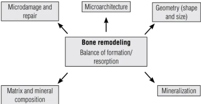
of bone renewal in which old or damaged bone is re-sorbed and new bone is formed to replace it producing a mechanically sound bone and maintaining calcium homeostasis. Bone structural properties include its geometry (size and shape) and microarchitecture (tra-becular/cancellous architecture and cortical thickness/ porosity), while bone material properties include its mineral and collagen composition as well as microdam-age and its repair (Figure 1).
While there is no precise measure of bone strength, BMD has been widely used as a noninvasive surrogate of this parameter as well as an accurate predictor of frac-ture risk. Besides using DXA, BMD can be measured by quantitative computer tomography (QCT)
relect-Microarchitecture Geometry (shape and size) Microdamage and
repair
Matrix and mineral composition
Mineralization
Bone remodeling
Balance of formation/ resorption
Figure 1. Determinants of bone quality.
ing volumetric BMD instead of an areal projection and therefore allowing the determination of the actual vol-umetric density of bone independent of its size.
However, using bone biopsy or autopsy specimens, a number of approaches have been developed provid-ing better comprehension of how bone quality contrib-utes to bone strength in untreated and treated disease states. The most popular of these approaches is histo-morphometry, but newer imaging techniques such as microcomputed tomography (µCT) and magnetic res-onance microimaging (µMRI) allow the measurement of three-dimensional (3D) trabecular microarchitecture in bone specimens in a non-destructive way. Although MRI and µCT analysis are reliable, they should be preferably used in combination to obtain valid conclu-sions. Nevertheless, the use of these techniques is still restricted due to their limited availability, high cost and relatively high radiation exposure.
Bone turnover or remodeling
Bone turnover coordinates the various factors that con-tribute to bone quality (Figure 1). The balance between bone resorption and formation is the key component in preserving bone quality, repairing microarchitectural damage, maintaining BMD and reducing fracture risk.
Accelerated bone turnover leads to the irreversible loss of some trabeculae, resulting in weaker bone and increased fracture risk. Since it is not possible to rou-tinely assess trabecular connectivity in patients with os-teoporosis, bone turnover is most commonly assessed in clinical practice by the measurement of biochemical markers of bone turnover.
Detectable in blood or urine, bone turnover mark-ers are products of osteoblasts/osteocytes and osteo-clasts type I collagen breakdown relecting bone for-mation and bone resorption, respectively. For instance, CTx (bone type I collagen C-telopeptide) is a product of collagen degradation and therefore mirrors bone re-sorption; elevated CTx levels generally mean acceler-ated bone turnover.
Cop
yright
© ABE&M t
odos os dir
eit
os r
eser
vados
.
Bone turnover can also be assessed by bone histo-morphometry using tetracycline labeling prior to biop-sy. The extent of tetracycline-labeled surfaces indicates bone turnover, provided that bone remodeling is in a steady state and that bone resorption and formation are coupled.
Histomorphometry consists of the quantitative analysis of bone resorption parameters, formation and structure on histological sections, and is largely con-sidered the gold standard for assessing bone turnover since it is the only available method for direct in situ
analysis of bone cells and their activity. In addition, this technique can assess 2D bone microarchitecture allow-ing measurements such as thickness and connectivity of trabeculae. Moreover, computerized analysis of biopsy specimens can assess resorption cavity characteristics in quantitative terms such as mean and maximum eroded depth, and eroded area. Nevertheless, bone turnover in iliac crest biopsies may not relect changes at other skeletal sites and the invasive nature of this procedure may pose as a disadvantage to its widespread use (6).
Biochemical markers and histomorphometry differ in their assessment of bone turnover particularly in re-spect to the degree of suppression of bone turnover by anti-resorptive agents, which is generally greater when assessed by the latter technique.
Bone geometry
The external diameter and cortical thickness of bone play crucial roles in determining bone strength. Bone geometry takes into account the distribution of bone mass and the ability of bone to resist torsion and bend-ing (i.e., the wider the external diameter of a cylinder, the higher its resistance to bending). Therefore,for the same areal BMD, a wider bone has greater bending strength and axial strength because its mass is distrib-uted further away from the center (Figure 2A).
Considerable evidence indicates that age-related de-clines in the material properties of bone tissue are ac-companied by a redistribution of cortical and trabecu-lar bone. Speciically in the appendicutrabecu-lar skeleton these changes involve endosteal resorption within the bone combined with periosteal apposition on the external sur-face. This leads to an age-related increase in the diameter of long bones but a decrease in cortical thickness. This increase in the outer diameter helps to maintain the re-sistance to bending and torsional loads. For many years, it has been suggested that men undergo this pattern of favorable geometric adaptation to a greater extent than women, providing one possible explanation for lower fracture rates in elderly men than women. However, re-cent data has shown that both men and women undergo favorable geometric changes with aging (Figure 2B).
Figure 2.Influence of bone geometry on bone strength. A.for the same areal BMD, bone C has progressively greater bending strength and axial strength than bone B and bone A because the mass of bone C is distributed further away from the center – adapted from Bouxsein(63).B.Sex and aging differences in periosteal apposition and endocortical resorption in tubular bones. Adapted from Seeman (64).
A
A
Periosteal surface Before puberty
During puberty
Aging
Male
Female
Endocortical surface
Neutral axis
B
C
Cop
yright
© ABE&M t
odos os dir
eit
os r
eser
vados
.
Bone microarchitecture
Changes in bone microarchitecture make an important contribution to bone strength that may not always be captured by bone mineral density measurements. Corti-cal and cancellous architecture are both important in this respect. In cancellous bone, the number and thickness of trabeculae and their connectivity and orientation (an-isotropy) contribute to bone strength, whilst in cortical bone its width and porosity are the main determinants.
Altered bone microarchitecture in untreated and treated disease states result from underlying changes in bone remodeling. High turnover states and increased os-teoclast activity predispose to trabecular penetration, loss of connectivity, cortical thinning and increased cortical porosity, whereas low bone turnover states and reduced bone formation are associated with trabecular thinning and relative preservation of bone micro architecture.
Although some of these architectural features can be assessed by bone histomorphometry, more sophis-ticated methods have now been developed that enable 3D visualization and quantiication.
The development of µCT scanning has enabled 3D evaluation of trabecular bone specimens at resolutions of 14-50 µm. With the use of µCT, bone volume (BV), trabecular number and thickness, and connectivity can be assessed. Strain on bone tissue can be measured by comparing structural elements with and without me-chanical load. This technique is also potentially useful for studying molecular aspects of bone physiology as it can be performed at low temperatures, preserving RNA and therefore allowing the investigation of interactions be-tween genetic proiles and biomechanical properties (6). Microinite element analysis by combining bone geometry with material characteristics to predict bone strength is a promising technique for fracture assess-ment based on the calculation of the mechanical prop-erties of trabecular sites from high-resolution images. The combined use of these two methods provided an
in vivo assessment of apparent BV to total volume (BV/ TV) and quantiied changes in horizontal elements of trabecular bone of the distal radius in postmenopausal women in terms of both structure and strength (6).
Finally, noninvasive, nonionizing high-resolution MRI can be used to evaluate trabecular bone. It has been employed to compare the trabecular structure of the calcaneus in women with and without hip fractures related to osteoporosis, allowing clear distinction be-tween the two groups (9). More recently, the effects of calcitonin on parameters of trabecular microarchitecture
were analyzed comparing both noninvasive MRI at mul-tiple skeletal sites (radius, hip and calcaneous) and µCT/ histomorphometry acquired by iliac crest bone biopsy, in the QUEST (Qualitative Effects of Salmon-Calcitonin Treatment) trial. Although the results supported the use of MRI for assessment of trabecular microarchitecture in clinical research trials, the authors have highlighted site-speciic differences in response to antiresorptive thera-pies and theneed for suficiently large sampling in order to reliably assess bone architecture (10).
Bone matrix composition
Bone matrix has essentially two constituents, mineral and collagen. The majority of evidence suggests that in normal bone the mineral provides stiffness and strength whereas collagen affords bone its ductility and ability to absorb energy (toughness) before fracturing.
Bone collagen is continuously renewed and its i-bers are stabilized posttranslationally through enzy-matic cross-linking (pyridinoline and deoxypyridino-line), non-enzymatic glycation generating AGEs (aged glycation product ends) such as pentosidine, and β-isomerization of the CTx epitope.
The ratio of pyridinoline/deoxypyridinoline (PYD/ DPD) has been shown to be positively associated with strength and stiffness in bone, but appears to have little correlation with toughness. Non-enzymatic col-lagen cross-links (AGEs) make the tissue more brittle and susceptible to fractures. In vitro studies of fetal bo-vine cortical bone have recently shown that changes in cross-link (PYD, DPD and AGEs) and in the degree of
β-isomerization of carboxy telopeptide of type I colla-gen accompany changes in bone mechanical properties resultant from aging (11).
The excess of AGEs leads to a decrease in tough-ness and strength due to increased stiffness. Moreover, AGE-receptors present on certain cells (osteoblasts, for instance) may downregulate these cells. Interestingly, AGEs are increased in diabetes and may participate in the accelerated aging experienced by diabetic subjects (12).
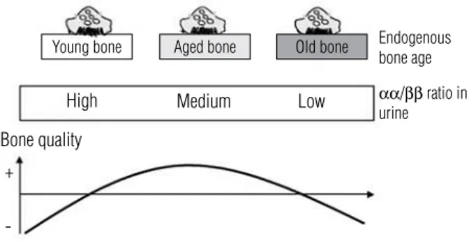
CTx originates from recently synthesized type I col-lagen as αα CTx and undergoes isomerization generat-ing ββ CTx, which is released from aged type I collagen. Therefore, the assessment of αα and ββ CTx fragments by enzyme-linked immunosorbent assay relects the age of endogenous bone collagen. The calculated αα/ββ
Cop
yright
© ABE&M t
odos os dir
eit
os r
eser
vados
.
scopic techniques. The degree of mineralization is cap-tured by BMD measurements but its contribution, in relation to other factors inluencing BMD, cannot be directly deduced.
Microdamage
Bone undergoes repeated cyclic loading and the result-ing fatigue damage in bone matrix is expressed through microcracks or microdamage. Microdamage is gener-ally deined as linear cracks detectable by light micros-copy, and although the optimal method to quantify microdamage in bone is under debate, numerous stud-ies have now shown that the accumulation of damage weakens the bone. Moreover, it appears that micro-damage triggers remodeling, presumably to repair the damaged tissue (15). On the other hand, accumulation of microdamage may result from increased mineraliza-tion secondary to suppression of remodeling, making the bone more brittle.
There is ongoing debate regarding the optimal level of bone turnover to prevent architectural deterioration while preserving the ability of bone to maintain calcium homeostasis, respond to altered mechanical loading, and to repair microdamage. The role of microdamage in age-related fragility fractures has yet to be estab-lished. Microdamage accumulation has been proposed to be a factor that may contribute to increased skeletal fragility with age (16).
Techniques to detect microcracks require expensive technologies and training. Most data have been ob-tained in animal models, and the applicability of some experimental results to the humans remains unclear. Studying microdamage in humans, speciically to de-termine the impact of bone drugs, relies on iliac crest bone biopsies, which may not be appropriate because of the low numerical crack density on this site in com-parison to others. Noninvasive techniques need to be developed to improve assessment of microdamage in vivo (17).
EFFECTS OF OSTEOPOROSIS MEDICATIONS ON
BONE QUALITY
Drugs for osteoporosis treatment can be classiied in two groups: antiresorptive or stimulating of bone for-mation. However, it is increasingly clear that they act not only by improving BMD but also by increasing bone strength, modifying bone quality, and therefore reducing the risk of fracture.
Young bone Aged bone Old bone
High
Bone quality
+
-Medium
Endogenous bone age
αα/ββ ratio in urine Low
Figure 3. Schematic overview of the bone collagen age profile measured as the ratio between ααCTx and ββCTx. Adapted from Leeming and cols.(13).
has been shown that the mean age of bone assessed by αα/ββ CTx is higher in accelerated bone turnover (tra-becular bone) and conversely lower in reduced bone turnover (cortical bone).
Mineralization
Mineralization of bone matrix consists of two succes-sive steps. Primary mineralization occurs when the new collagen matrix begins to mineralize quickly and rep-resents 50% to 60% of the mineralization maximum. Subsequently, the rate of mineralization slows down and secondary mineralization proceeds for a number of years. Typically, mineralization stabilizes around 90% to 95% of maximum. The degree of secondary miner-alization is dependent on bone turnover; when this is low, there is more time for mineralization to proceed whereas in high turnover states, recently formed bone is removed before there is time for appropriate second-ary mineralization.
As mineralization increases, the tissue becomes more brittle and requires less energy to fracture. Therefore, it is possible for a bone that is hypermineralized to be more fragile than a bone with a lower degree of miner-alization. This effect may partially explain the indings of Riggs and cols. (14), who demonstrated that despite dramatic increases in BMD with luoride treatment there was a signiicant increase in the number of pa-tients with nonvertebral fractures in the luoride group as compared with placebo. Conversely, when bone re-sorption begins before completion of mineralization, a cumulative deicit in mineralization arises leading to decreased bone stiffness and strength.
spectro-Cop
yright
© ABE&M t
odos os dir
eit
os r
eser
vados
.
Bisphosphonates (BPs)
Bisphosphonates (BPs) have great afinity for bone min-eral and inhibitory effects on osteoclasts. The attach-ment of BPs to crystalline hydroxyapatite is determined by their structure, mainly the P–C–P coniguration. Ni-trogen-containing bisphosphonates have become the standard of care for osteoporosis. Current oral BPs are: alendronate (daily or weekly dosing), risedronate (daily or weekly dosing) and ibandronate (monthly dosing) whereas intravenous regimens are available with pami-dronate and zoledronic acid (quarterly or yearly) (18).
Besides signiicantly increase BMD in patients with osteoporosis, BPs also act by modifying bone quality to improve bone strength. Although it is dificult to assess the actions of BPs on bone quality in general practice, ev-idence in that direction has accumulated in recent years.
Effects on bone mineralization
BPs decrease bone turnover and increase the duration of the secondary mineralization, thus improving the degree of mineralization. Boivinand cols. (19), treating postmenopausal women with osteoporosis with three-year alendronate therapy, have shown an increase in mean mineralization at the iliac bone of approximately 11% both in cancellous and cortical bone. Further, Bo-rah and cols. (20), using µCT to assess biopsies from patients at baseline and after 3 and 5 years of risedro-nate therapy, showed an increased homogeneity of mineralization. There was no signiicant difference in average or homogeneity mineralization values between 3 and 5 years of risedronate therapy, neither hypermin-eralization of the bone matrix was observed (21). Moreover, Recker and cols. (22) have shown that an-nual IV administration of zoledronic acid during three years is effective in reducing bone remodeling and pre-serving bone structure in postmenopausal women with osteoporosis. However, differences between zoledronic acid and oral BPs on mineralization have been veriied as highlighted by Ebeling and Burr (23): zoledronic acid stimulates osteoblast activity resulting in acceler-ated mineral apposition and increases bone volume but not secondary mineralization.
Role in microdamage
Bone remodeling aims to repair fatigue microdamage, nevertheless an excessive reduction of bone turnover may result in inadequate microdamage repair leading to a fracture. Animal bones overtreated with BPs exhibit
suppressed trabecular bone turnover associated with increased vertebral strength, even with signiicant mi-crodamage accumulation and reduced intrinsic energy absorption capacity (toughness) (24,25).
The mechanism of bisphosphonate-induced micro-damage accumulation has not been fully established. BPs suppress turnover, reducing both targeted and sto-chastic remodeling, and allowing microdamage to per-sist for longer in comparison to untreated bone. More, increased mineralization and increased tissue homoge-neity, both of which resultant from BP treatment, are permissive to the formation and accumulation of mi-crodamage (26).
Action on type I collagen
Limited data is available concerning BP effects on the organic component of bone.
Saito and cols. (27) and Allen and cols. (28) have documented changes in enzymatic (PYD/DPD), non-enzymatic cross-linking (AGEs) and collagen isomeri-zation of the organic matrix in bisphosphonate-treated animals. Following treatment with a wide range of BP doses, the ratio PYD/DPD in the trabecular bone of lumbar vertebrae was signiicantly increased compared to vehicle-treated animals as well as the level of pento-sidine (AGEs) in a dose-dependent fashion; conversely,
the ratio of αα/ββ CTx was decreased when compared to untreated animals.
In human bone, pentosidine markedly increases with age, and its content in bone from patients with hip fracture is signiicantly higher than that of non-frac-tured age-matched controls (29).
As the organic matrix is known to contribute to bio-mechanical properties, these data suggest that changes in the non-mineral component with BP treatment may inluence mechanical properties and therefore fracture risk. However, changes in the organic matrix may have some effect on tissue strength and stiffness, even if these properties are predominantly determined by min-eralization.
Effects on bone geometry and microarchitecture
Each of the changes obtained with BPs treatment has a signiicant effect on material-level biomechanical prop-erties, independent of changes in bone mass, but their speciic individual contribution is dificult to assess ex-perimentally. Probably, effects on bone mineralization
stiff-Cop
yright
© ABE&M t
odos os dir
eit
os r
eser
vados
.
ness and strength, whereas the increased microdamage
tends to decrease both. The relevance of these changes remains enigmatic, however, considering that BPs also modify bone geometry and microarchitecture.
In this way,Davison and cols. (30) reviewed reports of reduced cortical porosity following treatment with BP and changes in parameters of trabecular 3D archi-tecture after one-year risedronate therapy assessed by µCT. In the placebo group, bone volume, trabecular number and trabecular connectivity decreased whereas no such deterioration was observed in the risedronate group, with similar results even after 3 years of risedro-nate therapy.
In the DIVA (Dosing IntraVenous Administra-tion) study, conducted in postmenopausal women, around 12 mg in a year of IV ibandronate provided signiicantly greater gains in BMD than 2.5 mg daily oral ibandronate, with equivalent eficacy and similar safety. Following 2 years of ibandronate treatment, tra-becular bone maintained its normal lamellar structure with no evidence of woven bone, marrow ibrosis, cel-lular toxicity, or other qualitative abnormalities. His-tomorphometric analysis of 89 transiliac bone biopsies demonstrated normal micro-structure of newly formed bone with normal mineralization and reduced remod-eling after oral or IV ibandronate (31). Because the IV ibandronate regimen in DIVA has an annual cumulative exposure similar to the 150 mg monthly oral ibandro-nate, it is possible that the positive histomorphometric and bone safety proiles observed may also relect the effects of its current oral use.
To date, it appears these macro-level changes drive the anti-fracture eficacy of BPs, and can adequately compensate for reduced material properties. Neverthe-less, research on long-term BP treatment is certainly warranted since structural-level beneits of BPs could potentially be overridden with time (26).
Recombinant human parathyroid hormone peptide 1-34: teriparatide (TPT)
The effects of PTH on the skeleton are complex and differ between states of elevated endogenous and exog-enous administration of PTH.
The amino-terminal fragment of human parathy-roid hormone (PTH 1-34), also known as teripara-tide (recombinant DNA origin), has an anabolic effect on bone if administrated intermittently serving as a bone-forming agent for the treatment of osteoporosis.
A daily subcutaneous injection of teriparatide (TPT) produces a rapid increase in markers of bone forma-tion, followed by a more delayed increase in markers of bone resorption. It also signiicantly increases BMD measured by DXA reducing the risk of new vertebral and nonvertebral fractures by 65% and 53%, respective-ly, in postmenopausal women with osteoporosis. That increase in BMD accounts for no more than 40% of its anti-fracture eficacy, suggesting that TPT can also modify bone quality (32).
Improvement in both trabecular and cortical bone microarchitecture has been suggested in patients using TPT. To elucidate these issues, investigations have been conducted to evaluate whether changes in BMD cor-relate with bone structural improvements observed in patients treated with TPT.
Bone turnover
Assessment with bone histomorphometry (33) has shown that TPT changes bone remodeling by increas-ing bone formation rates and turnover, securincreas-ing a large number of active bone multicellular units (BMUs) lay-ing down new bone. Stimulation of bone remodellay-ing by TPT at both cancellous and endosteal surface reach-es a maximum after 6 months of treatment. Subse-quently, bone turnover returns toward levels measured in untreated postmenopausal women, with formation still exceeding resorption. In addition, there is direct evidence that 12 to 24-months TPT therapy induce modeling bone formation at quiescent surfaces (34). These mechanisms may contribute to the improvement of trabecular and cortical architecture seen after TPT treatment.
Microarchitecture and geometry
Cop
yright
© ABE&M t
odos os dir
eit
os r
eser
vados
.
However, cortical porosity in patients with hyper-parathyroidism has raised the concern that intermittent PTH given to treat osteoporotic patients may weaken cortical bone by increasing its porosity. To evaluate this potential effect of TPT therapy, Burr and cols. (38) treat-ed ovariectomiztreat-ed monkeys for up to 18 months with TPT and observed increased dose-dependent intracorti-cal porosity in their humerus without a signiicant effect on bone strength. Most porosity was concentrated near the endocortical surface where its mechanical effect is small. Further, Sato and cols. (39) conducted a detailed quantitative analysis of the effects of TPT on the proxi-mal femur of ovariectomized monkeys and concluded that TPT had beneicial effects on the proximal femur despite increasing cortical porosity. Cortical porosity did not adversely affect the mechanical integrity of the proximal femur because enhanced cortical area (prob-ably due to increased periosteal bone apposition) and trabecular bone volume more than compensated for the porosity. Of note, much of the beneicial effects of TPT were retained 6 months after treatment.
Mineralization
Misof and cols. (40) have evaluated BMD distribution of iliac crest bone biopsies before and after TPT treat-ment for 18-36 months in men and women with os-teoporosis using qBEI. In cortical bone, pairwise com-parison of biopsies before and after treatment revealed a reduction in the typical calcium concentration in men but no change in women, with an increase in the het-erogeneity of mineralization in both. In cancellous bone, there was no change in the typical calcium concentra-tion, but there was a greater heterogeneity of mineral-ization in both men and women due to newly formed bone matrix. Small angle X-ray scattering performed on a subgroup of subjects revealed normal collagen/min-eral structure. These indings conirm the observations that PTH stimulates skeletal remodeling, resulting in an increased percentage of newly formed bone matrix of lower mineral density.
Collagen and microdamage
Garnero and cols. (41) have studied the effects of PTH (1-84) and alendronate (ALN) on urinary αα/ββ CTx ratio in postmenopausal women with osteoporosis. During the irst year, there was no signiicant change in the αα/ββ CTx ratio with PTH or ALN treatment, suggesting that type I collagen degradation products
excreted in the urine during the irst year of PTH may arise predominantly from resting bone that was formed before initiating therapy. Alternatively, isomerization of collagen formed during PTH treatment reached equi-librium before being degraded. At 24 months, howev-er, there was a marked increase in the αα/ββ CTx ratio in women who had received PTH during the irst year followed by a second year of placebo or ALN whereas the ratio only slightly increased after 2 yr of continu-ous ALN, suggesting that PTH therapy may result in decreased bone collagen maturation. The authors con-clude that treatment with PTH (1-84) for 1 yr followed by 1 yr of placebo or ALN may be associated with de-creased type I collagen isomerization. The inluence of these biochemical changes of type I collagen on bone fracture resistance remains to be studied.
Analogous results were obtained previously by Pas-chalis and cols. (42) analyzing bone biopsies from pa-tients following placebo or TPT. In the TPT group, a trend towards an increase in divalent crosslinks was observed after treatment, corresponding to increased formation of newly synthesized collagen.
Suppression of bone turnover by BPs is associ-ated with increased bone microdamage accumulation in animal models but effects of TPT on microdamage accumulation had not been reported. Hence Dobnig and cols. (43) have studied the effect of increased bone turnover and improved bone structure on microdam-age accumulation in 66 postmenopausal women with osteoporosis who started with TPT (20 µg/d) as irst treatment (38 women) and in those who switched from long-term ALN (10 mg/d or 70 mg/wk) to TPT treatment (28 women). Iliac crest bone biopsies were collected and analyzed for microstructure and micro-damage accumulation at baseline and after 24 months of TPT administration.
TPT treatment reduced microdamage accumulation in osteoporotic patients who had been previously treat-ed with ALN and rtreat-eductreat-ed crack length regardless of prior treatment. Moreover the authors highlighted that an intact microarchitecture is essential for maintaining microdamage accumulation at physiologically normal levels in osteoporotic patients.
Strontium ranelate (SR)
osteo-Cop
yright
© ABE&M t
odos os dir
eit
os r
eser
vados
.
blastic differentiation and raising the osteoprotegerin/ RANK-L ratio. Aside from these effects on osteoblasts, SR decreases bone resorption by inhibiting osteoclast activity and osteoclastic differentiation (44). Recent studies have shown that the activation of osteoblast replication is partly mediated by the calcium sensing re-ceptor (CaR) which is also involved in the SR-induced osteoclast apoptosis (45).
Because strontium is a heavier element than calci-um, its incorporation into bone inluences BMD mea-surements. In addition, it has been shown that SR is distributed in calciied matrix and is easily exchange-able from bone mineral, being slightly linked to mature crystals through ionic substitution. The combined ef-fects of strontium distribution in bone and increased X-ray absorption of strontium compared with calcium lead to ampliication of BMD measurement by DXA. These effects account for approximately 50% of the measured changes in BMD. However, an algorithm for adjustment of BMD involves a number of assumptions and cannot be used for individual patients (46). Over-all, BMD changes in patients treated with SR account for 75% of the reduction in fracture risk suggesting that this therapy also interferes on bone quality (45).
Microarchitecture and geometry
In vivo studies indicate that SR decreases bone resorp-tion and promotes bone formaresorp-tion, preventing bone loss. This positive uncoupling between bone formation and resorption results in bone gain and improvements in bone geometry and microarchitecture in growing animals. When administered to female rats during 2 years, SR induced a dose-dependent increase in bone mechanical properties at the level of the vertebral body and midshaft femur (47). In addition, treatment with SR prevents the deleterious effect of ovariectomy on bone strength. After one year of exposure to SR, bone mechanical properties of vertebrae of ovariectomized rats were signiicantly preserved in association with a partial preservation of the trabecular microarchitecture: a dose-dependent effect on the bone volume/trabecular volume ratio and trabecular number and thickness (48).
Iliac crest bone biopsies of postmenopausal osteopo-rotic women analyzed by 3D µCT have demonstrated some improvement of both trabecular and cortical bone microstructure after 3 years of treatment with SR. In comparison to placebo, patients treated with SR for 3 years showed signiicant increase in the number of tra-beculae (+14%), decrease of trabecular separation (-16%)
and increase in cortical thickness (+18%). Furthermore, a signiicant decrease (-22%) of the model index struc-ture suggested a shift in trabecular strucstruc-ture from rod-like to plate-rod-like coniguration resulting in stronger bone. These changes in trabecular and cortical structure may explain the anti fracture eficacy of SR (49).
Mineralization
Boivin and cols. reported histomorphometric evalua-tion of osteoporotic women treated with SR and found no effect on crystal characteristics or mean degree of mineralization of bone (MDMB) over a large range of doses (0.5, 1, and 2 g/d) (30).
Estrogens: hormonal replacement therapy (HRT)
The Women’s Health Initiative (WHI) demonstrated the effect of hormone therapy on BMD and on osteo-porotic fracture reduction at several sites, including the hip. Even though the beneits of postmenopausal hormone replacement therapy (HRT) on fracture were clear, adverseeffects such as increased risk of stroke and deep vein thrombosis were concerning, aggravated by later indings of increased risk of breast cancer and lack of cardio-protective beneits (50). For these reasons, in spite of its effectiveness for preventing postmenopausal osteoporosis, HRT should only be considered in wom-en at signiicant risk of osteoporosis who cannot take non-estrogenic medications.
Bone turnover and microarchitecture
HRT is an effective prophylactic treatment for early postmenopausal bone loss since it reduces bone re-modeling, which may be accelerated by progressive osteoclastic hyperactivity. Besides, estrogens are also capable of exerting an anabolic effect in women with osteoporosis, even when started well into menopause as shown by Khastgir and cols. (51). Histomorphometric studies of iliac bone biopsies performed on 22 older postmenopausal females with low BMD, before and 6 yr after HRT, revealed BMD improvement at both lumbar spine and proximal femur sites. Furthermore, they observed an increase in cancellous bone volume and an increment of wall thickness after 6 yr of HRT, indicating net bone gain.
Cop
yright
© ABE&M t
odos os dir
eit
os r
eser
vados
.
women, Vedi and cols. (52) conducted histomorpho-metric studies plus image analysis of the iliac bone at baseline and 2 yr after HRT. High-dose estrogen-treat-ed women showestrogen-treat-ed the highest cortical width while the proportion of canals with an eroded surface was signii-cantly lower than in women before or after conventional HRT. Their bone formation rate was signiicantly lower than in untreated women and intermediate values were found in women treated with conventional HRT. These results provided evidence that estrogen induces sup-pression of bone turnover in iliac crest cortical bone of postmenopausal women, in a dose-dependent manner.
Mineralization and collagen
To assess whether HRT modiies the degree of bone mineralization and collagen cross-linking, Paschalis and cols. (53) analyzed bone samples from early postmeno-pausal women at baseline and 2 years after HRT by a combination of hystomorphometric and infrared im-aging techniques. They observed a shift in the degree of bone mineralization and collagen cross-links ratio (PYD/DPD ratio) toward higher values after HRT, suggesting that the bone was more mature, as might be expected from suppressed osteoclastic activity.
Selective estrogen receptor modulators (SERMs)
During the past decade, considerable attention has been focused on SERMs as an alternative to postmenopausal estrogen therapy. These agents act as estrogen agonists in some tissues and antagonists in others due to their speciic actions on estrogen receptors. Beneits with ral-oxifene (a nonsteroidal SERM) therapy arose from the MORE (Multiple Outcomes of Raloxifene Evaluation) trial, in which 7705 postmenopausal women with os-teoporosis were studied. BMD increased in the spine and femoral neck and the risk of vertebral fractures was signiicantly reduced, without any signiicant effect on non-vertebral fractures. The risk of endometrial hy-perplasia was unaltered and the risk of invasive breast cancer was signiicantly reduced while there was an in-creased risk of venous thromboembolism (54). Even though the risk of coronary events was unaltered by therapy, LDL cholesterol levels were reduced and HDL levels were raised.
Only 4%-5% of the fracture reduction observed with raloxifene (RLX) results from increments in lumbar spine or femoral neck BMD, suggesting that RLX may also improve bone matrix properties (55).
Bone turnover
To compare raloxifene therapy to HRT, Weinstein and cols. (56) analyzed the impact of these drugs on bone turnover after one year of treatment. The frequency of remodeling events on cancellous bone and rate of bone formation in both cancellous and endocortical bone increased in the placebo group, while these measure-ments decreased in both drug treatment groups. BMD increased from baseline at the lumbar spine (in the HRT group) and in the total body (for both RLX and HRT). Compared with RLX group, the increase in BMD was greater in the HRT group at the lumbar spine but not in the total body. Bone markers signiicantly decreased in both active treatment groups, changes signiicantly different from those seen with placebo. Overall, the authors suggest that RLX preserves bone mass by re-ducing the elevated bone turnover found in postmeno-pausal women by mechanisms similar to those operative in postmenopausal women receiving HRT.
Microarchitecture and geometry
Although there are similarities between raloxifene and BPs in preventing decreases in maximal load, bone mass, and microarchitecture in ovariectomized rats, the raise in BMD is more pronounced after BP than RLX (57). This inding parallels observations from canine studies that showed a signiicant improvement of the mechanical properties at the material level after 1-year treatment with raloxifene even if aBMD, vBMD, BV/ TV, and percent ash remained unaltered in comparison with control. These material-level improvements have been detected at both cortical and trabecular bone sites.
In the clinical setting, a relatively mild effect of RLX on BMD has also been observed despite a very noteworthy vertebrae antifracture eficacy. The ob-served improvements in microarchitecture may, at least in part, underlie the ability of RLX to increase bone strength and reduce fracture risk, yet it seems that the substantial beneicial action on intrinsic bone material quality, both at the cortical and trabecular level, plays an important role in the mechanism by which this agent is effective in osteoporosis treatment. In addition, it should also be noted that SERMs are known to have a protective effect on osteocytes in vitro; however, there is no convincing evidence of this effect in humans (57).
Mineralization
Cop
yright
© ABE&M t
odos os dir
eit
os r
eser
vados
.
also investigated in a prospective longitudinal study of a subset of 64 patients enrolled in the MORE trial. Quan-titative microradiography analysis of iliac crest biopsies taken at baseline and after 2 years of treatment revealed a signiicant dose-dependent increase on MDMB com-pared with baseline. The observed increase in mineral content with preserved heterogeneity of mineral distri-bution is a result of the ability of RLX to decrease bone turnover, therefore extending the duration of second-ary mineralization of bone basic structural units (BSUs) and allowing new bone to achieve a higher degree of mineralization. This mechanism has been shown to im-prove thebiomechanical properties of bone and should contribute to the reduction in fracture riskobserved af-ter treatment with RLX. On the other hand, increased skeletal mineral can improvestructural rigidity and too much mineral may lead to an increase in brittleness (58).
Collagen and microdamage
It has been hypothesized that RLX may alter the or-ganic matrix (26) and, in particular, collagen, which is known to contribute to the biomechanical and intrinsic properties of bone. To answer this question, Byrjalsen and cols.(11) evaluated the effect of different anti-re-sorptive treatments (BPs, HTR and RLX) on bone col-lagen maturation measured by the αα/ββ CTx ratio. It was found that these anti-resorptive therapies induce differences in the maturation proile of bone collagen, i. e. αα/ββ CTx ratio is greater after treatment with HRT than RLX whereas αα/ββ CTx ratio after RLX therapy is greater than BPs.
There is considerable debate on deining normal re-modeling rate and the potential deleterious effects of an excessive suppression by antiresorptive agents, given the potential impairment on microdamage repair and on the replacement of old bone by fresh new units. Although no fracture data support this highly contro-versial theory, SERMs do not suppress bone turnover to an extent that would cause such concerns. In fact, the data show that RLX restores bone turnover to pre-menopausal levels (59) and experimental data demon-strate that the drug actually reduces microcrack density in bone tissue.
Calcitonin (CTN)
Calcitonin (CTN) is an endogenous polypeptide hor-mone secreted by thyroid C cells that inhibits bone resorption by osteoclasts. Nevertheless, there is also
concern that prolonged exposure to CTN may down-regulate calcitonin receptors on osteoclasts, which could allow the osteoclasts to recover from the sup-pressive action of CTN. Its intermittent administration has been recommended as a strategy to avoid clinical resistance (60).
A number of randomized trials have suggested that injectable or intranasal salmon CTN is effective in pre-vention of trabecular bone loss in late menopause. One classical trial evaluated intranasal administration of 200 IU of calcitonin and showed a vertebral fracture reduc-tion of 33-36% without substantial effects on BMD (61). A 30% reduction in hip fracture was observed by Kanis in patients treated with injectable CTN but data with usual doses of nasal CTN in nonvertebral sites are controversial. In addition, CTN may have an analgesic effect in women with acute vertebral fractures, which appears to be independent of its effect on osteoclastic resorption (60).
Clinical studies addressing the effects of CTN on bone quality are scarce but at least one should be point-ed out. The authors postulatpoint-ed that CTN therapy may be associated with improvements in bone microstructure
that are not detected by BMD. To address this hypoth-esis, a 2-year randomized placebo-controlled study of daily dose of 200 IU salmon calcitonin nasal spray (CT-NS) was carried out including approximately 45 postmenopausal osteoporotic women per group, using the noninvasive MRI technology at multiple skeletal sites and µCT/histomorphometry from iliac crest bone biopsies to assess trabecular microarchitecture (10).
MRI assessmentof distal radius and lower trochan-ter of the hip revealed preservation of parametrochan-ters of tra-becular microarchitecture in the CT-NS group, where-as signiicant deterioration wwhere-as observed in the placebo group. Combined µCT/histomorphometric analysis of iliac crest bone biopsies as well as BMD measured by DXA did not reveal consistent differences in architec-ture between CT-NS and placebo groups.
Cop
yright
© ABE&M t
odos os dir
eit
os r
eser
vados
.
SUMMARY AND CONCLUSIONS
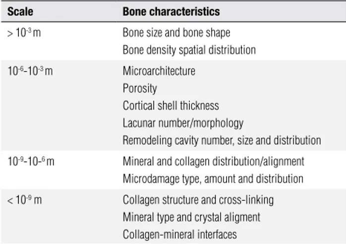
Despite genuine advance, therapy for osteoporosis still represents a considerable research challenge. BMD is a useful clinical tool for diagnosing and monitoring os-teoporosis but has limitations that need to be recog-nized and addressed. Since drugs and bone disorders have a spectrum of effects ranging from the microscop-ic to the macroscopmicroscop-ic level (Table 1) that may change depending on the underlying physiology of a particular patient and BMD may not detect all anticipated effects.
Bone turnover markers may be a useful comple-ment for solving this puzzle as they reveal effects on bone remodeling in the absence of changes on bone density tests. Unfortunately, no monitoring technique is absolutely accurate. Future prospects for in vivo bone strength analysis using CT or MRI have yet to be de-veloped to the point of clinical utility, but this may well be the way of the future. Finite element analyses can reproduce 3D constructs of bone through noninva-sive measurements, congregating all elements of bone strength into a useful clinical tool for assessing thera-peutic response (62).
Up to now, the main effects of osteoporosis medi-cations on bone quality can be summarized as follow. Antiresorptive therapy, specially the bisphosphonates, generally increases MDMB and homogeneity of miner-alization. It is suggested that most of the change in the aBMD induced by antiresorptive therapies results from the increase in the MDMB.
The effects of antiresorptive therapy on cortical bone
are limited, but suggestive of little impact on size albeit decreased porosity. The relatively modest changes in aBMD despite dramatic decreases in antifracture efica-cy observed in antiresorptive trials may be at least par-tially a consequence of reduced bone remodeling which leads to an increase in bone mass and to a decrease in the number of trabecular stress-risers.
On the other hand, TPT has been shown to in-crease trabecular thickness and bone turnover; however, because of its anabolic properties, it results in net bone gain, perhaps explaining some of its positive impact on fracture risk. Despite a dramatic increase in bone mass, the mineralization is actually decreased and the net re-sult is a greater aBMD following therapy. Strontium ranelate, however, induces a falsely elevated aBMD as a result of its incorporation into the apatite.
Regarding microdamage, microcrack accumulation
was observed in bone treated with BPs but its presence,
induced by other drugs used in osteoporosis therapy, was not conirmed. In any case, therapeutic doses of BPs do not negatively impact on the accumulation of microcracks to a level that would increase fracture sus-ceptibility in humans.
The future goal for fracture risk assessment is the development of a virtual biopsy to assess the material and structural properties of bone at clinically important sites simultaneously and noninvasively (30).
Table 1. Factors that determine bone quality categorized by physical scale Scale Bone characteristics
> 10-3 m Bone size and bone shape Bone density spatial distribution
10-6-10-3 m Microarchitecture Porosity
Cortical shell thickness Lacunar number/morphology
Remodeling cavity number, size and distribution
10-9-10-6 m Mineral and collagen distribution/alignment Microdamage type, amount and distribution
< 10-9 m Collagen structure and cross-linking Mineral type and crystal aligment Collagen-mineral interfaces
Adapted from Hernandez et al., 2006 (65).
Acknowledgments: we thank Dr. Bruno Ferraz de Souza for as-sisting the preparation of this manuscript.
Disclosure: no potential conlict of interest relevant to this article was reported.
REFERENCES
1. Christiansen C. Osteoporosis: diagnosis and management today
and tomorrow. Bone. 1995;17(5 Suppl):513S-6S.
2. Kanis JA. Diagnosis of osteoporosis and assessment of fracture risk. Lancet. 2002;359(9321):1929-36.
3. Sornay-Rendu E, Munoz F, Garnero P, Duboeuf F, Delmas PD. Iden-tiication of osteopenic women at high risk of fracture: the OFELY study. J Bone Miner Res. 2005;20(10):1813-9.
4. Cummings SR, Karpf DB, Harris F, Genant HK, Ensrud K, LaCroix AZ, et al. Improvement in spine bone density and reduction in risk of vertebral fractures during treatment with antiresorptive drugs. Am J Med. 2002;112(4):281-9.
5. Chen P, Miller PD, Delmas PD, Misurski DA, Krege JH. Change in lumbar spine BMD and vertebral fracture risk reduction in te-riparatide-treated postmenopausal women with osteoporosis. J Bone Miner Res. 2006;21(11):1785-90.
6. Chesnut CH, 3rd, Rosen CJ. Reconsidering the effects of antire-sorptive therapies in reducing osteoporotic fracture. J Bone Mi-ner Res. 2001;16(12):2163-72.
7. Watts NB. Bone quality: getting closer to a deinition. J Bone
Cop
yright
© ABE&M t
odos os dir
eit
os r
eser
vados
.
8. Felsenberg D, Boonen S. The bone quality framework: determi-nants of bone strength and their interrelationships, and implica-tions for osteoporosis management. Clin Ther. 2005;27(1):1-11. 9. Link TM, Majumdar S, Augat P, Lin JC, Newitt D, Lu Y, et al. In
vivo high resolution MRI of the calcaneus: differences in tra-becular structure in osteoporosis patients. J Bone Miner Res. 1998;13(7):1175-82.
10. Chesnut CH, 3rd, Majumdar S, Newitt DC, Shields A, Van Pelt J, Laschansky E, et al. Effects of salmon calcitonin on trabecular mi-croarchitecture as determined by magnetic resonance imaging: re-sults from the QUEST study. J Bone Miner Res. 2005;20(9):1548-61. 11. Byrjalsen I, Leeming DJ, Qvist P, Christiansen C, Karsdal MA. Bone
turnover and bone collagen maturation in osteoporosis: effects of antiresorptive therapies. Osteoporos Int. 2008;19(3):339-48. 12. Laurent Benhamou C. Bone ultrastructure: evolution during
oste-oporosis and aging. Osteoporos Int. 2009;20(6):1085-7.
13. Leeming DJ, Henriksen K, Byrjalsen I, Qvist P, Madsen SH, Garne-ro P, et al. Is bone quality associated with collagen age? Osteopo-ros Int. 2009;20(9):1461-70.
14. Riggs BL, O’Fallon WM, Lane A, Hodgson SF, Wahner HW, Muhs J, et al. Clinical trial of luoride therapy in postmenopausal oste-oporotic women: extended observations and additional analysis. J Bone Miner Res. 1994;9(2):265-75.
15. Ruppel ME, Burr DB, Miller LM. Chemical makeup of microdama-ged bone differs from undamamicrodama-ged bone. Bone. 2006;39(2):318-24. 16. Burr DB, Forwood MR, Fyhrie DP, Martin RB, Schafler MB, Turner
CH. Bone microdamage and skeletal fragility in osteoporotic and stress fractures. J Bone Miner Res. 1997;12(1):6-15.
17. Chapurlat RD, Delmas PD. Bone microdamage: a clinical perspec-tive. Osteoporos Int. 2009;20(8):1299-308.
18. Sunyecz J. Optimizing dosing frequencies for bisphosphonates in the management of postmenopausal osteoporosis: patient consi-derations. Clin Interv Aging. 2008;3(4):611-27.
19. Boivin GY, Chavassieux PM, Santora AC, Yates J, Meunier PJ. Alendronate increases bone strength by increasing the mean degree of mineralization of bone tissue in osteoporotic women. Bone. 2000;27(5):687-94.
20. Borah B, Dufresne TE, Ritman EL, Jorgensen SM, Liu S, Chmie-lewski PA, et al. Long-term risedronate treatment normalizes mi-neralization and continues to preserve trabecular architecture: se-quential triple biopsy studies with micro-computed tomography. Bone. 2006;39(2):345-52.
21. Zoehrer R, Roschger P, Paschalis EP, Hofstaetter JG, Durchschlag E, Fratzl P, et al. Effects of 3- and 5-year treatment with risedrona-te on bone mineralization density distribution in triple biopsies of the iliac crest in postmenopausal women. J Bone Miner Res. 2006;21(7):1106-12.
22. Recker RR, Delmas PD, Halse J, Reid IR, Boonen S, Garcia-Her-nandez PA, et al. Effects of intravenous zoledronic acid once ye-arly on bone remodeling and bone structure. J Bone Miner Res. 2008;23(1):6-16.
23. Ebeling PR, Burr DB. Positive effects of intravenous zoledronic acid on bone remodeling and structure: are different effects on osteoblast activity to other oral bisphosphonates responsible? J Bone Miner Res. 2008;23(1):2-5.
24. Mashiba T, Turner CH, Hirano T, Forwood MR, Johnston CC, Burr DB. Effects of suppressed bone turnover by bisphosphonates on microdamage accumulation and biomechanical properties in cli-nically relevant skeletal sites in beagles. Bone. 2001;28(5):524-31. 25. Allen MR, Iwata K, Sato M, Burr DB. Raloxifene enhances verte-bral mechanical properties independent of bone density. Bone. 2006;39(5):1130-5.
26. Allen MR, Burr DB. Mineralization, microdamage, and matrix: how bisphosphonates inluence material properties of bone. Bo-nekey Osteovision. 2007;4(2):49-60.
27. Saito M, Mori S, Mashiba T, Komatsubara S, Marumo K. Collagen maturity, glycation induced-pentosidine, and mineralization are increased following 3-year treatment with incadronate in dogs. Osteoporos Int. 2008;19(9):1343-54.
28. Allen MR, Gineyts E, Leeming DJ, Burr DB, Delmas PD. Bisphos-phonates alter trabecular bone collagen cross-linking and isome-rization in beagle dog vertebra. Osteoporos Int. 2008;19(3):329-37. 29. Saito M, Fujii K, Soshi S, Tanaka T. Reductions in degree of mi-neralization and enzymatic collagen cross-links and increases in glycation-induced pentosidine in the femoral neck cortex in cases of femoral neck fracture. Osteoporos Int. 2006;17(7):986-95. 30. Davison KS, Siminoski K, Adachi JD, Hanley DA, Goltzman D,
Hodsman AB, et al. The effects of antifracture therapies on the components of bone strength: assessment of fracture risk today and in the future. Semin Arthritis Rheum. 2006;36(1):10-21. 31. Recker RR, Ste-Marie LG, Langdahl B, Czerwinski E, Bonvoisin B,
Masanauskaite D, et al. Effects of intermittent intravenous iban-dronate injections on bone quality and micro-architecture in wo-men with postwo-menopausal osteoporosis: The DIVA study. Bone. 2009 Nov 10.
32. Chen P, Miller PD, Recker R, Resch H, Rana A, Pavo I, et al. Increa-ses in BMD correlate with improvements in bone microarchitec-ture with teriparatide treatment in postmenopausal women with osteoporosis. J Bone Miner Res. 2007;22(8):1173-80.
33. Arlot M, Meunier PJ, Boivin G, Haddock L, Tamayo J, Correa-Rotter R, et al. Differential effects of teriparatide and alendronate on bone remodeling in postmenopausal women assessed by histomor-phometric parameters. J Bone Miner Res. 2005;20(7):1244-53. 34. Ma YL, Zeng Q, Donley DW, Ste-Marie LG, Gallagher JC, Dalsky
GP, et al. Teriparatide increases bone formation in modeling and remodeling osteons and enhances IGF-II immunoreactivity in postmenopausal women with osteoporosis. J Bone Miner Res. 2006;21(6):855-64.
35. Jiang Y, Zhao JJ, Mitlak BH, Wang O, Genant HK, Eriksen EF. Re-combinant human parathyroid hormone (1-34) [teriparatide] im-proves both cortical and cancellous bone structure. J Bone Miner Res. 2003;18(11):1932-41.
36. Paritt AM. Parathyroid hormone and periosteal bone expansion. J Bone Miner Res. 2002;17(10):1741-3.
37. Zanchetta JR, Bogado CE, Ferretti JL, Wang O, Wilson MG, Sato M, et al. Effects of teriparatide [recombinant human parathyroid hormone (1-34)] on cortical bone in postmenopausal women with osteoporosis. J Bone Miner Res. 2003;18(3):539-43.
38. Burr DB, Hirano T, Turner CH, Hotchkiss C, Brommage R, Hock JM. Intermittently administered human parathyroid hormone(1-34) treatment increases intracortical bone turnover and porosity wi-thout reducing bone strength in the humerus of ovariectomized cynomolgus monkeys. J Bone Miner Res. 2001;16(1):157-65. 39. Sato M, Westmore M, Ma YL, Schmidt A, Zeng QQ, Glass EV, et al.
Teriparatide [PTH(1-34)] strengthens the proximal femur of ova-riectomized nonhuman primates despite increasing porosity. J Bone Miner Res. 2004;19(4):623-9.
40. Misof BM, Roschger P, Cosman F, Kurland ES, Tesch W, Messmer P, et al. Effects of intermittent parathyroid hormone administra-tion on bone mineralizaadministra-tion density in iliac crest biopsies from patients with osteoporosis: a paired study before and after treat-ment. J Clin Endocrinol Metab. 2003;88(3):1150-6.
Cop
yright
© ABE&M t
odos os dir
eit
os r
eser
vados
.
42. Paschalis EP, Glass EV, Donley DW, Eriksen EF. Bone mineral and collagen quality in iliac crest biopsies of patients given teriparati-de: new results from the fracture prevention trial. J Clin Endocri-nol Metab. 2005;90(8):4644-9.
43. Dobnig H, Stepan JJ, Burr DB, Li J, Michalska D, Sipos A, et al. Teriparatide reduces bone microdamage accumulation in post-menopausal women previously treated with alendronate. J Bone Miner Res. 2009 May 19.
44. Reginster JY, Bruyere O, Sawicki A, Roces-Varela A, Fardello-ne P, Roberts A, et al. Long-term treatment of postmenopausal osteoporosis with strontium ranelate: results at 8 years. Bone. 2009;45(6):1059-64.
45. Cortet B. Strontium ranelate: new perspectives for the ma-nagement of osteoporosis. Rheumatology (Oxf). 2009;48 Suppl 4:iv1-2.
46. Bruyere O, Roux C, Detilleux J, Slosman DO, Spector TD, Far-dellone P, et al. Relationship between bone mineral density chan-ges and fracture risk reduction in patients treated with strontium ranelate. J Clin Endocrinol Metab. 2007;92(8):3076-81.
47. Ammann P, Shen V, Robin B, Mauras Y, Bonjour JP, Rizzoli R. Strontium ranelate improves bone resistance by increasing bone mass and improving architecture in intact female rats. J Bone Mi-ner Res. 2004;19(12):2012-20.
48. Ammann P. Strontium ranelate: a physiological approach for an improved bone quality. Bone. 2006;38(2 Suppl 1):15-8.
49. Arlot ME, Jiang Y, Genant HK, Zhao J, Burt-Pichat B, Roux JP, et al. Histomorphometric and microCT analysis of bone biopsies from postmenopausal osteoporotic women treated with strontium ra-nelate. J Bone Miner Res. 2008;23(2):215-22.
50. Nelson HD, Humphrey LL, Nygren P, Teutsch SM, Allan JD. Post-menopausal hormone replacement therapy: scientiic review. JAMA. 2002;288(7):872-81.
51. Khastgir G, Studd J, Holland N, Alaghband-Zadeh J, Fox S, Chow J. Anabolic effect of estrogen replacement on bone in postmeno-pausal women with osteoporosis: histomorphometric evidence in a longitudinal study. J Clin Endocrinol Metab. 2001;86(1):289-95. 52. Vedi S, Bell KL, Loveridge N, Garrahan N, Purdie DW, Compston
JE. The effects of hormone replacement therapy on cortical bone in postmenopausal women. A histomorphometric study. Bone. 2003;33(3):330-4.
53. Paschalis EP, Boskey AL, Kassem M, Eriksen EF. Effect of hormone replacement therapy on bone quality in early postmenopausal women. J Bone Miner Res. 2003;18(6):955-9.
54. Ettinger B, Black DM, Mitlak BH, Knickerbocker RK, Nickelsen T, Genant HK, et al. Reduction of vertebral fracture risk in post-menopausal women with osteoporosis treated with raloxifene: results from a 3-year randomized clinical trial. Multiple Outco-mes of Raloxifene Evaluation (MORE) Investigators. JAMA. 1999;282(7):637-45.
55. Sarkar S, Mitlak BH, Wong M, Stock JL, Black DM, Harper KD. Rela-tionships between bone mineral density and incident vertebral frac-ture risk with raloxifene therapy. J Bone Miner Res. 2002;17(1):1-10. 56. Weinstein RS, Paritt AM, Marcus R, Greenwald M, Crans G, Mu-chmore DB. Effects of raloxifene, hormone replacement therapy, and placebo on bone turnover in postmenopausal women. Oste-oporos Int. 2003;14(10):814-22.
57. Brennan TC, Rizzoli R, Ammann P. Selective modiication of bone quality by PTH, pamidronate, or raloxifene. J Bone Miner Res. 2009;24(5):800-8.
58. Boivin G, Lips P, Ott SM, Harper KD, Sarkar S, Pinette KV, et al. Contribution of raloxifene and calcium and vitamin D3 sup-plementation to the increase of the degree of mineralization of bone in postmenopausal women. J Clin Endocrinol Metab. 2003;88(9):4199-205.
59. Johnell O, Scheele WH, Lu Y, Reginster JY, Need AG, Seeman E. Additive effects of raloxifene and alendronate on bone densi-ty and biochemical markers of bone remodeling in postmeno-pausal women with osteoporosis. J Clin Endocrinol Metab. 2002;87(3):985-92.
60. Cranney A, Tugwell P, Zytaruk N, Robinson V, Weaver B, Shea B, et al. Meta-analyses of therapies for postmenopausal osteoporosis. VI. Meta-analysis of calcitonin for the treatment of postmenopau-sal osteoporosis. Endocr Rev. 2002;23(4):540-51.
61. Chesnut CH, 3rd, Silverman S, Andriano K, Genant H, Gimona A, Harris S, et al. A randomized trial of nasal spray salmon calcitonin in postmenopausal women with established osteoporosis: the prevent recurrence of osteoporotic fractures study. PROOF Study Group. Am J Med. 2000;109(4):267-76.
62. Licata AA. Clinical perspectives on bone quality in osteoporosis: effects of drug therapy. Drugs Aging. 2007;24(7):529-35. 63. Bouxsein ML. Determinants of skeletal fragility. Best Pract Res
Clin Rheumatol. 2005;19(6):897-911.
64. Seeman E. Bone quality: the material and structural basis of bone strength. J Bone Miner Metab. 2008;26(1):1-8.