528
Revista da Sociedade Brasileira de Medicina Tropical 44(4):528-530, jul-ago, 2011
1. Laboratório de Virologia, Instituto de Pesquisas em Patologias Tropicais, Porto Velho, RO. 2. Laboratório de Virologia, Centro de Pesquisas em Medicina Tropical, Porto Velho, RO.
Address to: Dr. Weber Cheli Batista. Caixa Postal 76812-329. Av. Guaporé 215, Bairro Lagoa, 76812-329 Porto Velho, RO, Brasil.
Phone/Fax: 55 69 3219-6012 e-mail: [email protected] Received in 01/12/2009 Accepted in 22/03/2011
Case Report/Relato de Caso
INTRODUCTION
CASE REPORT
Notiication of the irst isolation of Cacipacore virus in a human in
the State of Rondônia, Brazil
Notiicação do primeiro isolamento do vírus Cacipacoré em um ser humano, no Estado de
Rondônia, Brasil
Weber Cheli Batista
1, Glauciane da Silva Bifano Tavares
1, Deusilene Souza Vieira
1, Eduardo Rezende Honda
1,2,
Soraya Santos Pereira
1and Mauro Shugiro Tada
2ABSTACT
Flavivirus is a genus of arthropod-transmitted viruses of the family Flaviviridae, and in Brazil, up to eleven diferent Flavivirus have been isolated. We collected blood from farmers in the municipality of heobroma, which is located 320km from the City of Porto Velho, the former capital of the Brazilian state of Rondônia. For viral isolation, we used newborn mouse brain, followed by RT-PCR with speciic universal Flavivirus primers. We obtained fragments 958bp and 800bp in length. Based on BLAsT, these sequences were 91% similar to a sequence of Cacipacore virus.
Keywords: Cacipacore virus. RT-PCR. Flavivirus.
RESUMO
Flavivirus é um gênero dos vírus transmitidos por artrópode da família
Flaviviridae e, no Brasil, são isolados onze Flavivirus diferentes. Foi coletado o sangue de um agricultor, no município de heobroma situado a 320km de distância da Cidade de Porto Velho, capital do Estado Brasileiro, Rondônia. Para isolamento viral, foi usado cérebro de camundongos recém-nascido, seguido por RT-PCR com primers universais especíicos de Flavivirus. Nós obtivemos fragmentos com 958bp e 800bp de comprimento. Ao Blast das sequências obtivemos 91% de similaridade com uma sequência do vírus de Cacipacoré.
Palavras-chaves: Vírus Cacipacoré. RT-PCR. Flavivirus.
he genus Flavivirus is composed of arthropod-transmited viruses belonging to the Flaviviridae family1. Flavivirus comprises
more than 70 viruses sharing common antigenic determinants and contains the irst human virus to be isolated, yellow fever virus, which is also the prototype virus of the genus. Eleven laviviruses are known to occur in Brazil: Bussuquara virus (BsGV), Cacipacore virus (CPCV), dengue virus (DENV-1, 2, 3, and 4), Iguape virus (IGUV), Ilheus virus (ILHV), Rocio virus (ROCV), saint Louis encephalitis virus (sLEV) and yellow fever virus (YFV). Most of the Brazilian laviviruses are maintained in nature as sylvatic zoonoses that occasionally infect humans and domestic animals in rural and
periurban areas2. he diferential clinical diagnosis among arboviruses
can be diicult, principally in the acute phase of the infection where disease symptoms are similar. Traditionally, the diagnosis of Flavivirus infection has been done by virus isolation or serological testing. To overcome such problems, reverse transcription-polymerase chain reaction (RT-PCR) methods using universal primers have been described for certain laviviruses3-7.
he patient under study was afarmworker thirty three years of age, originating from the municipality of heobroma, which is located 320km from the City of Porto Velho, the former capital of the Brazilian state of Rondônia. He was admited to the Intensive Treatment Unit of the Dr. Ary Pinheiro Hospital with suspected diagnoses of both leptospirosis and yellow fever.
Blood samples were collected in the intensive treatment unit and sent to Institute for Research in Tropical Diseases to be used in viral isolation using newborn mouse brain. Ater isolation RT-PCR with speciic universal Flavivirus primers would be conducted for the identiication of the viruses via the electrophoretic migration patern of the amplicons, nucleotide sequencing and comparison with published sequences.
Viral isolation
Five microliters of the patient’s serum was inoculated into the brains of six newborn mice. he mice were observed for approximately seven days for signs of encephalitis. Diseased mice were stored at -80oC.
he brain was eventually macerated and diluted into Leibovitz’s-15 media in a 1/20 dilution. Next, 300μL of this mixture was inoculated in a monolayer of C6/36 cultures of Aedes albopictus cells8. Uninfected
cell culture supernatant was used as a negative control. strains of yellow fever virus and dengue virus type 3 were used as positive controls to test the speciicity of the assays and were grown and maintained with Leibovitz’s-15 medium (Leibovitz) containing 10% fetal bovine serum and 100mg/ml of antibiotic-antimycotic. he infected cultures were observed for seven days, and 500μl of supernatant was collected for viral RNA extraction. Cultures were maintained in a humidity controlled atmosphere at 28oC until the supernatants were collected.
Viral ribonucleic acid (RNA) extraction
529
Batista WC et al - First isolation of Cacipacore virus in a human in Rondonia, Brazil
Reverse transcription - polymerase chain reaction
As previously described by De Moraes Bronzoni et al.10, PCR
was performed using genus-speciic rapid detection of the Flavivirus genus, which amplifies the Ns5 gene, and using the mosquito-borne Flavivirus universal primer pair, selected by Tanaka et al.7, for
maximum homology with six species of non-American laviviruses based on the original sequence of the yellow fever virus (17D vaccine).
Puriication, cloning and nucleotide sequencing
Amplicons were puriied from a 2% agarose gel using Perfectprep®
Gel cleanup Kit (Eppendorf, UsA). he DNA fragments were cloned using the pGEM T Easy cloning kit (Promega-UsA). he cloned DNA fragment was sequenced using the DYEnamic ET DYE
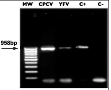
FIGURE 1 - Image of a 2% agarose gel stained with ethidium bromide showing the NS5 gene amplicon with universal primers for Flavivirus, FG1/FG2. Line 1: MW: 100bp molecular weight marker . Line 2: 958bp fragment corresponding to ampliication of the Cacipacore virus (CPCV) genome. Line 3: C+ positive control fragment of 958bp corresponding to ampliication of the yellow fever 17DD vaccinal strain genome. Line 4: C+: positive control using dengue virus 3 genome isolated in Porto Velho. Line 5: C-: negative control with non-infected monolayer of C6/36 tissue.
FIGURE 2 - Image of a 2% agarose gel stained with ethidium bromide showing the NS5 and 3’NCR amplicons using universal primers for Flavivirus, FLAV1/FLAV2. Line 1: MW: 50bp molecular weight marker of. Line 2: 800bp fragment corresponding to amplification of the Cacipacore virus (CPCV) genome. Line 3: 600bp positive control fragment corresponding to ampliication of the Yellow Fever virus (YFV) 17DD vaccinal strain genome. Line 4: C- negative control with Oropouche virus (OPOV-Orthobunyavirus). Line 5: C-: negative control with non-infected monolayer of C6/36 tissue.
Cacipacore virus BeAn 4073 NS5 gene, partial cds Length=274
Score = 272 bits (137), Expect = 1e-69
Identities = 242/264 (91%), Gaps = 11/264 (4%) Strand=Plus/Plus
Query 378 GGAATATGGTAGTATTAAGTTAAGCAAATAGATAAGGAAATTGTTAGAATAT-ATATGTT 436
|||||||||||||||||||||| |||| ||||||||||||| |||||||||| |||||||
CACVP 18 GGAATATGGTAGTATTAAGTTA-GCAA-TAGATAAGGAAAT-GTTAGAATATTATATGTT 74
Query 437 AGTTAAGAAGGTATTTG-TAGATTATATAGTATGGTTAAGTTAAGAATAAGGGATTTGTA 495 |||||||||| |||||| |||||||||||||||||||| ||| | || |||| ||||||
CACVP 75 AGTTAAGAAG-TATTTGGTAGATTATATAGTATGGTTA--TTACGTATTAGGGTTTTGTA 131
Query 496 TAAATCCAAATGTAAATAAGGGACAATTCTTATATTTAGTGGTTTAGAGACTTATTAGTC 555 ||||| |||||||||||| ||| |||| || ||||||||||||||||||||||||||||
CACVP 132 -AAATC-AAATGTAAATAAAGGAAAATTTTT-TATTTAGTGGTTTAGAGACTTATTAGTC 188
Query 556 AGGCCAGAACTGCCACCGGATGCTGGGTAAACGGTGCTGACTGTAACCTAGCCCCAGGAG 615 ||||||||| ||||||||||||||||||||||||||||| ||||||||||||||||||||
CACVP 189 AGGCCAGAAATGCCACCGGATGCTGGGTAAACGGTGCTGCCTGTAACCTAGCCCCAGGAG 248
Query 616 GACTGTGTGAACAAACTTACGTAA 639 ||||| |||||||||||| |||||
CACVP 249 GACTGGGTGAACAAACTTCCGTAA 272
terminator cycle sequencing kit in a mega BACE 1000 sequencer (Amersham Pharmacia Biotech, UsA).
Amplicons of 958bp and 800bp were obtained by RT-PCR using the genus-speciic, Flavivirus primer pairs FG1/FG2 and (Figure 1) FLAV1/FLAV2, respectively (Figure 2). Results revealed that the virus isolated from the patient showed 91% sequence similarity to other isolates of Cacipacore BeAn 4073when using BLAsT and the sequences available in GenBank (Figure 3).
530
Rev Soc Bras Med Trop 44(4):528-530, jul-ago, 2011
DISCUSSION
ACKNOWLEDGMENTS
REFERENCES
In Brazil, arboviruses (arthropod born viruses) have a wide geographic distribution and cause several diseases, thus constituting an important health problem. Eicient and continued epidemiological surveillance programs are necessary to monitor viral activity in regions where they are endemic or enzootic10. hese
programs should help to restrict viral spreading and reduce the socioeconomic impact caused by these diseases. To achieve this, fast, sensitive, speciic, and low-cost methods of diagnosing arboviral diseases are essential. he region in which the state of Rondônia is located is favorable to the dissemination of arboviruses because it possesses a large border with Bolivia and because the state is crossed by the BR 364 road, which links southern Brazil to the state of Acre, to Peru via the Inter-ocean road, and to the southern state of Amazonas (and thus to newly expanded agricultural frontiers). he totality of these conditions is favorable for the dissemination of arboviruses in the state of Rondônia.
he use of molecular methodologies, such as RT-PCR with universal Flavivirus primers, is very important because it allows rapid diagnosis of viral infections, including outbreaks of emergent and reemergent of Flaviviruses in Rondônia.
We are grateful to Prof. Dr. Luiz Tadeu Moraes Figueiredo and Victor Hugo Aquino (Center of Virology Research of the Faculty of Medicine of Ribeirão Preto - são Paulo University) whom we consulted with in many aspects of the RT-PCR technique, Christian Julián Villabona (Molecular Evolution Laboratory and Bioinformatics - são Paulo University) for providing language help, writing assistance and proof reading the article and inally, Dr. spartaco Astoli Filho (Multidisciplinary support Center of Federal Amazon University) for nucleotide sequencing of the Cacipacore virus.
1. Monath TP, Heinz FX. Flaviviruses. In: Fields BM, Kinipe PM, editors. Virology. Philadelphia: Lippincot-Raven Publishers; 1996. p. 961-1034.
2. Figueiredo LTM. he Brazilian laviviruses. Microbes Infectol 2000; 2:1-7. 3. Eldadah ZA, Asher DM, Godee Ms, Pomeroy KL, Goldfarb LG, Feinstone sM,
et al. Detection of flavivirus by reverse-transcriptase polymerase chain reaction. J Med Virol 1991; 33:260-267.
4. Figueiredo LTM, Batista WC, Kashima s, Nassar Es. Identiication of Brazilian laviviruses by a simpliied reverse transcription polymerase chain reaction method using Flavivirus universal primers. Am J Trop Med Hyg 1998; 59:357-362.
5. Kuno G. Universal diagnostic RT-PCR protocol for arboviroses. J Virol Methods 1998;72:27-41.
6. scaramozzino N, Crance JM, Jouan A, Debriel DA, stoll F, Garin D. Comparison of Flavivirus universal primer pairs and development of a rapid, highly sensitive heminested reverse transcription-PCR assay for detection of laviviruses targeted to a conserved region of the Ns5 gene sequences. J Clin Microbiol 2001; 39:1922-1927.
7. Tanaka M. Rapid identiication of lavivirus using the polymerase chain reaction. J Virol Methods 1993; 41:311-322.
8. Figueiredo LTM. Uso de células de Aedes albopictus C6/36 na propagacão e classiicaçãoo de arbovírus das famílias Togaviridae, Bunyaviridae, Flaviviridae e Rhabdoviridae. Rev soc Bras Med Trop 1990; 23:13-18.
9. Chomczynski P, sacchi N. singer-step method of RNA isolation d acid guanidinium thiocyanate-phenol-chroform extraction. Anal Biochem 1987; 162:156-159.