Immunophenotypic characterization of acute leukemia
at a public oncology reference center in Maranhão,
northeastern Brazil
Caracterização imunofenotípica das leucemias agudas em um centro
oncológico de referência público no Maranhão, Nordeste do Brasil
Elda Pereira Noronha
I, Heliana Trindade Marinho
I, Erika Bárbara Abreu Fonseca Thomaz
II, Cintia Assunção Silva
III, Geni
Lourdes Ramos Veras
IV, Raimundo Antônio Gomes Oliveira
VClinical Research Center, University Hospital, Universidade Federal do Maranhão (UFMA), and Instituto Maranhense de Oncologia
Aldenora Bello (IMOAB), São Luís, Maranhão, Brazil
ABSTRACT
CONTEXT AND OBJECTIVES: The incidence of acute leukemia (AL) subtypes varies according to geographical distribution. The aim here was to determine the incidence of morphological and immunophenotypic AL subtypes in the state of Maranhão, Brazil, and to correlate the expression of aberrant phenotypes in children with acute lymphoblastic leukemia (ALL) with prognostic factors.
DESIGN AND SETTING: Single prospective cohort study at a public oncology reference center in Maranhão.
METHODS: Seventy AL cases were diagnosed between September 2008 and January 2010. For the diagnosis, complete blood cell counts, myelograms (at diagnosis and at the end of the induction phase), cytochemical analy-sis and immunophenotyping were performed.
RESULTS: Among adult patients (n = 22), the incidence of AL types was: ALL (22.7%) and acute myeloid leukemia (AML) (77.3%). The subtype AML M0 occurred most frequently (29.4%). In children (n = 48), the types were: AML (18.7%), most frequently subtype AML M4 (33.4%); biphenotypic acute leukemia (BAL) (4.2%); and ALL (77.1%), including the subtypes B-ALL (72.9%) and T-ALL (27.1%). Among the children with ALL, there were no statistically signiicant diferences between patients with and without aberrant phenotypes, in relation to hematological pa-rameters and treatment response.
CONCLUSION: This work demonstrates that the frequencies of AML M0 cases among adults and T-ALL cases among children in Maranhão were high. This suggests that there may be diferences in AML subtype incidence, as seen with ALL subtypes, in diferent regions of Brazil. No association was found between the expression of aberrant phenotypes and prognostic factors, in children with ALL.
RESUMO
CONTEXTO E OBJETIVOS: A incidência dos subtipos de leucemias agudas (LA) tem mostrado variações em rela-ção à distribuirela-ção geográica. Objetivou-se determinar a incidência dos subtipos morfológicos e imunofenotípicos de LA no estado do Maranhão, Brasil, e relacionar a expressão de fenótipos aberrantes em crianças com leucemia linfoblástica aguda (LLA) com fatores prognósticos.
TIPO DE ESTUDO E LOCAL: Estudo de coorte único prospectivo em um centro oncológico de referência público no Maranhão.
MÉTODOS: Diagnosticaram-se 70 casos de LA no período de setembro de 2008 a janeiro de 2010. No diagnóstico, realizaram-se hemogramas, mielogramas (no diagnóstico e ao inal da fase de indução), citoquímica e imunofe-notipagem.
RESULTADOS: Nos pacientes adultos (n = 22), as incidências dos tipos de LA foram: LLA (22,7%) e leucemia mieloi-de aguda (LMA) (77,3%), sendo o subtipo LMA M0 mais frequente (29,4%). Em crianças (n = 48): LMA (18,7%), sub-tipo LMA M4 mais frequente (33,4%), leucemia bifenotípica aguda (BAL) (4,2%) e LLA (77,1%), sendo os subsub-tipos LLA-B (72,9%) e LLA-T (27,1%). Na LLA, em crianças, não se encontrou diferença estatisticamente signiicante entre pacientes com e sem fenótipos aberrantes, em relação aos parâmetros hematológicos e resposta ao tratamento.
CONCLUSÃO: Esta pesquisa demonstra elevada frequência de casos de LMA M0 em adultos, bem como das LLA-T em crianças no Maranhão, sugerindo que podem haver diferenças na incidência dos subtipos das LMA, assim como dos subtipos de LLA, em diferentes regiões do Brasil. Não foi encontrada associação entre a expressão de fenótipos aberrantes e fatores prognósticos em crianças com LLA.
IMSc. Pharmacist in the Masters’ degree program
on Mother and Child Health, Universidade Federal do Maranhão (UFMA), and Clinical Research Center, University Hospital, UFMA, São Luís, Maranhão, Brazil.
IIPhD. Professor, Department of Public Health,
Universidade Federal do Maranhão (UFMA), São Luís, Maranhão, Brazil.
IIIMD. Pediatric Oncologist, Instituto Maranhense
de Oncologia Aldenora Bello (IMOAB), São Luís, Maranhão, Brazil.
IVMSc. Pediatric Oncologist, Instituto Maranhense
de Oncologia Aldenora Bello (IMOAB), São Luís, Maranhão, Brazil.
VPhD. Professor, Department of Pharmacy,
Universidade Federal do Maranhão (UFMA), and Pharmacist, Clinical Research Center, University Hospital, UFMA, São Luís, Maranhão, Brazil.
KEY WORDS:
Precursor cell lymphoblastic leukemia-lymphoma.
Leukemia, myeloid, acute. Prevalence.
Immunophenotyping. Prognosis.
PALAVRAS-CHAVE:
Leucemia-linfoma linfoblástico de células precursoras.
Leucemia mielóide aguda. Prevalência.
INTRODUCTION
Acute leukemias comprise a heterogeneous group of diseases characterized by rapid and uncontrolled clonal expansion of progenitor cells of the hematopoietic system.1 hey are the most
common form of childhood neoplasia, and acute lymphoblastic leukemia (ALL) represents 75% of all such cases. his percent-age is much lower in adults, in whom acute myeloid leukemias (AMLs) are more common. In children, the vast majority of ALL cases (80%–85%) are of precursor B-lineage and about 15% of all cases are of T-lineage.2,3 A small number of patients whose
blasts simultaneously present antigens of the myeloid and lym-phoid lineages are characterized as carriers of mixed, hybrid or biphenotypic acute leukemias (BALs).4,5
AML and ALL in which the blasts contain one or two anti-gens of another lineage, but do not meet the criteria for BAL, are known respectively as acute myeloid leukemias with anomalous lymphoid expression and acute lymphoblastic leukemias with anomalous myeloid expression.6,7 Occurrence of aberrant
phe-notypes (or anomalous expression) are reported to have variable frequency and their prognostic value is controversial.8
Early classiication systems for acute leukemias were based only on cytomorphological and cytochemical investigations. Morphology still plays a central role, but current classiication systems have incorporated immunophenotyping in order to achieve greater precision in delineating the hematopoietic lin-eage and diferentiation stage of particular leukemias. Immu-nophenotyping is fundamental for classifying lymphoid malig-nancies and is also essential for recognizing several subtypes of acute myeloid leukemia (e.g. AML-M0 and AML-M7) and biphenotypic acute leukemias; for monitoring the responses to treatment, including detection of minimal residual disease (MRD); and for identifying markers with prognostic implica-tion. he current World Health Organization (WHO) classii-cation of tumors of hematopoietic and lymphoid tissues incor-porates not only immunophenotyping but also cytogenetic and molecular characteristics that contribute towards deining bio-logically and clinically relevant leukemia subsets. However, it is neither necessary nor cost-efective to perform multiple studies on every specimen.9-11
Analysis of the incidence of leukemia subtypes across the world has revealed important variations in relation to geograph-ical distribution, sex, age, ethnicity and socioeconomic status, thus suggesting that several etiological factors exist.12,13
here-fore, further studies on the frequencies of diferent subtypes of acute leukemias are of great importance, especially in regions with diferent socioeconomic characteristics such as the north-east of Brazil, where such data are scarce.
Unfortunately, before our group created an immunopheno-typing service for the state of Maranhão, acute leukemias were not diagnosed through immunological markers, except in rare
cases of patients whose relatives had the inancial means to send their samples to a more advanced center. As a result, the vast majority of such patients were treated and given a prognosis on the basis of morphological and clinical indings alone, which may sometimes blur the choice of the most appropriate treatment.
OBJECTIVES
he aim of the present study was to determine the incidence of diferent morphological and immunophenotypic subtypes of acute leukemia among patients referred to an oncology reference center in the state of Maranhão for treatment of acute leukemias, and to correlate the expression of aberrant phenotypes in chil-dren with acute lymphoblastic leukemia (ALL) with prognostic factors.
METHODS
Sample and setting
he present investigation was approved by the Research Ethics Committee of the University Hospital of Universidade Federal do Maranhão (UFMA) (number 115/2008). It included all consecu-tive patients who had been referred to the Oncology Reference Center during the study period, consisting of 73 adults (≥ 18 years) and children (< 18 years). The Oncology Reference Center is a public institution in São Luís, Maranhão, Brazil. They were diagnosed with de novo acute leukemia between September 2008 and January 2010. The morphological and immunophe-notypic evaluations on the samples were performed at the Clinical Research Center of the University Hospital, Universi-dade Federal do Maranhão (UFMA). Patients who refused to take part or whose legal guardians did not allow their partici-pation (n = 1) and cases of blast crisis chronic myeloid leuke-mia (n = 2) were excluded, thus resulting in 70 participants in this study. The following patient data were gathered: sex, age and occurrences of deaths of children with ALL before the end of the induction phase.
We evaluated the association between prognostic factors (white blood cell count, platelet count, hemoglobin level, periph-eral blast percentage and response to treatment) and expression of aberrant phenotypes in children with ALL. his association could not be analyzed in cases of children (n = 9) and adults (n = 17) with AML, or among adults with ALL (n = 5), due to the small sample size.
Diagnosis
May-Grünwald-Giemsa. he analysis was performed by two morphologists and a diagnosis of acute leukemia was made when the bone mar-row blast count was greater than 20%. he myeloperoxidase cytochemical test was considered positive when 3% or more of the blasts showed positive staining granules. he periodic acid-Schif (PAS) test was considered positive when the blasts showed granular block and partial or complete ring-type PAS staining. For alpha-naphthyl acetate esterase, positive indings in more than 20% of the blasts deined the types AML-M4 or M5. AML was classiied in accordance with the French-American-British (FAB) criteria.14-16 he classiications of ALL and BAL were made
based on the criteria of the European Group for the Immunologi-cal Characterization of Leukemias (EGIL).6 Only the
morpholog-ical and immunophenotypic criteria for leukemia diagnosis were used in the present study, and cytogenetic data that are taken into considered by WHO were not used because this service is still unavailable in our state.
Samples of peripheral blood (n = 12) or bone marrow (n = 58) were collected in ethylenediaminetetraacetic acid (EDTA) for immunophenotyping. he samples were processed within four hours of collection.
For the immunophenotypic diagnosis of acute leukemias, a combination of two or three luorochrome-conjugated mono-clonal antibodies (MoAb) per tube was added to the samples. All the MoAbs were obtained from Becton Dickinson (San José, California, United States). hey were conjugated with luores-cein isothiocyanate (FITC), phycoerythrin (PE) or peridinin chlorophyll protein (PerCP), and were directed to antigens for T cells (CD1a, CD2, cytoplasmic (c) CD3, CD4, CD5, CD7 and CD8), B cells (CD10, CD19, cCD22, cCD79a, supericial and cIgM), myeloid cells [CD13, CD33, CD117 and myeloperoxidase (MPO)], monocytes (CD14 and CD64), erythroid cells (alpha-glycophorin), platelet cells (CD61 and CD41a), non-speciic lin-eage pan-leukocytes (CD45) and precursor cells [CD34, human leukocyte antigen-DR (HLA-DR) and terminal deoxynucleotidyl transferase (TdT)].
Membrane and intracytoplasmic labeling was performed using 1 x 10⁶ cells per tube. For membrane labeling, the samples
were incubated with each antibody for 20 minutes. he eryth-rocytes were lysed with 2 ml of FACS lysing solution (Becton Dickinson, San José, California, United States), diluted to 1:10 and then washed with 2 ml of phosphate-bufered saline (PBS; pH = 7.4). For intracytoplasmic labeling, FACS permeabilizing solution (Becton Dickinson, San José) was used in accordance with the manufacturer’s instructions.
Data acquisition and sample analysis was performed in a FACSCalibur low cytometer (Becton Dickinson, San José), using the CellQuest sotware (Becton Dickinson, San José), ater cali-bration with the Calibrite bead kit (Becton Dickinson, San José), using the FACSComp program (Becton Dickinson, San José).
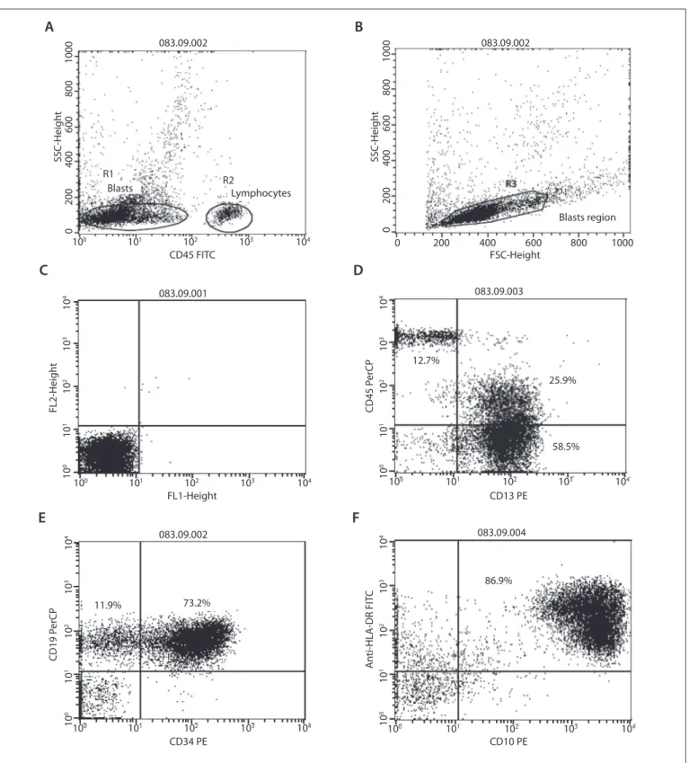
Lymphocyte labeling with CD4 FITC/CD8 PE/CD3 PerCP was used, to compensate for luorescence and eliminate overlap-ping before data acquisition. he blast gating strategy included using dot plots of CD45 expression versus intracellular com-plexity (side scatter angle, SSC) (Figure 1a) and also a second gate considering cell size (forward scatter angle, FSC) versus SSC (Figure 1b). A total of 10,000 events were acquired in the target gate. Negative controls using isotype IgG1 and IgG2a monoclo-nal antibodies were run in all cases. he criteria used for deter-mining antigen positivity included analysis of negative controls (Figure 1c) and expression of the marker by more than 20% of the gated cells (Figure 1d, 1e and 1f). Similarly, aberrant phe-notypes (Figure 1d) were deined when at least 20% of the blast cells expressed the particular aberrant marker. In ALL, aberrant expression of CD33 and CD13 was analyzed while in AML, aber-rant expression of CD2, CD7 and CD19 was analyzed.
Chemotherapy for children with ALL
Patients with ALL aged under 18 were treated in accordance with the protocol of the Brazilian Group for the Treatment of Childhood Leukemias (Grupo Brasileiro de Tratamento das Leucemias Infantis, GBTLI/9917). Patients with B-ALL
were treated during the induction phase with dexamethasone (6 mg/m2/day), three doses per day for four weeks; vincristine
(1.5 mg/m2/week) and daunorubicin (25 mg/m2/week) on days
0, 7, 14 and 21; L-asparaginase (5000 IU/m2/day), nine doses
beginning between days 3 and 5 and administered three times per week; and MADIT (combination of methotrexate, cytar-abine and dexamethasone administered intrathecally) with doses adjusted according to the patient’s age and administered on days 0, 14 and 28.17 Patients with T-ALL were treated with
the same drugs described above, although with the following modifications: daunorubicin (35 mg/m2/dose) on days 0, 28
and 42, and methotrexate (1 g/m2/dose) on days 7 and 21.
Remission criteria among children with ALL
he response to treatment among children with ALL was eval-uated by means of a bone marrow aspirate smear at the end of the induction phase and the presence of less than 5% blasts in the bone marrow was considered to be a criterion for remission. Patients who died before the end of the induction (n = 10) were considered to be patients who did not go into remission.
Statistical analysis
083.09.002 083.09.002
083.09.002 083.09.004
083.09.001 083.09.003
1000
800
600
400
200
0
1000 800
600 400
200 0
1000
800
600
400
200
0
R3
R1 R2
Blasts region Blasts
FSC-Height
SSC-Heigh
t
SSC-Heigh
t
FL2-Heigh
t
FL1-Height
Lymphocytes
CD45 FITC
100 101 102 103 104
100 101 102 103 104
10
0
10
1
10
2
10
3
10
4
CD45 P
erCP
CD13 PE
100 101 102 103 104
10
0
10
1
10
2
10
3
10
4
A
n
ti-HLA
-DR FIT
C
CD10 PE CD34 PE
101
100 102 103 104
10
0
10
1
10
2
10
3
10
4
101
100 102 103 104
10
0
10
1
10
2
10
3
10
4
12.7%
25.9%
58.5%
86.9%
CD19 P
erCP
11.9% 73.2%
Figure 1. Immunophenotyping of B-ALL with aberrant antigen expression of CD13 in a patient at the Oncology Reference Center, São Luís, Maranhão. a) Dot plot of CD45 expression versus intracellular complexity (side scatter angle, SSC), differentiating the blast region (R1), with partial loss of CD45 expression and lymphocyte region (R2); b) Blast region (R3) in dot plot considering cell size (forward scatter angle, FSC) versus SSC; c) Analysis on negative controls (isotype IgG1 and IgG2a monoclonal antibodies); d) Positivity for the myeloid marker CD13 (25.9% + 58.5% = 84.4%) and for CD45 (25.9%); e) Positivity for the immature cell marker CD34 (73.2%) and for the B cell marker CD19 (73.2%); f ) Positivity for the immature cell marker HLA-DR (86.9%) and for the B cell marker CD10 (86.9%).
A
C
E
B
D
using the Mann-Whitney test. he response to induction and the frequency of aberrant phenotypes compared between B-ALL and T-ALL, as well as between ALL and AML, were assessed by applying Fisher’s exact test and the chi-square test. he signii-cance level was taken to be 5% (P < 0.05) in all tests.
RESULTS
A total of 70 patients with de novo acute leukemia were studied. Table 1 shows the incidence of the types of leukemias in the pop-ulation studied. ALL was more frequent in children (77.1%) and AML was more common in adults (77.3%). BAL accounted for 2.9% of the cases of acute leukemias.
B-ALL was found in 72.9% (27/37) of the cases of ALL in chil-dren, while T-ALL accounted for 27.1% (10/37) of the cases. he
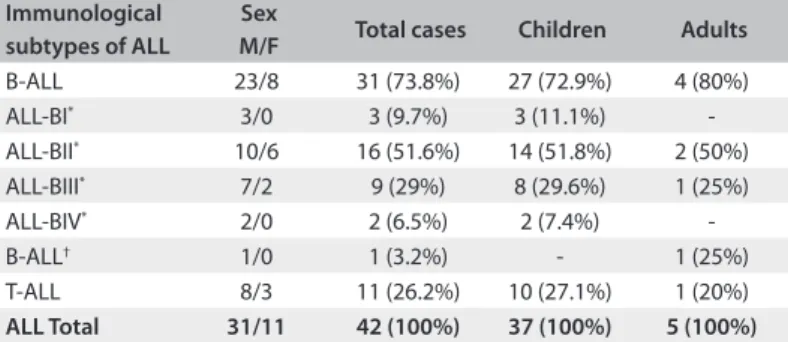
immunological subtype ALL-BII (common B) was the most fre-quent type (51.6%; 16/31) among the cases of B-ALL (Table 2).
he median age of the children with B-ALL was four years (range from one to 16 years), while that of children with T-ALL was eight years (range from two to 17 years). he peak incidence of ALL in children occurred between the ages of one and four years, representing 40.5% (22/37) of the cases of ALL diagnosed in children. Among the adults, the median age of those with ALL was 24 years (range from 19 to 56 years). Among the patients under the age of 18 years with ALL, the male-to-female ratio was 3.1:1.0. For B-ALL and T-ALL considered separately, the ratios were 3.5:1.0 and 2.3:1.0, respectively.
he hematological characteristics of the subtypes of ALL are presented in Table 3. It can be seen that T-ALL presented higher white cell counts, with a median greater than 50x 109/l.
Regard-ing the hemoglobin level, ALL-BI (pro-B) presented a lower median (6.4 g/dl). All of the subtypes exhibited thrombocytope-nia, with a median of less than 50 x 109 platelets/l.
Among the markers used to characterize B-ALL in cyto-plasm, CD79a and CD22 presented the highest positivity rate (100%), followed by CD19 in the cell membrane (86.6%). CD10 and the immature cell marker HLA-DR presented positivity rates of 83.3% and 95%, respectively. he marker IgM in cyto-plasm (cyt IgM) had a positivity rate of 33% and was positive in ten cases, which were classiied as BIII-ALL (pre-B). CD34, an immature cell marker, was expressed in 61.2% of the cases and CD45 in 87%. Of the myeloid markers expressed anomalously, CD13 was the most frequent (48.2%).
In the immunophenotypic characterization of T-ALL, the markers CD3 (cytoplasm), CD5, CD7 and CD8 exhibited pos-itivity rates of 100%. The markers CD2, CD1a, CD4, HLA-DR and CD10 showed positivity rates of 85.7%, 57.1%, 40%, 10% and 9.1%, respectively. CD34 was expressed in 54.5% of the cases and CD45 (with moderate to weak expression) was pres-ent in all cases. CD13 was the most frequpres-ent anomalous myel-oid marker (36.3%).
here were aberrant phenotypes in 48.4% of the cases of B-ALL (15/31) and 36.4% (4/11) of the cases of T-ALL. here was no statistically signiicant diference between B-ALL and T-ALL in relation to the frequency of aberrant phenotypes (Table 4).
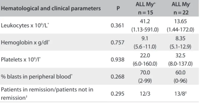
No statistically signiicant diference was found in relation to prognostic factors (white blood cell count, platelet count, hemo-globin level, peripheral blast percentage and response to induc-tion phase) in ALL in children with and without aberrant phe-notypes (Table 5), although the number of patients who went into remission was higher in the group with aberrant phenotypes (ALL My+) (80%), compared with the group without aberrant
phenotypes (ALL My-) (62%).
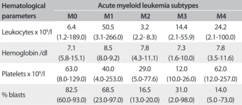
he distribution of AML according to the FAB criteria revealed that the AML M4 subtype (3/9; 33.4%) was the one most
Table 1. Frequency of each type of acute leukemia, according to age group, among patients at the Oncology Reference Center, São Luís, Maranhão
Type of acute leukemia Total cases
n = 70
Children n = 48 (68.6%)
Adults n = 22 (31.4%)
Acute lymphoblastic leukemia 42 (60%) 37 (77.1%) 5 (22.8%)
Acute myeloid leukemia 26 (37.1%) 9 (18.7%) 17 (77.3%)
Biphenotypic acute leukemia 2 (2.9%) 2 (4.2%)
-Table 2. Distribution of acute lymphoblastic leukemia (ALL) subtypes and patients’ sex at the Oncology Reference Center, São Luís, Maranhão
Immunological subtypes of ALL
Sex
M/F Total cases Children Adults
B-ALL 23/8 31 (73.8%) 27 (72.9%) 4 (80%)
ALL-BI* 3/0 3 (9.7%) 3 (11.1%)
-ALL-BII* 10/6 16 (51.6%) 14 (51.8%) 2 (50%)
ALL-BIII* 7/2 9 (29%) 8 (29.6%) 1 (25%)
ALL-BIV* 2/0 2 (6.5%) 2 (7.4%)
-B-ALL† 1/0 1 (3.2%) - 1 (25%)
T-ALL 8/3 11 (26.2%) 10 (27.1%) 1 (20%)
ALL Total 31/11 42 (100%) 37 (100%) 5 (100%)
M = male; F = female; *The value refers to the percentage of each subtype, calculated
from the total number of cases of ALL-B in each age group; †One case could not
be subclassiied in terms of the phase of maturation due to scarcity of material for evaluation.
Table 3. Hematological parameters of the complete blood cell counts of acute lymphoblastic leukemia (ALL) subtypes on diagnosis, among patients at the Oncology Reference Center, São Luís, Maranhão
Median values (range).
Hematological parameters
ALL subtypes
ALL-BI ALL-BII ALL-BIII ALL-BIV T-ALL
Leukocytes x 109/l
23.2 (2.35-102)
19.5 (1.44-157)
3.1 (1.13-135)
4.6 (2.3-6.9)
81.0 (9.1-591)
Hemoglobin g/dl 6.4
(6.0-6.6) 8.3 (3.4-12.9)
8.8 (7.0-10.9)
11 (9.3-12.7)
9.4 (6.5-12.5)
Platelets x 109/l 12.0
(12-13)
18.0 (6.0-111)
45.0 (22-160)
33.5 (29-38.1)
39.0 (14-157)
% blasts 70.0
(46-96) 65 (0-97)
21.0 (0-99)
5.0 (4-6)
frequently occurring in children. In the adults, the predominant subtype was AML-M0 (5/17; 29.4%) (Table 6).
he adult patients presented a median age of 35 years (range: 19-67 years), while the median age among the children was 8 years (range: 1-15 years). he ratio of males to females with AML was 1.3:1.
Table 7 shows the hematological characteristics of the AML subtypes. It can be seen that AML-M1 presented a higher white blood cell count with a median of 50.5x 10⁹/l. In relation to the
hemoglobin level, the subtypes of leukemias presented medi-ans with very similar values, varying between 7.1 g/dl (AML- M0) and 8.5 g/dl (AML-M1). All of the subtypes exhibited thrombocytopenia.
Among the markers used to characterize AML, CD117 and CD13 exhibited the highest positivity rates (100%), followed by CD33 (96.1%). MPO presented a positivity rate of 73.9%, while CD14 and CD64 presented positivity rates of 29.1% and 33.3%, respectively. he immature cell markers CD34, HLA-DR and CD117 had positivity rates of 69.2%, 63% and 100% respectively. Out of the ive cases diagnosed with AML-M3, only one expressed CD34, and none expressed HLA-DR. CD45 was expressed in 92.3% of the cases.
here were aberrant phenotypes in 27% of the diagnosed cases, and expression of CD7 occurred most frequently (19.2%). here was no statistically signiicant diference between ALL and AML in relation to the frequency of aberrant phenotypes (Table 8).
DISCUSSION
his work constitutes the irst study to be carried out in the state of Maranhão involving characterization of the immunopheno-typic proile of cases of acute leukemias, and thereby determin-ing the various immunological subtypes of these pathological conditions. he majority of ALL cases (88.1%) examined were diagnosed in children, and this matches the epidemiological data already described, which showed much lower frequencies of this neoplasia in adults.18,19 In the present investigation, the cases of
ALL in adults represented just 22.7% of all the cases of acute leu-kemias in this age group.
he incidence of ALL is higher among men than among women, independent of the age group analyzed. In a study involv-ing children from the states of Rio de Janeiro and Bahia and the Federal District, the ratio of males to females was 1.2:1.13 In the
state of Pernambuco, this ratio was 1.7:1,20 while in Ribeirão Preto
(city in the state of São Paulo), it was 1.8:1 for all subtypes of ALL, with an even higher predominance of males for T-ALL, at 4.2:1.21
In our work, for all subtypes of ALL, we found a higher proportion of males (3.1:1) than what has been described in other Brazilian state of, with a higher male-to-female ratio for B-ALL (3.5:1) than for T-ALL (2.3:1).
In relation to the subtypes of ALL, the present study revealed that 73.8% of the cases were classiied as B-ALL and 26.2% of the cases as T-ALL. Among the B-ALL subtypes, ALL-BII (common B) occurred most frequently. hese results are similar to those of other studies, except for the high frequency of T-ALL in children. In the population of Maranhão, the chil-dren presented a higher frequency of T-ALL (27%) than what is generally described in the literature (7.3% to 16%).13,21,22
Nev-ertheless, in another study involving children, Bachir et al.23
Table 4. Frequency of aberrant phenotypes in B-ALL and T-ALL patients at the Oncology Reference Center, São Luís, Maranhão
Subtype of ALL Aberrant phenotypes Total
Absent Present
B-ALL 16 (51.6%) 15 (48.4%) 31
T-ALL 7 (63.6%) 4 (36.4%) 11
Total 23 19 42
ALL = acute lymphoblastic leukemia; Fisher’s exact test was applied; P = 0.726.
Table 5. Assessment of aberrant phenotypes in acute lymphoblastic leukemia (ALL), in relation to hematological parameters and response to induction, among children treated at the Oncology Reference Center, São Luís, Maranhão
Hematological and clinical parameters P ALL My
+ n = 15
ALL My -n = 22
Leukocytes x 109/L* 0.361 41.2
(1.13-591.0)
13.65 (1.44-172.0)
Hemoglobin x g/dl* 0.757 9.1
(5.6 -11.0)
8.35 (5.1-12.9)
Platelets x 109/l* 0.938 22.0
(6.0-160.0)
32.5 (8.0-137.0)
% blasts in peripheral blood* 0.268 70.0
(2-99)
60.0 (0-96) Patients in remission/patients not in
remission† 0.295 12/3 13/8
‡
ALL My+ = acute lymphoblastic leukemiawith aberrant phenotypes; ALL My- =
acute lymphoid leukemiawithout aberrant phenotypes; hematological parameters = values expressed as medians (range); *Mann-Whitney test applied; †Fisher’s exact
test applied; ‡one patient abandoned the treatment before the end of induction
and was excluded from this analysis.
FAB subtypes of AML
Sex
M/F Total cases Children Adults
AML-M0 5/1 6 (23.1%) 1 (11.1%) 5 (29.4%)
AML-M1 0/4 4 (15.4%) 2 (22.2%) 2 (11.8%)
AML-M2 3/2 5 (19.2%) 1 (11.1%) 4 (23.5%)
AML-M3 1/4 5 (19.2%) 1 (11.1%) 4 (23.5%)
AML-M4 5/0 5 (19.2%) 3 (33.4%) 2 (11.8%)
AML-M6 1/0 1 (3.9%) 1 (11.1%)
-Total 15/11 26 (100%) 9 (100%) 17 (100%)
M = male; F = female.
observed a frequency of 21.1% for T-ALL in Morocco, while in a study carried out in India by Rajalekshmy et al.,24 it was
found that the frequency of T-ALL in children was 45.9%. In Recife (Brazilian state of Pernambuco), this frequency was 18.5%,20 similar to what was found in the state of Minas Gerais,
Brazil (18%).25
A high frequency of T-ALL is associated with poor socio-economic conditions, as demonstrated by research conducted in Brazil, which found a direct association between poor socio-economic status (low per capita income) and the T phenotype.25
Another factor associated with increased frequency of T-ALL is ethnic origin, since the T phenotype has been shown to be more frequent among nonwhites.13 Taking into consideration
that the state of Maranhão is the second poorest in Brazil,26
and that the population is predominantly nonwhite,27 this
may explain the high incidence of T-ALL found in the pres-ent investigation.
In relation to AML, our results show that this disease was much more frequent among adults (65.4%) than in children (34.6%), and this inding is in agreement with reports in the literature.28
With regard to another aspect of our indings, taking into consideration all of the patients, our results indicate a much lower frequency of AML (37.1%) than of ALL (60%). A similar result was described by Rego et al.,18 who found that in the state
of Piauí, the frequency of AML was half that of ALL.
In relation to the age of the patients with AML, the median obtained for the population of Maranhão was lower than that of patients with AML in developed countries,29,30 but was similar to
that observed in other studies conducted in Brazil.18,31,32 his can
be explained by the fact that, in developed countries, life expec-tancy is higher and elderly individuals constitute a greater pro-portion of the population. his difers from Brazil, especially in the northeast of the country, where life expectancy is low and, consequently, the elderly population is much smaller, as reported by Rego et al.18 Since the highest incidence of AML is seen in
peo-ple over 60 years of age,28 this may explain the low incidence of
the disease in the population of Maranhão.
Analysis on the distribution of the FAB subtypes revealed pre-dominance of AML- M4 followed by subtype AML-M1 in chil-dren. his result is similar to that found in research performed in São Paulo,33 while a higher frequency of subtypes M2 and M3
was reported in Minas Gerais.34 In contrast, work by the
Berlin-Frankfurt-Münster (BFM) group in Germany showed higher fre-quency of subtypes M4 and M5.35
Among the adults, the most frequent subtype was M0, fol-lowed by subtypes M2 and M3, which presented equal frequency. Aside from the M0 subtype, which is considered to be a less com-mon form of leukemia comprising approximately 2% to 3% of myeloid leukemias,36 the M2 and M3 subtypes are very frequent
in the Brazilian population.18,31,32,37 According to Rego et al.,18
the distribution of FAB subtypes is irregular, showing large geo-graphical variations possibly as a result of ethnic and environ-mental factors.
Other studies carried out in Brazil have indicated difer-ences with regard to the distribution of morphological subtypes of acute leukemias. In comparison with rates in Campinas (state of São Paulo) and Teresina (state of Piauí), it was found that the most common subtype in Teresina was M2, followed by M4 and M5 with equal frequencies, while in Campinas there was higher frequency of the M4 subtype followed by the M3 subtype.18 In
São José dos Campos (state of São Paulo), there was higher preva-lence of the M1 subtype, followed by M2.37 In Rio de Janeiro, there
was higher frequency of M2, followed by M3 and M4, which were equally frequent,31 while in Rio Grande do Sul, higher frequency
of M2 was found, followed by M1.32
In the present study, the increased incidence of the FAB sub-type M0 was based on samples obtained from a single institution. It should be emphasized that the patients studied may not be rep-resentative of the whole region. Similar problems have afected other studies.31,32,37 In addition, the majority of the studies18,31,32
that have described the distribution of FAB morphological sub-types were retrospective, carried out by analyzing the medical records, many of which do not include immunophenotyping as a diagnostic technique for AML. In other cases, the immu-nophenotypic diagnosis was introduced a long time ater the
Values expressed as medians (range).
Table 7. Hematological parameters of the complete blood cell counts for acute myeloid leukemia subtypes among patients at the Oncology Reference Center, São Luís, Maranhão
Hematological parameters
Acute myeloid leukemia subtypes
M0 M1 M2 M3 M4
Leukocytes x 109/l 6.4
(1.2-189.0) 50.5 (3.1-266.0)
3.2 (2.2- 8.3)
14.4 (2.1-55.9)
24.2 (2.1-100.0)
Hemoglobin /dl 7.1
(5.8-15.1) 8.5 (8.0-9.2)
7.8 (4.3-11.1)
7.3 (1.6-10.0)
7.8 (3.5-11.6)
Platelets x 109/l 63.0
(8.0-129.0) 40.0 (4.0-253.0)
29.0 (5.0-77.6)
12.0 (10.0-26.0)
62.0 (12.0-257.0)
% blasts 82.5
(60.0-93.0) 68.5 (23.0-97.0)
16.5 (13.0-20.0)
31.0 (2.0-98.0)
14.0 (5.0 -73.0)
Table 8. Frequency of aberrant phenotypes in acute lymphoblastic leukemia and acute myeloid leukemia among patients at the Oncology Reference Center, São Luís, Maranhão
The chi-square test was applied; P = 0.199.
ALL subtype Aberrant phenotypes Total
Absent Present
Acute lymphoblastic leukemia 23 (54.7%) 19 (45.3%) 42
Acute myeloid leukemia 19 (73%) 7 (27%) 26
beginning of data collection, which probably means that the fre-quency of FAB M0 subtypes in the Brazilian population has been underestimated.
Numerous studies in developed countries, which used not only morphology and cytochemistry but also immunopheno-typing, found relatively higher frequencies of the M0 subtypes among AML cases, as reported for example by Suárez et al.38 in
Europe, Kaleem et al.39 in the United States and Chang et al.29
in Canada, who found frequencies of 15%, 17.6% and 9.5%, respectively.
he frequency of abnormal expression of an antigen from a given lineage in another lineage (aberrant phenotypes) is very variable in acute leukemias.8 In our study, 45.2% of the cases
of ALL exhibited aberrant phenotypes, and the most frequent marker was CD13. his was similar to the results of Putti et al.,40
Den Boer et al.41 and Bachir et al.23 he frequency of myeloid
coexpression was higher in B-ALL cases (48.4%) than in T-ALL cases (36.4%), although the diference was not signiicant. Den Boer et al.41 and Abdelhaleem42 also found a higher frequency
in B-ALL cases, but only the latter found the diference to be signiicant. In AML cases, there was anomalous expression in 26.9% of them, and the most frequent marker was CD7, thus conirming the indings of Zheng et al.43
he association between prognostic factors and aberrant phenotypes in ALL in children remains controversial. Putti et al.40 and Pui et al.44 did not ind any signiicant association
with adverse prognostic factors. Riley et al.45 stated that
aber-rant phenotypes in ALL, both in adults and in children, were sig-niicantly associated with short duration of event-free survival, short duration of irst remission and high relapse rates within the treatment phase.
In our analysis of cases of ALL in individuals under the age of 18 years, there was no diference between patients with and without aberrant phenotypes in relation to prognostic factors, although the number of patients who achieved remission was greater in the group with anomalous expression. his is similar to the results found by Bhushan et al.8
he literature shows great variation of data in relation to the frequency of anomalous expression in acute leukemia cases, and this may explain the lack of consistency with regard to the prog-nostic value of aberrant phenotypes in acute leukemia. Such vari-ation may have multiple causes, including the use of diferent luorochromes, the use of diferent clones of monoclonal anti-bodies, the sample characteristics, the technique and form of analysis used, the numbers and characteristics of patients, and the treatment protocol adopted.
CONCLUSION
he use of immunophenotyping in our study made it possi-ble to diagnose cases of minimally diferentiated acute myeloid
leukemia (AML-M0) and to diferentiate B-ALL from T-ALL. he study showed that T-ALL and AML-M0 occurred at higher frequency in the population studied, thus suggestingthat there may be diferences in the incidence of the FAB subtypes of AML, as well as in the subtypes of ALL, in diferent regions of Brazil. Furthermore, in all of these cases, the lack of immunophenotypic analysis could have compromised the diagnosis and, as a result, compromised the choice of the most appropriate treatment. We did not ind any association between aberrant phenotypes and the prognostic factors and clinical outcomes. he evaluations on the parameters examined in this work, such as aberrant pheno-types, leukemia subtypes and patient survival, can be improved; however, this requires longer duration of observation, and this should be borne in mind in future investigations.
REFERENCES
1. McCulloch EA. Stem cells in normal and leukemic hemopoiesis
(Henry Stratton Lecture, 1982). Blood. 1983;62(1):1-13.
2. Onciu M. Acute lymphoblastic leukemia. Hematol Oncol Clin North
Am. 2009;23(4):655-74.
3. Pui CH, Relling MV, Downing JR. Acute lymphoblastic leukemia. N
Engl J Med. 2004;350(15):1535-48.
4. Killick S, Matutes E, Powles RL, et al. Outcome of biphenotypic acute
leukemia. Haematologica. 1999;84(8):699-706.
5. Lee PS, Lin CN, Liu C, Huang CT, Hwang WS. Acute leukemia with
myeloid, B-, and natural killer cell diferentiation. Arch Pathol Lab
Med. 2003;127(2):E93-5.
6. Bene MC, Castoldi G, Knapp W, et al. Proposals for the immunological
classiication of acute leukemias. European Group for the
Immunological Characterization of Leukemias (EGIL). Leukemia.
1995;9(10):1783-6.
7. The value of c-kit in the diagnosis of biphenotypic acute leukemia.
EGIL (European Group for the Immunological Classiication of
Leukaemias). Leukemia. 1998;12(12):2038.
8. Bhushan B, Chauhan PS, SalujaS, et al. Aberrant phenotypes in
childhood and adult acute leukemia and its association with
adverse prognostic factors and clinical outcome. Clin Exp Med. 2010;
10(1):33-40.
9. Szczepański T, van der Velden VH, van Dongen JJ. Classiication
systems for acute and chronic leukaemias. Best Pract Res Clin
Haematol. 2003;16(4):561-82.
10. Craig FE, Foon KA. Flow cytometric immunophenotyping for
hematologic neoplasms. Blood. 2008;111(8):3941-67.
11. Jafe ES, Harris NL, Stein H, Vardiman JW. Pathology and genetics of
tumours of haematopoietic and lymphoid tissues. Lyon: World Health
Organization/IARC Press; 2001.
12. Groves FD, Linet MS, Devesa SS. Patterns of occurrence of the
leukaemias. Eur J Cancer. 1995;31A(6):941-9.
13. Pombo-de-Oliveira MS, Cordoba JC, Alencar DM, et al. Biological
of immunophenotypic proiles to epidemiological studies. Rev Bras
Hematol Hemoter. 2005;27(1):21-6.
14. Bennett JM, Catovsky D, Daniel MT, et al. Proposals for the classiication
of the acute leukaemias. French-American-British (FAB) co-operative
group. Br J Haematol. 1976;33(4):451-8.
15. Bennett JM, Catovsky D, Daniel MT, et al. Proposed revised criteria
for the classiication of acute myeloid leukemia. A report of the
French-American-British Cooperative Group. Ann Intern Med.
1985;103(4):620-5.
16. Bennett JM, Catovsky D, Daniel MT, et al. Proposal for the recognition
of minimally diferentiated acute myeloid leukaemia (AML-MO). Br J
Haematol. 1991;78(3):325-9.
17. oncopediatria.org. Protocolo GBTLI LLA-99. Objetivos. Available
from: http://www.oncopediatria.org.br/portal/artigos/proissionais/
protocolos/lla99.jsp. Acessed in 2011 (Apr 6).
18. Rego MFN, Pinheiro, GS, Metze K, Lorand-Metze I. Acute leukemias in
Piauí: comparison with features observed in other regions of Brazil.
Braz J Med Biol Res = Rev Bras Pesqui M D Biol. 2003;36(3):331-7.
19. Knox-Macaulay HH, Brown LC. Descriptive epidemiology of de
novo acute leukaemia in the Sultanate of Oman. Leuk Res. 2000;
24(7):589-94.
20. Leite EP, Muniz MTC, Azevedo ACAC, et al. Fatores prognósticos em
crianças e adolescentes com Leucemia Linfóide Aguda [Prognostic
factors in children and adolescents with Acute Lymphoblastic
Leukemia]. Rev Bras Saúde Matern Infant. 2007;7(4):413-21.
21. Rego EM, Garcia AB, Viana SR, Falcão RP. Characterization of acute
lymphoblastic leukemia subtypes in Brazilian patients. Leuk Res.
1996;20(4):349-55.
22. Pombo de Oliveira MS, Koifman S, Vasconcelos GM, et al. Development
and perspective of current Brazilian studies on the epidemiology of
childhood leukemia. Blood Cells Mol Dis. 2009;42(2):121-5.
23. Bachir F, Bennani S, Lahjouji A, et al. Characterization of acute
lymphoblastic leukemia subtypes in moroccan children. Int J Pediatr.
2009;2009:674801.
24. Rajalekshmy KR, Abitha AR, Pramila R, Gnanasagar T, Shanta V.
Immunophenotypic analysis of T-cell acute lymphoblastic leukaemia
in Madras, India. Leuk Res. 1997;21(2):119-24.
25. Paes CA, Viana MB, Freire RV, et al. Direct association of socio-economic
status with T-cell acute lymphoblastic leukaemia in children. Leuk
Res. 2003;27(9):789-94.
26. Barreto FA, Manso CA, França JM, Maros PF, Santos A. Relatório
de Pesquisa no 6. Quais os estados brasileiros que obtiveram os
melhores desempenhos? Fortaleza: Universidade Federal do Ceará;
2009. Available from: http://www.caen.ufc.br/~lep/relatorios/rp6.
pdf. Acessed in 2011 (Apr 29).
27. Brasil. Ministério do Planejamento, Orçamento e Gestão. Instituto
Brasileiro de Geograia e Estatística - IBGE. Diretoria de Pesquisas.
Coordenação de População e Indicadores Sociais. Estudos e Pesquisas.
Informação Demográica e Socioeconômica. Síntese de Indicadores
Sociais. Uma análise das condições de vida da população brasileira
2010. Rio de Janeiro: Instituto Brasileiro de Geograia e Estatística; 2010.
Available from: http://www.ibge.gov.br/home/estatistica/populacao/
condicaodevida/indicadoresminimos/sinteseindicsociais2010/
SIS_2010.pdf. Acessed in 2011 (Apr 29).
28. Estey E, Döhner H. Acute myeloid leukaemia. Lancet. 2006;
368(9550):1894-907.
29. Chang H, Salma F, Yi QL, et al. Prognostic relevance of
immunophenotyping in 379 patients with acute myeloid leukemia.
Leuk Res. 2004;28(1):43-8.
30. Al-Mawali A, Gillis D, Hissaria P, Lewis I. Incidence, sensitivity, and
speciicity of leukemia-associated phenotypes in acute myeloid
leukemia using speciic ive-color multiparameter low cytometry.
Am J Clin Pathol. 2008;129(6):934-45.
31. Pulcheri W, Spector N, Nucci M, et al. The treatment of acute
myeloid leukemia in Brazil: progress and obstacles. Haematologica.
1995;80(2):130-5.
32. Bittencourt R, FogliatoL, Daudt L, et al. Leucemia Mielóide Agura:
peril de duas décadas do Serviço de Hematologia do Hospital das
Clínicas de Porto Alegre - RS [Acute Myelogenous Leukemia: two
decades overview - Hematology Service Hospital de Clínicas de
Porto Alegre – RS]. Rev Bras Hematol Hemoter. 2003;25(1):17-24.
33. Zanichelli MA, Cristófani LM, AlmeidaMTA, Maluf Júnior PT, Odone
Filho V. Perspectivas para a leucemia mielóide aguda na infância após
a observação de um grupo de pacientes tratados convencionalmente
[Pediatric acute myeloid leukemia outcomes after conventional
treatment]. Rev Bras Hematol Hemoter. 2006;28(4):246-52.
34. Viana MB, Cunha KCCMS, Ramos G, Murao M. Leucemia mielóide
aguda na criança: experiência de 15 anos em uma única instituição
[Acute myeloid leukemia in childhood: a ifteen-year experience in a
single institution]. J Pediatr (Rio J). 2003;79(6):489-96.
35. Creutzig U, Harbott J, Sperling C, et al. Clinical signiicance of surface
antigen expression in children with acute myeloid leukemia: results
of study AML-BFM-87. Blood. 1995;86(8):3097-108.
36. Kotylo PK, Seo IS, Smith FO, et al. Flow cytometric immunophenotypic
characterization of pediatric and adult minimally diferentiated acute
myeloid leukemia (AML-M0). Am J Clin Pathol. 2000;113(2):193-200.
37. Callera F, Mulin CC, Rosa ES, Melo DB, Melo CM. High prevalence
of morphological subtype FAB M1 in adults with de novo acute
myeloid leukemia in São José dos Campos, São Paulo. Sao Paulo Med
J. 2006;124(1):45-7.
38. Suárez L, Vidriales MB, García-Laraña J, et al. CD34+ cells from acute
myeloid leukemia, myelodysplastic syndromes, and normal bone
marrow display diferent apoptosis and drug resistance-associated
phenotypes. Clin Cancer Res. 2004;10(22):7599-606.
39. Kaleem Z, Crawford E, Pathan MH, et al. Flow cytometric analysis of
acute leukemias. Diagnostic utility and critical analysis of data. Arch
Pathol Lab Med. 2003;127(1):42-8.
lacks prognostic impact in children treated for acute lymphoblastic
leukemia: Italian experience in AIEOP-ALL 88-91 studies. Blood.
1998;92(3):795-801.
41. Den Boer ML, Kapaun P, Pieters R, et al. Myeloid antigen co-expression
in childhood acute lymphoblastic leukaemia: relationship with in
vitro drug resistance. Br J Haematol. 1999;105(4):876-82.
42. Abdelhaleem M. Frequent but nonrandom expression of myeloid
markers on de novo childhood acute lymphoblastic leukemia. Exp
Mol Pathol. 2007;83(1):138-41.
43. Zheng J, Wang X, Hu Y, et al. A correlation study of immunophenotypic,
cytogenetic, and clinical features of 180 AML patients in China.
Cytometry B Clin Cytom. 2008;74(1):25-9.
44. Pui CH, Behm FG, Singh B, et al. Myeloid-associated antigen
expression lacks prognostic value in childhood acute lymphoblastic
leukemia treated with intensive multiagent chemotherapy. Blood.
1990;75(1):198-202.
45. Riley RS, Massey D, Jackson-Cook C, Idowu M, Romagnoli G.
Immunophenotypic analysis of acute lymphocytic leukemia.
Hematol Oncol Clin North Am. 2002;16(2):245-99, v.
Sources of funding: This research was supported by the Fundação de Amparo à Pesquisa e ao Desenvolvimento Cientíico e Tecnológico do
Maranhão (Fapema), procedural number 357/08. Elda Pereira Noronha and
Heliana Trindade Marinho were recipients of fellowships from Coordenação
de Aperfeiçoamento de Pessoal de Nível Superior (Capes)
Conlict of interest: None
Date of irst submission: November 22, 2010
Last received: April 19, 2011
Accepted: May 3, 2011
Address for correspondence: Elda Pereira Noronha
Raimundo Antonio Gomes Oliveira
Centro de Pesquisa Clínica do Maranhão - Hospital Universitário da
Universidade Federal do Maranhão (HUUFMA)
Rua Almirante Tamandaré, 1
Centro — São Luís (MA) — Brasil
CEP 65020-600
Tel. (+55 98) 2109-1294
E-mail: [email protected]