The
The
The
The
The use
use
use
use
use of
of
of
of
of double-J
double-J
double-J
double-J catheter
double-J
catheter
catheter
catheter
catheter decreases
decreases
decreases
decreases
decreases complications
complications
complications
complications
complications of
of
of
of
of
retroperitoneoscopic
retroperitoneoscopic
retroperitoneoscopic
retroperitoneoscopic
retroperitoneoscopic ureterolithotomy
ureterolithotomy
ureterolithotomy
ureterolithotomy
ureterolithotomy
O
O
O
O
O emprego
emprego do
emprego
emprego
emprego
do
do
do
do cateter
cateter
cateter duplo
cateter
cateter
duplo
duplo
duplo
duplo JJJJJ diminui
diminui
diminui
diminui
diminui as
as
as
as complicações
as
complicações
complicações
complicações na
complicações
na
na
na
na ureterolitotomia
ureterolitotomia
ureterolitotomia
ureterolitotomia
ureterolitotomia
retroperitoneoscópica
retroperitoneoscópica
retroperitoneoscópica
retroperitoneoscópica
retroperitoneoscópica
ALEXANDRE CAVALHEIRO CAVALLI1; RENATO TAMBARA FILHO2; LUIZ EDISON SLONGO3; RAFAEL CAVALHEIRO CAVALLI4; LUIZ CARLOSDE ALMEIDA
ROCHA5
A B S T R A C T A B S T R A C T A B S T R A C T A B S T R A C T A B S T R A C T
Objective: Objective: Objective: Objective:
Objective: To evaluate the retroperitoneoscopic ureterolithotomy in the treatment of ureteral calculi and the need for double-J catheter to reduce the procedure-related complications. Methods:Methods:Methods:Methods:Methods: We conducted a retrospective study with 47 patients submitted to retroperitoneoscopic ureterolithotomy, of which 31 were selected and divided into two groups: Group 1, whose patients did not have double-J catheter placement, and Group 2, who underwent perioperative double-J catheter implantation. Data collected comprised pre-and post-operative excretory urography, operative time, postoperative analgesia, length of hospital stay and catheter removal. Results:Results:Results:Results:Results: The groups were similar as for age and gender, degree of dilation of the urinary tract, position and average size of the calculi (Group 1 = 15.5 ± 6.6 mm, Group 2 = 16.3 ± 6.1 mm). Operative time was also not significantly different (Group 1 = 130 ± 40.3 min, Group 2 = 136.3 ± 49.3 min). Group 1 had six patients (37.5%) with early (four cases of urinary fistula) and late complications (one case of stenosis of the ureter, one case of functional exclusion of the operated kidney), while Group 2 had no complications, This difference was statistically significant (p = 0.011). Conclusion:Conclusion:Conclusion:Conclusion:Conclusion: The use of double-J catheter was associated with significantly fewer complications in retroperitoneoscopic ureterolithotomy. Surgical time, postoperative analgesia and length of stay were similar between groups with and without catheter.
Key words: Key words: Key words: Key words:
Key words: Ureteral calculi. General surgery. Urinary catheterization. Retroperitoneal space. Laparoscopy.
From the Post-graduation Program in Surgery and discipline of Urology, Department of Surgery of Health Sciences Center, Federal University of Paraná, Curitiba, PR, Brazil.
1. Assistant Professor of Urology, State University of Ponta Grossa; 2.Associate Professor of Urology, Federal University of Paraná (UFPR); 3. Associate Professor of Urology, UFPR; 4. Resident in Urology at the Hospital de Clinicas da Universidade Federal do Paraná; 5. Professor of Urology, UFPR
INTRODUCTION
INTRODUCTION
INTRODUCTION
INTRODUCTION
INTRODUCTION
U
rinary lithiasis is one of the most common diseases of mankind. It is estimated that 12% of men and 6% of women present at least one episode of urinary calculi during their lifetime 1. In addition, around 50% will have a newepisode within 10 years 2
The treatment of ureteral calculi has evolved over the past 25 years with the advances of endourology, lithotripters and extracorporeal shock wave, making the approach of ureterolithiasis less invasive and replacing open surgery as the first therapy choice 3, 4.
With technological advance, there was also an increase in treatment costs and the consequent restriction in its use, especially in developing countries 5.
In 1979, there was the first description of a case of ureteral stones treated by retroperitoneoscopy 6 and on
the 90’s it was considered a minimally invasive technique that reproduces the open operation7.
Many authors advocate laparoscopic ureterolithotomy as the primary treatment of proximal ureteral calculi, especially in stones larger than 1.5 cm, in obstructive impacted calculi, as well as second-line treatment to refractory calculi from either ureteroscopy or extracorporeal lithotripters wave shock 8, 9, 10.
The procedure-related complications were reported by some authors, but the number is small and usually easily treated. These are mainly the prolonged urine leakage through the drain and postoperative ureteral stenosis 11, 12, 13.
The double-J catheter use in this circumstance is not a consensus. Many authors advocate that the minimally invasive approach with careful handling of the ureter may be sufficient for proper healing of the body, without complications and without the use of a ureteral stent 12, 14, 15, 16.
the use of ureteral stent, with few complications reported.
In series of the Hospital de Clínicas, Federal University of Paraná in Curitiba, Brazil, the ureterolithotomy through retroperitoneoscopy was done with and without the use of double-J catheter. Due to the lack of consensus on the criteria to indicate the use of the catheter, this study was developed with the objective to evaluate the early and late complications occurring after in retroperitoneoscopic ureterolithotomy done into two groups: 1) surgical patients using double-J catheter, and 2) patients submitted to surgery without its use. We compared operative time, postoperative analgesia and length of hospital stay between the two groups.
METHODS
METHODS
METHODS
METHODS
METHODS
This study was approved by the Ethics in Research Committee of Hospital de Clínicas, Federal University of Paraná, No. 1297-145/2006-10.
Forty-seven patients underwent ureterolitotomy via retroperitoneoscopy and data were retrospectively collected. They were separated into two groups: Group 1, in which no drainage was used; and Group 2, in which
a double-J catheter was routinely implanted during surgery.
The Inclusion criterion was the existence of proximal ureteral calculi treated by retroperitoneoscopic ureterolithotomy.
Exclusion criteria were patients not submitted to this method, impossibility to collect data from pre, intra or postoperative periods, as well as absence of follow-up (at least one visit) and / or excretory urography not performed in follow-up.
The surgical technique used was described by Gaur 17 with some modifications, with general anesthesia
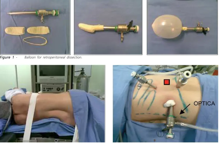
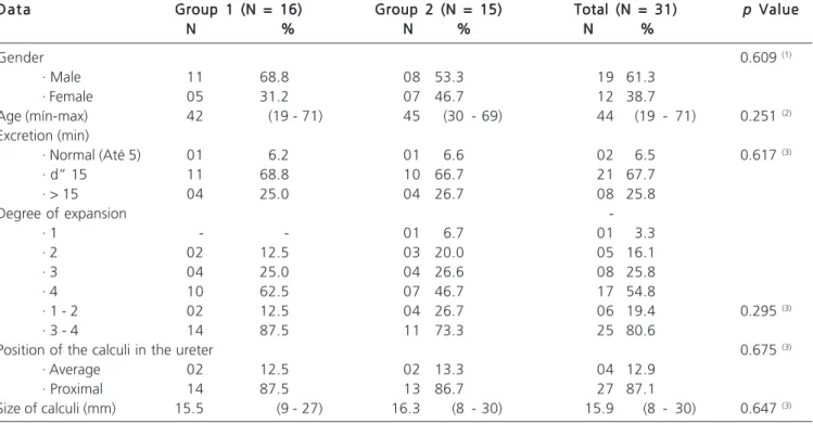
and lateral position (position for lumbotomy). Open access in the lumbar region allowed reaching the retroperitoneum. A special craft balloon was inserted (Figure 1) using a two fingers of a latex glove fixed at the distal end of a 10-mm trocar, which was filled with 500 ml of saline solution in order to create a retroperitoneal space to work on. The portals were positioned (Figure 2) to permit ureteral dissection, ureteral opening and removal of calculi.
In cases in which a double-J catheter was used, the insertion of the guide was done by percutaneous renal puncture in a contraincision to the ureteral opening, positioning the double-J catheter anterogradely oriented by
Figure 1 Figure 1 Figure 1 Figure 1
Figure 1 - Balloon for retroperitoneal dissection.
Figure 2 Figure 2 Figure 2 Figure 2
the guide (Figure 3). The proximal end of the catheter was placed into the renal pelvis in retrograde fashion. In cases of lower ureteral incision due to a lower position of calculi we performed another incision in the proximal ureter, through which the device was introduced. Afterwards we closed the ureter with poligalactine 4-0 (Figure 3) sutures and left a laminar drainage of the retroperitoneal space and calculi removal from cavity.
All patients had negative urine culture.
Pre and postoperative serum creatine was measured. Pre and postoperative excretory urography was performed according to hospital radiology protocol and interpretation of the renal collecting system dilatation followed the principles proposed by Talner, O’Reilly and Roy 18, graduated from 1 to 4. The postoperative
examinations were obtained at six and 12 months. Pain control was achieved with sodium dipyrone to all patients postoperatively until the end of the first day.
Ketoprofen or tramadol hydrochloride were used as intermediate analgesics. After the 2nd day analgesics were kept on demand and by oral intake. Morphine sulfate was necessary for cases of refractory pain.
Statistical analysis was performed using the Student’s t test and the nonparametric Mann-Whitney, chi-square and Fisher exact tests. The significance level was less than 5% (p <0.05).
RESULTS
RESULTS
RESULTS
RESULTS
RESULTS
We evaluated 31 patients with mean age of 45.1 ± 14 years, ranging from 19 to 71; 61.3% were men and 38.7% women (Table 1).
In the evaluation of preoperative intravenous urography we predominantly observed excretion time d” 15 minutes (67.7%), degree of expansion equal to four
Table 1 Table 1Table 1 Table 1
Table 1 - Data of patients and results of excretory urography.
D a t a D a t aD a t a D a t a
D a t a Group 1 (N = 16)Group 1 (N = 16)Group 1 (N = 16)Group 1 (N = 16)Group 1 (N = 16) Group 2 (N = 15)Group 2 (N = 15)Group 2 (N = 15)Group 2 (N = 15)Group 2 (N = 15) Total (N = 31)Total (N = 31)Total (N = 31)Total (N = 31)Total (N = 31) ppppp Value Value Value Value Value N
NN
NN %%%%% NNNNN %%%%% NNNNN %%%%%
Gender 0.609 (1)
· Male 11 68.8 08 53.3 19 61.3
· Female 05 31.2 07 46.7 12 38.7
Age (mín-max) 42 (19 - 71) 45 (30 - 69) 44 (19 - 71) 0.251 (2)
Excretion (min)
· Normal (Até 5) 01 6.2 01 6.6 02 6.5 0.617 (3)
· d” 15 11 68.8 10 66.7 21 67.7
· > 15 04 25.0 04 26.7 08 25.8
Degree of expansion
-· 1 - - 01 6.7 01 3.3
· 2 02 12.5 03 20.0 05 16.1
· 3 04 25.0 04 26.6 08 25.8
· 4 10 62.5 07 46.7 17 54.8
· 1 - 2 02 12.5 04 26.7 06 19.4 0.295 (3)
· 3 - 4 14 87.5 11 73.3 25 80.6
Position of the calculi in the ureter 0.675 (3)
· Average 02 12.5 02 13.3 04 12.9
· Proximal 14 87.5 13 86.7 27 87.1
Size of calculi (mm) 15.5 (9 - 27) 16.3 (8 - 30) 15.9 (8 - 30) 0.647 (3)
(1) chi-square, (2) Mann-Whitney test, (3) Fisher. Figure 3
Figure 3Figure 3 Figure 3
(54.8%) and calculi position on the proximal ureter (87.1%).
The calculi size was on average 15.9 ± 6.3 mm (range 8 to 30 mm) and there was no significant difference between groups. The affected side showed a slight predominance for the right ureter (54.8%).
The average value of preoperative creatinine was 1.07 ± 0.24 mg/dl.
All patients in both groups showed ureteral impaction of the calculi for more than two months.
The operating time was 130 ± 40.3 min in Group 1 and 136.3 ± 49.3 min in Group 2 (Table 2).
Group 1 patients were discharged on average 4.1 ± 2.3 days after the operation, while in Group 2, 3.6 ± 0.9 days postoperatively. Although the difference was not statistically significant, patients in Group 1 had in average one extra day in hospital (Table 2).
All studied patients received dipyrone postoperatively. In Group 1, five also received ketoprofen, other five tramadol (62.5%). In Group 2, two patients received ketoprofen and tramadol in other three (33.3%). Morphine sulfate was needed in eight patients in Group 1 and in seven in Group 2. On average these patients received 18.5 ± 6.5 mg morphine, with no difference between the two groups in this parameter.
The removal of the calculi was done in 100% of the cases.
In Group 1, four patients required implantation of a double-J catheter in the postoperative period due to massive and prolonged urinary drainage (more than 500 ml of urine / day for longer than two days). In these patients, the catheter was implanted between 3 and 7 days after surgery and removed on average 6.3 ± 2.2 weeks postoperatively. In Group 2 it was removed on average 7.6 ± 5.5 weeks after the procedure.
Group 1 had four cases of early complication, all with urinary fistulas and two cases of late complications (one mild ureteral stenosis and one case of renal exclusion due to obstruction). In Group 2, there were no complications. The groups complications were not significant, however it is noteworthy that all the early complications were reported in Group 1 (p = 0.058) (Table 3).
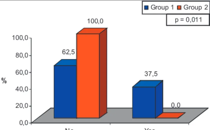
When assessing the overall complications (early + late), we noted that all of them occurred in Group 1 (37.5%) (p = 0.011), this difference being significant (Figure 4).
DISCUSSION
DISCUSSION
DISCUSSION
DISCUSSION
DISCUSSION
The use of minimally invasive techniques for treatment of ureteral stones is currently the gold standard, regardless of the situation. The intracorporeal ureterolithotripsy and the extracorporeal lithotripsy are the
Tabela 2 Tabela 2 Tabela 2 Tabela 2
-Tabela 2 - Dados do tratamento e pós-operatório.
V a r i a b l e s V a r i a b l e s V a r i a b l e s V a r i a b l e s
V a r i a b l e s nnnnn AverageAverageAverageAverageAverage Standard DeviationStandard DeviationStandard DeviationStandard DeviationStandard Deviation M í n i m u mM í n i m u mM í n i m u mM í n i m u mM í n i m u m M a x i m u mM a x i m u mM a x i m u mM a x i m u mM a x i m u m pp value ppp value value value value (1)(1)(1)(1)(1)
Operating time (min) 31 133.1 44.2 60.0 220.0 0.736 (2)
· Group 1 16 130.0 40.3 70.0 210.0
· Group 2 15 136.3 49.3 60.0 220.0
Removal of the drain in days 31 3.1 1.1 2.0 6.0 0.584 (2)
· Group 1 16 3.1 1.4 2.0 6.0
· Group 2 15 3.0 0.7 2.0 4.0
Hospitalization in days 31 3.9 1.8 2.0 10.0 0.885 (2)
· Group 1 16 4.1 2.3 2.0 10.0
· Group 2 15 3.6 0.9 2.0 6.0
(1) Student t, (2) Mann-Whitney test.
Table 3 Table 3 Table 3 Table 3
Table 3 - Evaluation of early and late complications.
V V V V
V ariariariariaria b l e sa b l e sa b l e sa b l e sa b l e s GroGrGrGrGrooooup 1(N = 16)up 1(N = 16)up 1(N = 16)up 1(N = 16)up 1(N = 16) GrGrGroGrGrooooup 2(N = 15)up 2(N = 15)up 2(N = 15)up 2(N = 15) Total (N = 31)up 2(N = 15) Total (N = 31)Total (N = 31)Total (N = 31)Total (N = 31) ppp Valpp ValValValValueueueueue N
NN
NN %%%%% NNNNN %%%%% NNNNN %%%%%
Early complications 0.058 (2)
· Yes (urinary fistulae) 04 25.0 - - 04 12.9
Late complications 0.258 (2)
· Yes (1 ureteral stenosis. 1 exclusion of one kidney) 02 12.5 - - 02 6.5
Complications (general) 0.011 (2)
· No 10 62.5 15 100.0 25 80.6
· Yes 06 37.5 - - 06 19.4
first options in most cases 19. Some situations, however,
are not resolved with these procedures and require the classical approach (open surgery). In the past decade, laparoscopy has replaced open surgery in the treatment of this disease 20. Its results are reproducible and have the
advantages of minimally invasive treatment, such as better analgesy control, shorter hospital stay and earlier return to usual activities21.
Retroperitoneoscopic ureterolithotomy was the first choice for middle ureter calculi in 39 cases (83%), and after lithotripsy with extracorporeal shock wave in eight (17%). The choice was due to the size of the calculus and time of obstruction, the position and anatomy of the urinary tract, and especially the fact that there was no usable flexible ureteroscopy and / or lithotripsy device, due to low socioeconomic status of the population. Gaur et al. 12 and
Matthias et al.22 selected the retroperitoneoscopic
ureterolithotomy as primary treatment in 59% and 55%, respectively. Both reinforce the socioeconomic status as a major factor in the therapeutic option.
The size of the calculus, of 16 mm on average, showed no difference between groups, and all cases had impacted stones in the ureter for more than two months. Other authors have reported that large calculus (> 15 mm) and especially those who are impacted for long periods are prime candidates for this technique, due to the high failure rate of other methods 23, 24, 25.
The operative time was 133 minutes in the global average, not statistically different between groups (a = 130 min, 2 = 136 min). Bove et al. 26 and Fan et al. 27 published
similar operation time in their series. Both point out that the reduced space in the retroperitoneum and lack of experience in early cases substantially contribute to the prolongation of the operation, and that the gain of skill and experience provide a reduction in operative time.
The patients stayed on average 3.9 days after the procedure and had the drain removed on the 3rd day on average. Compared with the findings of Basir et al. 24
and El-Moula et al. 10, who reported lengths of stay of 5.8
and 6.4 days, respectively, it can be seen that this series
had a better hospitalization time. Also compared to Kijvikai Patcharatrakul and 15 and Bove et al. 26, the time of drain
removal of 2.8 and three days was similar to the one found here.
The use of common analgesics was the routine. Additional analgesy with ketoprofen and tramadol was required in 15 patients and morphine sulfate in 15, averaging 18.5 mg / patient. Other authors reported the use of analgesics in 52% to 66% (16, 23). Kijvikai Patcharatrakul and 15 used 5.6 mg of morphine in their cases. Although
there is no uniformity in the literature regarding the type and dose of the analgesics used, and considering that in this study we used greater amounts of analgesy than in the series in the literature, the authors consider the effective control of pain the reason for shorter hospitalization time20, 28.
The removal of double-J catheter was on average seven weeks after the procedure, which is near the four to six weeks recommended by most authors 25, 29.
Finally, the group without catheter had prolonged drainage of urine through the wound on four of 16 of the patients (25%), which were considered as early complications. All these patients required double-J catheter, which meant a new invasive procedure. In contrast, in the catheters group no case of early complication was observed. Similarly, there were two cases of late complications, both in Group 1. One patient presented with ureteral stenosis, which was treated with ureteroscopy and dilatation, and remained four weeks with the double-J catheter. Another case involved a patient who had functional kidney exclusion seen by the post-operative urography, with ultrasound revealing hydronephrosis and narrowing of the large renal parenchyma; he underwent laparoscopic nephrectomy.
Thus, the rate of complications in the group without catheter was significantly higher than the group with it, considering overall complications. It should be noted that, although not statisticaly significant in separated evaluation, there is a tendency for more complications when the double-J catheter is not used, which probably would be confirmed with a greater number of patients.
Reviewing the literature, we found only three relevant publications that compare the use or absence of ureteral catheterization in this procedure. They did not employ double-J catheters in their series. Sinha and Sharma30
concluded that the use of the device decreases the time of drainage and promotes earlier discharge.
On the other hand, Goel and Hemal 11 concluded
that meticulous suture can replace the use of ureteral stent, except when there is reduced renal function and calculi impacted by more than three months, situations in which the stent must be used. Gaur et al. 12 described the largest
series operated by retroperitoneoscopic approach. The authors stressed that the use of ureteral stent reduces the leakage of urine, but did not recommend this type of drainage routinely, only in special situations, such as chronic inflammation, marked edema, friability and ureter calculus
Figure 4 Figure 4Figure 4 Figure 4
impacted for a long period.
Among the papers in the literature there are descriptions of two methods, with or without double-J catheter insertion. However, there is no clarity in comparing the results between them. Thus, in this study we attempted to assess whether the use of the catheter is advantageous for the patient. As for complications, there are similarities with the literature 12,14,13, 23,24, 10,25 ,22, pointing to the fact that the use
of the catheter likely reduces the appearance of complications. The retroperitoneoscopic ureterolithotomy procedure is still under debate, but it has obtained space as a minimally invasive treatment of ureteral calculi, either as a second-line option or even as the first choice in selected cases.
This study shows that the number of early and late complications was lower in patients operated with the use of double-J catheter. In relation to operative time, analgesy and length of hospitalization, both groups were similar.
So, the authors of this paper recommend the routine use of double-J catheter in order to reduce complications, but new studies should clarify the situations in which the double-J catheter is really essential.
In conclusion, the use of double-J catheter was associated with significantly fewer complications in retroperitoneoscopic ureterolithotomy. Surgical time, postoperative analgesia and length of stay were similar between groups, with and without catheter.
R E S U M O R E S U M O R E S U M O R E S U M O R E S U M O
Objetivo: Objetivo: Objetivo: Objetivo:
Objetivo: Avaliar os resultados da ureterolitotomia retroperitoneoscópica no tratamento do cálculo ureteral e a necessidade do cateter duplo J para reduzir complicações relacionadas ao procedimento. Métodos:Métodos:Métodos:Métodos:Métodos: Estudo retrospectivo comparativo de 47 pacientes operados pela técnica de ureterolitotomia retroperitoneoscópica, dos quais 31 foram selecionados e divididos em dois grupos: Grupo 1, cujos pacientes não receberam cateter duplo J, e Grupo 2, que foram submetidos ao implante de cateter duplo J transoperatório. Foram coletados dados de urografia excretora pré e pós-operatória, tempo cirúrgico, analgesia pós-operatória, tempo de internação e retirada do dreno. Resultados:Resultados:Resultados:Resultados:Resultados: Os grupos foram semelhantes quando comparados na idade e sexo, grau de dilatação do trato urinário, posição e tamanho médio do cálculo (Grupo 1= 15,5 ± 6,6mm; Grupo 2= 16,3 ± 6,1mm). O tempo operatório também não teve diferença significativa (Grupo 1= 130 ± 40,3min; Grupo 2= 136,3 ± 49,3min). O Grupo 1 apresentou seis pacientes (37,5 %) com complicações precoces (quatro casos de fístula urinária) e tardias (um caso de estenose de ureter, um caso de exclusão funcional do rim operado), enquanto o Grupo 2 não teve complicações, sendo esta diferença estatisticamente significativa (p=0,011). Conclusão:Conclusão:Conclusão:Conclusão:Conclusão: O emprego do cateter duplo J foi associado a um número significativamente menor de complicações na ureterolitotomia retroperitoneoscópica. Tempo cirúrgico, analgesia pós-operatória e tempo de internação foram semelhantes entre os grupos com e sem cateter.
Descritores: Descritores: Descritores: Descritores:
Descritores: Cálculos ureterais. Cirurgia geral. Cateterismo ureteral. Espaço retroperitoneal. Laparoscopia.
REFERENCES
REFERENCES
REFERENCES
REFERENCES
REFERENCES
1. Curhan GC. Epidemiology of stone disease. Urol Clin North Am. 2007;34(3):287-93.
2. Uribarri J, Oh MS, Carroll HJ. The first kidney stone. Ann Intern Med. 1989;111(12):1006-9.
3. Arrabal-Martín M, Pareja-Vilches M, Gutiérrez-Tejero F, Miján-Ortiz JL, Palao-Yago F, Zuluaga-Gómez A. Therapeutic options in lithiasis of the lumbar ureter. Eur Urol. 2003;43(5):556-63. 4. Preminger GM, Tiselius HG, Assimos DG, Alken P, Buck AC, Gallucci
M, et al. 2007 Guideline for the management of ureteral calculi. Eur Urol. 2007;52(6):1610-31.
5. Ansari MS, Gupta NP. Impact of socioeconomic status in etiology and management of urinary stone disease. Urol Int. 2003;70(4):255-61.
6. Wickham JEA. Urinary calculous disease. New York: Churchill Livingstone; 1979. The surgical treatment of renal lithiasis; p.145-98.
7. Abdelmaksoud A, Biyani CS, Bagheri F, Janetschek G. Laparoscopic approaches in urology. BJU Int. 2005;95(2):244-9.
8. Feyaerts A, Rietbergen J, Navarra S, Vallancien G, Guillonneau B. Laparoscopic ureterolithotomy for ureteral calculi. Eur Urol. 2001;40(6):609-13.
9. Nouira Y, Kallel Y, Binous MY, Dahmoul H, Horchani A. Laparoscopic retroperitoneal ureterolithotomy: initial experience and review of literature. J Endourol. 2004;18(6):557-61.
10. El-Moula MG, Abdallah A, El-Anany F, Abdelsalam Y, Abolyosr A, Abdelhameed D, et al. Laparoscopic ureterolithotomy: our experience with 74 cases. Int J Urol. 2008;15(7):593-7.
11. Goel A, Hemal AK. Upper and mid-ureteric stones: a prospective unrandomized comparison of retroperitoneoscopic and open ureterolithotomy. BJU Int. 2001;88(7):679-82.
12. Gaur DD, Trivedi S, Prabhudesai MR, Madhusudhana HR, Gopichand M. Laparoscopic ureterolithotomy: technical considerations and long-term follow-up. BJU Int. 2002;89(4):339-43.
13. Demirci D, Gülmez I, Ekmekçioðlu O, Karacagil M. Retroperitoneoscopic ureterolithotomy for the treatment of ureteral calculi. Urol Int. 2004;73(3):234-7.
14. Hemal AK, Goel A, Goel R. Minimally invasive retroperitoneoscopic ureterolithotomy. J Urol. 2003;169(2):480-2.
15. Kijvikai K, Patcharatrakul S. Laparoscopic ureterolithotomy: its role and some controversial technical considerations. Int J Urol. 2006;13(3):206-10.
16. El-Feel A, Abouel-Fettouh H, Abdel-Hakim AM. Laparoscopic transperitoneal ureterolithotomy. J Endourol. 2007;21(1):50-4. 17. Gaur DD. Laparoscopic operative retroperitoneoscopy: use of a
new device. J Urol. 1992;148(4):1137-9.
18. Talner LB, O’Reilly PH, Roy C. Urinary obstruction. In: Pollack HM, McClennan BL, editors. Clinical urography. 2nd ed. Philadelphia: WB Saunders; 2000. p.1846-1966.
20. Skrepetis K, Doumas K, Siafakas I, Lykourinas M. Laparoscopic versus open ureterolithotomy. A comparative study. Eur Urol. 2001;40(1):32-6; discussion 37.
21. Bishoff JT, Kavoussi LR. Laparoscopic surgery of the kidney. In: Kavoussi LR, Novick AC, Partin AW, Peters CA, Wein AJ, editors. Campbell-Walsh Urology. 9th ed. Philadelphia: Saunders; 2007; p. 1759-1809.
22. Matias DB, Alvim RG, Ribas M, de Oliveira BP, Chaves OH. Laparoscopic treatment of ureterolithiasis: our experience. Actas Urol Esp. 2009;33:667-9.
23. Flasko T, Holman E, Kovacs G, Tallai B, Toth C, Salah MA. Laparoscopic ureterolithotomy: the method of choice in selected cases. J Laparoendosc Adv Surg Tech A. 2005;15(2):149-52. 24. Basiri A, Simforoosh N, Ziaee A, Shayaninasab H, Moghaddam
SM, Zare S. Retrograde, antegrade, and laparoscopic approaches for the management of large, proximal ureteral stones: a randomized clinical trial. J Endourol. 2008;22(12):2677-80. 25. Khaladkar S, Modi J, Bhansali M, Dobhada S, Patankar S. Which is
the best option to treat large (>1.5cm) midureteric calculi ? J Laparoendosc Adv Surg Tech A. 2009;19(4):501-4.
26. Bove P, Micali S, Miano R, Mirabile G, De Stafani S, Botteri E, et al. Laparoscopic ureterolithotomy: a comparison between the transperitoneal and the retroperitoneal approach during the learning curve. J Endourol. 2009;23(6):953-7.
27. Fan T, Xian P, Yang L, Liu Y, Wei Q, Li H. Experience and learning
curve of retroperitoneal laparoscopic ureterolithotomy for upper ureteral calculi. J Endourol. 2009;23(11):1867-70.
28. Dunn MD, Terri GM. Anesthetic considerations. In: Smith AD, editor. Smith’s textbook of endourology. 2nd ed. Hamilton: BC Deker; 2007; p.385-94.
29. Wen X, Li X, Situ J, Fang Y, Chen X, Ruan X, et al. Application of a temporary ureter clamp for retroperitoneal laparoscopic ureterolithotomy. World J Urol. 2010;28(1):99-102.
30. Sinha R, Sharma N. Retroperitoneal laparoscopic management of urolithiasis. J Laparoendosc Adv Surg Tech A. 1997;7(2):95-8.
Received on 06/07/2011
Accepted for publication 16/09/2011 Conflict of interest: none
Source of funding: none
How to cite this article: How to cite this article:How to cite this article: How to cite this article:How to cite this article:
Cavalli AC, Tambara Filho R, Slongo LE, Cavalli RC, Rocha LCA. The use of double-j catheter decreases complications of retroperitoneoscopic ureterolithotomy. Rev Col Bras Cir. [periódico na Internet] 2012; 39(2). Disponível em URL: http://www.scielo.br/rcbc