The genus Colletotrichum is widely distributed throughout coffee (Coffea arabica) producing regions in the world, occurring sometimes as a saprophyte and sometimes as a plant pathogen (12). Three species of Colletotrichum are known: C. kahawae (geographically restricted to African coffee species), C. gloeosporioides and C. acutatum.
The predominant species in Brazil is C. gloeosporioides, which is associated with a complex of symptoms that include: anthracnose in fruits, tip blight and blister spot. Symptoms related to blister spot are characterized by the appearance of oily green spots throughout the plant. Although the pathogen is associated with a range of other symptoms, its importance has still been questioned due its various endophytic strains, consequently nonpathogenic, raising doubts as to the real value of this pathosystem.
RESUMO
Palavras-chave adicionais: Patogenicidade, Colletotrichum, Colonização. Colletotrichum gloeosporioides no Brasil está associado a um complexo de sintomas na cultura do café. Apesar da observação e da identificação da patogenicidade deste, o patógeno ainda tem sua importância questionada, devido às várias formas endofíticas do mesmo, levantando dúvidas sobre a real importância do patossistema. O presente trabalho teve como objetivo demonstrar por meio da utilização de um isolado transformado com o gene gfp a capacidade de infecção e colonização de C. gloeosporioides
Armesto, C.; Martins-Maia, F.G.; Monteiro, F.P.; Abreu, M.S. A proteína fluorescente GFP no estudo da mancha manteigosa em plântulas de café. Summa Phytopathologica, v.41, n.2, p.89-93, 2015.
em plântulas de café. Pode-se observar após o quarto dia da inoculação a exteriorização de sintomas como necrose pontuais as quais progrediram durante o período de avaliação, culminando na morte das plântulas. Por meio de microscopia de epifluorescencia pode-se confirmar a presença do patógeno nas plântulas, assim como visualizar a colonização interna do tecido, a formação de acérvulos e produção de conídios, confirmando que o mesmo foi o responsável pelos sintomas observados.
ARTIGOS
GFP-fluorescent protein on the study of blister spot in coffee plants
Cecilia Armesto
1; Fernanda Gonçalves Martins-Maia
2; Fernando Pereira Monteiro
1; Mário Sobral de Abreu
11Departamento de Fitopatologia, Universidade Federal de Lavras, Campus Universitário da Ufla, Lavras, MG - Brasil. 2Instituto de Ciências
Agrárias, Universidade Federal de Uberlândia, Campus Uberlândia, Uberlândia, MG - Brasil. Autor para correspondência: Cecilia Armesto ([email protected]).
Data de chegada: 03/11/2014. Aceito para publicação em: 15/04/2015.
10.1590/0100-5405/2047
According to Orozco et al. (14), Colletotrichum x coffee pathosystem is composed of Colletotrichum species that cause several damages or invasively colonize the plant tissue without manifesting symptoms. These authors also report that there may be pathogenic races within the population of this pathogen in coffee. This can be associated with the high genetic variability of this genus, which constitutes one of the factors that have hindered the identification of strains and even species, leading to doubts as to the pathogenicity of Colletotrichum spp.
Transgenic or molecularly tagged isolates have been an alternative to this situation. Genetic manipulation, by means of gene insertion, using filamentous fungi capable of producing fluorescent proteins became an important tool in studies for tracking pathogens in In Brazil, Colletotrichum gloeosporioides is associated with a
complex of symptoms in coffee culture. Although this pathogen had
its pathogenesis observed and identified, its importance has still been
questioned due to its several endophytic forms, raising doubts as to the real importance of the pathosystem. The aim of this study was to demonstrate, by using an isolate transformed with the gene gfp, the infection and colonization capability of C. gloeosporioides in
Armesto, C.; Martins-Maia, F.G.; Monteiro, F.P.; Abreu, M.S..GFP-fluorescent protein on the study of blister spot in coffee plants. Summa Phytopathologica, v.41, n.2, p.89-93, 2015.
Additional keywords: Pathogenicity, Colletotrichum, Colonization.
ABSTRACT
coffee seedlings. After the fourth day of inoculation, manifestation of symptoms as punctual necrosis could be observed, which progressed during the evaluation period, culminating in the death of seedlings.
Epifluorescence microscopy confirmed the presence of the pathogen
in the seedlings, as well as the visualization of internal colonization of
tissues, acervulus formation and conidium production, confirming that
plant tissues, particularly in studies involving living cells (6). There are numerous advantages of using genetic transformation technique with the GFP gene. Its expression can be visualized in vivo by observing individual cells and population of cells interacting with symbionts or the environment in real time. It has low toxicity, is extremely stable and maintains its activity when associated with different cellular and extracellular proteins (8, 18).
GFP also allows differentiation between the inoculated fungus and other possible parasites present in the plant because the former is seen by fluorescence microscopy at a determined wave length, which means that only the marked isolate can be observed.
In addition to the mentioned advantages, GFP has not shown any interference with the normal development of fungal cells, not affecting the morphology of the fungus, colony growth, pigmentation and formation of conidia, and the transgenic isolates, which remain as pathogenic as the wild type. Thus, this technique allows monitoring details of infection, penetration and colonization inside and outside tissues (16).
Based on the numerous GFP applications for microorganisms, the present study aimed to monitor a C. gloeosporioides isolate transformed with gfp protein, when it was associated with coffee plants. The pathogenic capacity of this fungus, as well as its development in the tissues of seedlings, was evaluated. Data about the progress of symptoms were collected to prove the importance of this pathogen for coffee crops.
MATERIAL AND METHODS
Colletotrichum gloeosporioides strain
The transformed C. gloeosporioides isolate (IS2-P) was selected from the mycological collection of the Laboratory of Diagnosis and Plant Diseases Control at the Department of Plant Pathology of the Federal University of Lavras. It was obtained from plants presenting blister spot and symptoms of dry plant pointer. Single spore cultures were prepared to confirm its pathogenicity. The gfp gene was inserted by the method of polyethylene glycol (PEG), as described by Armesto et al. (1). The plasmid used in the transformation process was pCT74 (pSC001), containing sgfp fragment with the promoter gene PtOx (Aspergillus nidulans) and hygromycin-B resistance gene (HPR). The plasmid was provided by the researcher Dr. Theo van der Lee (Plant Research International, The Netherlands).
Selecting Colonies
After three days of growth, the plates containing the transformed isolates were observed under an epifluorescence microscope. Colonies that showed fluorescence were transferred to new plates containing
MEA medium (Malt Extract Agar 2%) plus 100μgmL-1
hygromycin B, and incubated at 25° C in the dark.
Inoculation of seedlings
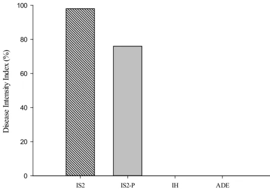
Inoculation was carry out by spraying 10 seedlings of the “Red Catuaí” coffee cultivar with conidial suspension at 106 conidia mL-1. Treatments were discriminated as follows: a) inoculation with the transformed isolate (IS2-P); b) inoculation with the pathogenic wild isolate (IS2); c) inoculation with the non-pathogenic isolate (IH); d) inoculation with sterile distilled water (ADE). Plants were maintained in the growth chamber at 25º C under a photoperiod of 12 hours for 15 days. Symptoms were assessed according to the scale of Van DenVossen et al. (19) and results were interpreted based on the disease intensity
index (DII = ∑( no. of hypocotyls in each class × numeric value of each class) / (no. of hypocotyls in all classes × 4)).
Epifluorescence Microscopy Observation
After the onset of symptoms, transversal and longitudinal sections of the stem and the leaves were collected by removing thin sections of the infected tissue, which were used for observation under an epifluorescence (Zeiss Axio Observer Z.1) microscope with filter of 470-490 nm and peak transmission at 510-560 nm.
RESULTS AND DISCUSSION
The transformation of C. gloeosporioides isolate (IS2-P) was successful by using antibiotic resistant hygromycin B and green fluorescence protein. As the wild isolate and the control do not emit fluorescence when subjected to epifluorescence microscopy, it was possible to differentiate them from transformed ones. Another confirmation procedure was performed by using the specific primers proposed by Botelho, Barrocas and Machado (2), based on the nucleotide sequences of sGFP.
At five days after inoculation, the first symptoms could be observed on the seedlings inoculated with the wild isolate (IS2). The transformed isolate (IS2-P) showed symptoms only on the seventh day after inoculation. Complete necrosis of the stem was seen in all inoculated seedlings after 15 days for both isolates. The control inoculated with sterile water (ADE) and the non-pathogenic isolate (IH) did not exhibit any symptoms over the 15 days of evaluation (Figure 1).
Tests on seedlings and fruits are the most used method to evaluate the pathogenicity and aggressiveness of C. gloeosporioides, evidencing that the interaction between the pathogen and the plant is very variable. According to Nechet & Abreu (11), the period between inoculation and expression of symptoms can range from 15 to 30 days. Rodrigues et al. (15) stated that the first lesions on hypocotyls may occur from five to seven days after the inoculation period, and the latency period may range from two to three weeks, depending on the aggressiveness of the isolate.
Visualization of fluorescence by transformed isolate on tissue plant demonstrated that the symptoms in coffee seedlings were related to internal colonization of C. gloeosoporiodes.
Although it showed a delay in the first symptoms, the transformed isolate showed a high rate of disease (Figure 1). However, the aggressiveness of the transformed isolate and that of the wild type were different. Thus, even after the introduction of an exogenous DNA fragment to isolate IS2, it still retained its capability of infecting and causing disease, but the virulence of the strain changed.
According to Welsh and Kay (20), GFP does not cause interference in cellular activities, evidencing thus that it does not affect the pathogenicity of the pathogen. Horowitz, Freeman and Sharon (7) reported that C. acutatum isolates transformed with gfp remained infective, as well as the wild types, and could be detected in infected tissues at three days after inoculation, highlighting the rapid development of the fungus, filling the mesophyll with dense mycelium that invaded the cells and causing tissue necrosis. Colletotrichum graminicola, modified with gfp gene, was also capable of systemically infecting corn seedlings, colonizing the surface of roots, invading the epidermis, the cortex and the vascular tissues (17).
starting with a small necrotic spot at the site of inoculation and progressing to necrosis around the site, reaching the entire stem and apical buds, and finally causing constriction of the stem, consequently leading to the seedling damping-off.
The infection process was observed in samples collected at seven days after inoculation through the presence of hyphae. Internal tissue colonization was initiated by cortical parenchyma, reaching the vascular tissues, since the symptoms were progressing towards the apex of seedlings. Similar results were observed by Pereira et al. (13), using scanning electron microscopy. They visualized the colonization of xylem vessels and parenchyma of coffee branches and also differentiated them from unaffected tissue. Ferreira et al. (4) noted the presence of mycelium of C. gloeosporioides in all organs of coffee plants, systemically colonizing tissues of xylem, phloem, cortex and endosperm cells, consequently causing the death of branches, fruit mummification and fall of leaves. Those authors also report colonization of exocarp and mesocarp in coffee fruits for different varieties (5). In the present study, the cortex colonization could not be observed due to a strong emission of autofluorescence from the tissue, which made it impossible to differentiate the fluorescence between fungal mycelium and tissue autofluorescence.
At ten days after inoculation with mutant isolate, acervulus formation was observed in the inoculated tissue, as well as consequent production of conidia (Figure 2). The acervuli were located in the epidermis, breaking the tissue surface, where the development of conidiophores and arrows could be noted. The formation of acervuli in hypocotyls was previously reported in an article written by Lins, Abreu and Alves (10), showing that conidiophores were formed on a subcuticular stroma, breaking the cuticle after inoculation. Ferreira (4) also observed acervulus formation and conidiophore emergence
through the stomata at 96 hours after inoculation of different C. gloeosporioides strains.
Species of the genus Colletotrichum may have different strategies for tissue colonization, which range from intracellular hemibiotrophic in C. lindemuthianum, C. gloeosporioides, C. graminicola, C. orbiculare and C. truncatum to subcuticular intramural, reported for C. capsici C. circinans C. gloeosporioides, and C. phlomoides; there are also species that can exhibit both habits such as C. gloeosporioides (9). Intracellular hemibiotrofic pathogens invade the cells for a short period, where they remain, apparently for their nutrition, then convert to the necrotrophic phase and killing the cell. Using the subcuticular-intramural strategy, the fungus grows under the cuticle and epidermal wall cells (3).
In the present study, intracellular hemibiotrofic strategy could be noted; during the first 96 hours, the fungus invaded the tissues searching for nutritional resources, but without symptoms, which highlights the biotrophic phase. At seven days after inoculation, reproductive structures were exteriorized on the surface of the inoculated plants. On the tenth day, cellular death of tissues was observed, characterizing the necrotrophic phase.
After 13 days of inoculation, the wild and the transformed isolate could be isolated in amid in malt extract- agar medium, kept for ten days in a growth chamber at 25°C, proving the stability of the transformed strain.
This paper presents strong evidence of the pathogenicity of C. gloeosporioides isolated from coffee seedlings with symptoms of blister spot through the use of GFP molecular marker. Tissue colonization, conidial germination, primary and secondary hyphal development and acervulus production were perfectly studied with this molecular marker. This is an important step for the pathosystem
Isolates of
Colletotrichum gloeosporioides
IS2 IS2-P IH ADE
D
is
ea
se
I
nt
ens
ity
Ind
ex
(%
)
0 20 40 60 80 100
IS2-P: Isolated from C. gloeosporioides transformed by PEG IS2: C. gloeosporioides wild type
in question since this study demonstrated that there is a species or possibly a pathogenic strain of the fungus as an etiologic agent of blister spot.
ACKNOWLEDGEMENTS
To “Fundação de Amparo a Pesquisa de Minas Gerais (FAPEMIG)” and “Conselho Nacional de Desenvolvimento Científico e Tecnológico (CNPq)” for research funding. We are also thankful to the laboratory of electron microscopy and ultra-structural Analysis of Federal University of Lavras (UFLA) and to Prof. Dr. Eduardo Alves for the microscopic analysis.
REFERENCES
01. Armesto, C.; Martins-Maia, F.G.; Abreu, M.S.; Figueira, A.R.; Silva, B.M.; Alencar, N.E. Genetic transformation with the gfp gene of Colletotrichum gloeosporioides isolates from coffee with blister spot. Brazilian Journal of Microbiology, São Paulo, v. 43, n. 3, p. 1222-1229, 2012.
02. Botelho, S.L.; Barrocas E.N.; Machado, J.C. Uso da técnica de PCR na confirmação da presença de marcadores tipo GFP (Green Fluorescent Protein) em isolados de Sclerotinia sclerotiorum transformadas em Cuiabá, MT. Fitopatologia Brasileira, Cuiabá, v. 35, supl., p. 250, 2010. (Resumo). 03. Diéguez-Uribeondo J, Förster, H; Soto-Estrada, A.; Adaskaveg, J.E.
development of Colletotrichum acutatum on almond. Phytopatology, St. Paul, v. 95, n. 7, p. 751-758, 2005.
04. Ferreira, J.B.; Abreu, M.S.; Alves, E.; Pereira, I.S.; Fernandes, K.D. Eventos do processo de infecção de Colletotrichum gloeosporioides inoculados em folhas de Coffea arabica L. Summa Phytopathologica, Botucatu,v.35, n.4, p.273-281, 2009.
05. Ferreira, J.B.; Abreu, M.S.; Pereira, I.S. Incidência de Colletotrichum spp. em frutos de Coffea arabica L. em diferentes estádios fisiológicos e tecidos do fruto maduro. Ciência & Agrotecnologia, Lavras, v. 29, n. 4, p. 880-885, 2005.
06. Freitag, M.; Hickey, P.C.; Raju, N.B.; Selker, E.U.; Read, N.D. GFP as a tool to analyze the organization, dynamics and function of nuclei and microtubules in Neurospora crassa. Fungal GeneticsBiology. v. 41, p. 897–910, 2004.
07. Horowitz, S.; Freeman, S.; Sharon, A. Use the green fluorescent protein-transgenic strains to study pathogenic and nonpathogenic lifestyles in Colletotrichum acutatum. Phytopatology, St. Paul, v.92 n.7, 743-749. 2002 08. Lagopodi, A.L.; Ram, A.F.J.; Lamers, G.E.M.; Punt, P.J.; Hondel,
C.A.M.J.J.van den; Lugtenberg, B.J.J.; Bloemberg, G.V. Novel aspects of tomato root colonization and infection by Fusarium oxysporum f. sp. radicis-lycopersici revealed by confocal laser scanning microscopic analysis
using the green fluorescent protein as a marker. Molecular Plant-Microbe Interactions, St. Paul, v. 15, n. 2, p. 172-179, 2002.
09. Lopez, A.M.Q. Taxonomia, patogênese e controle de espécies do gênero Colletotrichum. Revisão Anual de Patologia de Plantas, Passo Fundo, v. 9, p. 291-338, 2001.
10. Lins, S.R.O.; Abreu, M.S.; Alves, E. Estudos histopatológicos de Colleto-trichum spp. em plântulas de cafeeiro. Fitopatologia Brasileira, Brasília, v. 32, n.6, p. 488-495, 2007.
11. Nechet, K.L.; Abreu, M.S. Caracterização morfológica e testes de patoge-nicidade de isolados de Colletotrichum sp. obtidos de cafeeiro. Ciência &
Agrotecnologia, Lavras, v.26, n.6, p.1135-1142, 2002.
12. Paresqui, L. Patogenicidade de Colletotrichum gloeosporioides penz. ao cafeeiro (Coffea arabica L.). 2003. 44f. Tese (Doutorado em Fitopatologia) – Departamento de Fitopatologia – Universidade Federal de Viçosa, Viçosa. 13. Pereira, I.S.; Abreu, M.S.; Alves, E.; Ferreira, J.B. Estudos histopatológil-cos da interação Colletotrichum gloeosporioides – Cafeeiro. Bragantia, Campinas, v.68, n.1, p.117-123, 2009.
14. Orozco, E.F.M; Juliatti, F.C.; Dorizzotto, A.; Abreu, M.S. Determinação da existência de raças fisiológicas no patossistema café x Colletotrichum no Estado de Minas Gerais em Uberlândia, MG. Tropical Plant Pathology, Uberlândia, v. 28 p.222, 2003. (Resumo).
15. Rodriguez, C.J.; Varzea, V.M.P.; Hindorf, H.; Medeiros, E.F. Strain of Col-letotrichum coffeanum Noack in republic centrafricaine. Café Cacao Thé, Paris, v.13. n.3. p.221-230, 1991.
16. Siqueira, C.S. Transformação de Stenocarpella maydis com os genes marcadores GFP e DsRed e patogenicidade dos transformados em sementes de milho. 2009. 53f. Dissertação (Mestrado em Fitopatologia) – Departamento de Fitopatologia – Universidade Federal de Lavras, Lavras. 17. Sukno, S.A.; García, V.M,; Shaw, B.D.; Thon, M.R. Root infection
and systemic colonization of maize by Colletotrichum graminicola. Applied and Environmental Microbiology, New York, v. 74, n. 3, p. 823-832, 2008.
18. Valdivia, R.; Hromockyj, A.; Monack, D.; Ramakrishnan, L.; Falkow, S.
Ap-plications for green fluorescent protein (GFP) in the study of host-pathogen
interactions. Gene, Amsterdam, v. 173, n. 1, p. 47- 52, 1996.
19. Vossen, H.A.M. van der; Cook, R.T.A.; Murakuro, N.W. Breeding for resistance to coffee berry disease caused by Colletotrichum coffeanum Noack (sensu Hindorf) in Coffea arabica L.: I., methods of preselection for resistance. Euphytica, Wageningen, v. 25, n. 1, p. 733-745, 1976. 20. Welsh, S.; Kay, S.A. Reporter gene expression for monitoring gene transfer.