CYTOPATHOLOGY OF CALLUS CELLS INFECTED WITH GRAPEVINE
LEAFROLL-ASSOCIATED VIRUS 3*
SANDRA M. M. SCAGLIUSI1**, JORGE VEGA1** & HUGO KUNIYUKI2**
1Departamento de Fisiologia Vegetal, Instituto de Biologia, Universidade Estadual de Campinas (UNICAMP), Cx. Postal 6109, CEP 13083-970, Campinas, SP, Brazil, Fax: (19) 3289-3124, e-mail: [email protected];
2Centro de Fitossanidade, Instituto Agronômico Campinas, Cx. Postal 28, CEP 13020-902, Campinas, SP, Brazil
(Accepted for publication 22/03/2002)
Corresponding author: Sandra M.M. Scagliusi
SCAGLIUSI, S.M.M., VEGA, J. & KUNIYUKI, H. Cytopathology of callus cells infected with grapevine leafroll-associated virus 3. Fitopatologia Brasileira 27:384-388. 2002.
ABSTRACT
The cytopathology of grapevine (Vitis spp.) callus tissue
infected with Grapevine leafroll-associated virus 3 (GLRaV-3), genus Vitivirus was studied in order to investigate the usefulness
of callus cultures to study grapevine leafroll-associated viruses. Ultrathin sections were made from in vitro callus obtained from
stems and shoots of GLRaV-3 infected grapevine plants. Callus was composed of two types of tissue. Translucent, soft callus was formed and composed of large loosely arranged cells, containing big vacuoles and a thin layer of cytoplasm. Other parts of the callus were brown-coloured and composed of small compactly arranged cells, which showed flexuous and rod-shaped closterovirus-like particles, with 10-12 nm in diameter, at higher
magnifications. Groups of vesicles formed by a single membrane were also observed, with sizes ranging from 50-200 nm, containing fine fibrillar material, also typical of closterovirus infections. Virus concentration was monitored by Immunosorbent electron microscopy (ISEM) tests, which showed that in vitro culture of callus tissue from grapevine infected plants, could be used to study the GLRaV viruses through many successive generations, despite the decline in virus concentration after repeated transfers. No virus particles were observed in callus tissue obtained from healthy grapevines.
Additional keywords: C losterovirus, Vitis spp.,
transmission electron microscopy.
RESUMO
Citopatologia de tecido caloso infetado pelo vírus do enrolamento da folha da videira 3
Análises citopatológicas do vírus do enrolamento da folha da videira 3 (Grapevine leafroll-associated virus 3,
GLRaV-3), gênero Vitivirus foram feitas utilizando-se tecido caloso obtido in vitro, para investigar seu uso no estudo do vírus
associado ao enrolamento da folha da videira (EFV). Secções ultrafinas foram feitas a partir de cultura de calos obtidos de hastes e brotos de videiras (Vitis spp.) infetadas pelo GLRaV-3. Dois tipos de calos foram observados. Um tipo transparente composto de células grandes e altamente vacuoladas contendo uma fina camada de citoplasma. Um outro tipo, de coloração marrom, composto de um grupo de células pequenas, compac-tamente arranjadas, cujo citoplasma era bem mais abundante. Nestas células foram observadas, em maior aumento, massas
formadas por fibras flexuosas com 10-12 nm de diâmetro, cujo aspecto corresponde ao das partículas de closterovírus em cortes de tecido. Grupos de vesículas também foram observados, com tamanhos variando entre 50-200 nm, formadas por uma única membrana e contendo material fibrilar muito fino, caracterís-tico de infecções causadas por closterovírus. A concentração do vírus foi monitorada por testes de Immunosorbent electron microscopy (ISEM), os quais mostraram que a cultura de calos obtidos de videiras infetadas pôde ser usada para estudar os vírus associados ao EFV durante várias gerações, apesar da diminuição da concentração de vírus após repetidas transfe-rências. Não foram observadas partículas de vírus em tecido caloso obtidos de videiras sadias.
INTRODUCTION
Grapevine Leafroll Disease (GLR) is one of the most important graft-transmissible diseases affecting grapevines (Vitis spp.) and occurs world-wide (Goheen, 1988). Symptoms include downward rolling and interveinal reddening or
* Part of the MS Thesis of the first author. Universidade Estadual de Campinas (UNICAMP)
**CNPq fellowships
yellowing of leaves in dark and white fruit-bearing varieties, respectively, resulting in reduced yield, delayed ripening and reduced accumulation of sugar (Weber et al., 1993).
GLR, they are referred to as Grapevine Leafroll-Associated Viruses (GLRaVs). So far, eight closteroviruses have been reported from GLR affected plants and designated as GLRaV-1 to 8 (Hu et al., 1990; Boscia et al., 1995; Choueiri et al., 1996; Monis, 2000). Several antisera have been produced against the different GLR-associated closteroviruses (Zimmermann et al., 1990b) and mixed infections were frequently detected (Zimmermann et al., 1990a). More recently several of these closteroviruses have been partially sequenced, and many primers are available for use in RT-PCR tests (Saldarelli et al., 1998; Zhu et al., 1998; Martin et al., 2000).
The family Closteroviridae consists of two genera, a monopartite genus named Closterovirus with Beet yellow virus (BYV) as a type member and a bipartite genus named Crinivirus with Lettuce infectious yellows virus (LIYV) as a type member. In a recent review on the genetic diversity of closterovirus, Karasev (2000) suggests a modification on the current closterovirus classification based on the type of vector transmission. This classification would consist of three genera, as follows: Closterovirus (aphids), Vitivirus (mealy bugs) and Crinivirus (whiteflies). According to this review, the grapevine closteroviruses would be part of the Vitivirus genus, having Grapevine leafroll-associated virus 3 (GLRaV-3) as a type member. Grapevine closteroviruses are difficult to study, mainly because of their low concentration and uneven distribution of virus particles and also because of the presence of high concentration of phenolic compounds and tannins in Vitis spp. plants. These compounds are highly reactive with proteins, interfering in serological reactions and in virus purification. One way to overcome this difficulty is to use in vitro grown plants, which are regarded as having higher virus titer and lower tannin content (Barba et al., 1989, Monis & Bestwick, 1996). On the other hand, the culture of callus tissue, also used for its low phenolic content, has been generally associated with a decrease in virus concentration (Mori & Hosokawa, 1977; Omura & Wakimoto, 1978) with possible application for obtaining virus-free plants.
In this study, we present evidence that Grapevine leafroll-associated virus 3 (GLRaV-3) multiplies in the callus tissue obtained from infected grapevines, through several generations, showing the usefulness of callus tissue cultures for the study of grapevine viruses. A preliminary communication of these results was previously presented (Vega et al., 1989).
MATERIAL AND METHODS
Plant materials and virus source
Symptomatic grapevine plants of the varieties Pinot Noir, Seibel 2 and LN 33, which tested positively for GLRaV-3 by indexing and serological procedures, were kept in the greenhouse and used as virus source for the experiments. Grapevine plants from the same varieties, tested negatively for GLRaV by the same procedures, were used as negative controls. Healthy plants obtained from seeds were also used
as negative controls.
In vitro culture of callus tissue
Vegetatively growing stems and shoots (1-2 cm) from infected and healthy vines were harvested from the above-mentioned plants and were surface-sterilized by 70% alcohol and commercial sodium hypochlorite (0.4%). After surface sterilization, each end of the stems was trimmed to eliminate the bleach damaged tissue and rinsed in sterile distilled water. The stems and shoots were cultured in vials in propagation media, according to Murashige & Skoog (1962), containing 20.0 ml l-1 of MS inorganic salts, 30.0 g l-1 sucrose, 8.0 g l-1 agar, 0.1 g l-1 myo-inositol, 1.0 g l-1 casein, 0.3 mg l-1 IAA, 1.0 mg l-1 2,4-D and 0.3 mg l-1 kinetin. The cultures were maintained at 25 °C in the dark and transferred to fresh medium every month. After transferring to fresh medium, a new generation of callus tissue was formed.
In situ Electron Microscopy
Only callus tissue was used for cytopathological studies, avoiding explant tissue present in the initial callus generation. Samples were fixed with 4% paraformaldehyde and 1.25% glutaraldehyde, postfixed in 1% OsO
4
, both in cacodylate buffer (0.05 M, pH 7.0), then contrasted en bloc with 2% uranyl acetate, dehydrated in acetone and dimethoxypropane and embedded in Epoxy medium. Sections were stained with lead citrate (1%) and observed with a ZEISS EM 10C transmission electron microscope.
Serological methods
Plants were tested by F(ab’)
2 Enzime linked immunosorbent assay (ELISA) according to Rowhani (1992) using polyclonal antisera provided by Dr. A. Rowhani and Dr. D. Golino (University of California-Davis, USA). Immuno Sorbent Electron Microscopy (ISEM) tests were used to detect GLRaV through the callus generations. Tests were done according to Milne & Lesemann (1984) with some modifications, using GLRaV-3 antiserum provided by Dr. Gugerli (Nyon, Switzerland). Copper grids covered with Parlodion and carbon films were coated using [1:1000] dilutions of antisera, during 60 min at room temperature. Grapevine callus tissue was extracted in 0.06 M phosphate buffer, pH 7.3, containing 1% PEG (Polyethylene Glycol MW 6,000) and 2.5% PVP (Polyvinyl Pyrrolidone MW 40,000). Samples were prepared using 2.0 ml of buffer per gram of tissue and were incubated on the grids overnight at 4 °C. After each step the grids were rinsed with PBS buffer and distilled water and prior to observation they were negatively stained with 2% uranyl acetate.
RESULTS
Cytopathology of grapevine callus
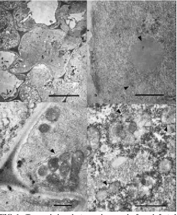
Portions of this callus tissue were brown-coloured, composed of small compactly arranged cells with more abundant cytoplasm (Figure 1A). Cells of this compact callus showed, at higher magnification, that a great part of the cytoplasm was occupied by large masses of flexuous closterovirus-like particles, ca. 10-12 nm in diameter (Figure 1B). Regular cell organelles, such as mitochondria in groups or isolated (Figure 1C), and lipid droplets (Figure 1B) were seen embedded in the virus-particle masses. Vesicular bodies were frequently seen within the virus particles masses. These bodies are formed by single membrane limited vesicles with sizes ranging from 50-200 nm, containing very fine fibrillar material (Figure 1D). The above-described alterations were not observed in callus tissue obtained from healthy grapevine plants.
Monitoring virus concentration from callus tissue by ISEM Closterovirus-like particles were detected by ISEM
using antibodies prepared for GLRaV-3 (Figure 2). The virus was detected by ISEM over approximately 15 generations and beyond that no virus particles were detected.
DISCUSSION
The cytopathology observed in the callus tissue obtained from GLRaV-3 infected grapevine plants is similar to that described for infected leaf-tissue (Faoro et al., 1992). The masses of long and flexuous virus particles, next to vesicles containing fibrillar material are characteristics of closterovirus infected tissue (Francki et al., 1985; Lesemann, 1988). The formation of characteristic vesicular bodies in grapevine plants infected by GLRaV-3 was described as starting from the vesiculation of modified mitochondria (Kim et al., 1989; Faoro & Carzaniga, 1995). Our results do not show mitochondria in the vesiculation process, but the vesicular bodies and the fibrillar material seen in the interior of some group of vesicles could correspond to the final stage of mitochondria vesiculation. The finely fibrillar content of these vesicles has been shown to be RNA, which are probably replication sites of the virus (Faoro & Carzaniga, 1995).
This study shows that GLRaV-3 is present in callus tissue growing in vitro and that the virus persists through many successive generations. The distribution of the virus particles in the callus tissue appeared to be uneven, since high concentrations occurred only in some groups of cells. These results are in contrast with the predominant idea in the literature, where it is generally assumed that callus tissue is not appropriate for virus replication, and in fact is a possible application for obtaining virus-free plants (Omura & Wakimoto, 1978; Xu et al., 2000). Our results indicate that
FIG. 1 - Transmission electron micrographs from infected grapevine (Vitis spp.) callus tissue. A) Groups of small cells of callus tissue from Grapevine leafroll disease (GLR) infected grapevines exhibiting a more abundant cytoplasm (Bar= 5 µµm); B) Higher magnification of the cytoplasm from the cells in A, showing large masses of closterovirus-like particles (arrowhead) (Bar = 500 nm); C) Cells showing mitochondria in groups (arrowheads) and isolated embedded in the virus-particle masses (Bar= 500 nm). D) Groups of vesicles (arrowheads) showing some granular material surrounded by masses of virus particles (Bar= 1 µµm).
callus may be useful for studying viruses in plants that have high contents of phenolic compounds, as in grapevine. Apparently, phenolics are in lower concentration in callus tissue, as suggested by the slower darkening of callus extracts, compared to leaf extracts. It is well known that phenolics readily oxidize and turn brown, generating products that form complexes with proteins, interfering in the recognition of viral proteins by antibodies (Croteau et al., 2000).
Another possibility for the easier detection of GLRaV-3 in callus is that during callus formation, cell proliferation occurs from some tissues more than others. Michaux-Ferrière & Carron (1989) observed in Hevea brasiliensis (Muell-Arg.) that a compact type of callus was formed from perivascular cells, indicating the possibility that this type of callus contains a higher proportion of vascular-parenchymatous cells. Since closterovirus are limited to the phloem parenchyma, the resulting callus could have a higher proportion of virus-containing cells.
ACKNOWLEDGEMENTS
Grateful thanks are expressed to Drs. D. Golino, A. Rowhani and P. Gugerli for supplying the antisera; to Prof. Ladaslav Sodek (UNICAMP) for critical review of the manuscript; and to Conselho Nacional de Desenvolvimento Científico e Tecnológico (CNPq, Brazil), for the award of a scholarship to S.M.M. Scagliusi.
LITERATURE CITED
BARBA, M., CUPIDI, A. & FAGGIOLI, F. In vitro culture of grapevine infected by closterovirus type III. Journal of Phytopathology 126:225-230. 1989.
BOSCIA, D. GREIF, C., GUGERLI, P., MARTELLI, G.P., WALTER, B. & GONSALVES, D. Nomenclature of grapevine leafroll-associated putative closteroviruses. Vitis 34:171-175. 1995.
CASTELLANO, M.A., MARTELLI, G.P. & SAVINO, V. Virus-like particles and ultrastructural modifications in the phloem of leafroll affected grapevines. Vitis 22:23-39. 1983. CHOUEIRI, E., BOSCIA, D., DIGIARO, M., CASTELLANO,
M.A. & MARTELLI, G.P. Some properties of a hitherto undescribed filamentous virus of the grapevine. Vitis 35:91-93.1996.
CROTEAU, R., KUTCHAN, T.M. & LEWIS N.G. Natural products (secondary metabolites). In: Buchanan, B., Gruissem, W. & Jones, R. (Eds.) Biochemistry & Molecular Biology of Plants. American Society of Plant Physiologists. 2000. pp. 1250-1318.
FAORO, F., TORNAGHI, S., CINQUANTA, G. & BELLI, G. Cytopathology of grapevine leafroll associated virus III (GLRaV-III). Rivista Patologia Vegetale 2:67-83. 1992. FAORO, F. & CARZANIGA, R. Cytochemistry and
immunocytochemistry of the inclusion bodies induced by grapevine leafroll-associated closteroviruses GLRaV-1 and GLRaV-3. Rivista Patologia Vegetale 5:85-94. 1995. FRANCKI, R.I.B., MILNE, R.G. & HATA, T. Closterovirus group.
In: Atlas of Plant Viruses. Vol. II (pp. 219-234) CRC press.
Boca Raton/FL.1985.
GOHEEN, A.C. Leafroll disease. In: Pearson, R.C. & Goheen, A.C. (Eds.) Compendium of Grape Diseases. (p.52 ) APS Press, Saint Paul/MN. 1988.
HU, J.S., GONSALVES, D. & TELIZ, D. Characterization of closterovirus-like particles associated with grapevine leafroll disease. Journal of Phytopathology 128:1-14. 1990. KARASEV, A.V. Genetic diversity and evolution of closteroviruses.
Annual Review Phytopathology 38:293-324. 2000. KIM, K.S., GONSALVES, D., TELIZ, D. & LEE, K.W.
Ultrastructure and mitochondrial vesiculation associated with closterovirus-like particles in leafroll-diseased grapevines. Phytopathology 79:357-360. 1989.
LESEMANN, D.E. Cytopathology of filamentous plant viruses. In: Milne R.G. (Ed.) The Plant Viruses. Vol. 4. Plenum Press, New York. 1988. pp.179-235.
MARTIN, R.R., JAMES, D. & LEVESQUE, C.A. Impacts of molecular diagnostic technologies on plant disease management. Annual Review Phytopathology 38:207-239. 2000.
MICHAUX-FERRIÈRE, N. & CARRON, M.P. Histology of early somatic embryogenesis in Hevea brasiliensis: The importance
of the timing of subculturing. Plant Cell, Tissue and Organ Culture 19:243-256. 1989.
MILNE, R.G. & LESEMANN, D.E. Immunosorbent electron microscopy in plant virus studies. Methods in Virology 8:85-101. 1984.
MONIS, J. & BESTWICK, R.K. Detection and localization of grapevine associated closteroviruses in greenhouse and tissue culture grown plants. American Journal of Enology and Viticulture 47/2:199-205. 1996.
MONIS, J. Development of monoclonal antibodies reactive to a new grapevine leafroll-associated closterovirus. Plant Disease 84:858-862.2000.
MORI, K. & HOSOKAWA, D. Localization of viruses in apical meristem and production of virus-free plants by means of meristem and tissue culture. Acta Horticulture 78:389-396. 1977.
MURASHIGE, T. & SKOOG, F. A revised medium for rapid growth and bioassays with tobacco tissue cultures. Physiologia Plantarum 15:473-497.1962.
NAMBA, S., YAMASHITA, S., DOI, Y. & YANO, R. Grapevine leafroll virus, a possible member of closteroviruses. Annual Phytopathology Society of Japan 45:497-502. 1979. OMURA, T. & WAKIMOTO, S. Differentiation ability of the
tobacco mosaic virus-eliminated tobacco tissue cultures. Annual Phytopathology Society of Japan 44:247-254.1978. ROWHANI, A. Use of F(ab’)
2 antibody fragment in ELISA for
detection of grapevine viruses. American Journal of Enology and Viticulture 43:38-40. 1992.
SALDARELLI, P,. ROWHANI, A., ROUTH, G. MINAFRA, A. & DIGIARO, M. Use of degenerate primers in a RT-PCR assay for the identification and analysis of some filamentous viruses, with special reference to clostero and vitiviruses of the grapevine. European Journal of Plant Pathology 104:945-950. 1998.
TANNE, E., SELA, I., KLEIN, M. & HARPAZ, I. Purification and characterization of a virus associated with the grapevine leafroll disease. Phytopathology 67:442-447. 1977. VEGA, J., OLIVEIRA, A.R., KUNIYUKI, H., BAPTISTA, C.R.
de closterovirus em calos de videira com enrolamento da folha obtidos in vitro. Fitopatologia Brasileira 14:137. 1989.
(Resumo).
WEBER, E., GOLINO, D.A. & ROWHANI, A. Leafroll disease in grapevines. Practical Winery Vineyard (march/april) 21-25. 1993.
XU, P.S., NIIMI, Y. & ARAKI, H. Production of virus-free bulblets from callus induced from scale culture of Lilium longiflorum
“Georgia”. Journal of the Japanese Society for Horticultural Science 69:97-102. 2000.
ZHU, H.Y., LING, K.S., GOSZCZYNSKI, D.E., MCCFERSON,J.R. & GONSALVES, D. Nucleotide sequence and genome organization of grapevine leafroll-associated virus 2 are
similar to Beet Yellows Virus, the closterovirus type member. Journal of General Virology 79:1289-1298.1998.
ZIMMERMANN, D., BASS, P., LEGIN, R. & WALTER, B. Characterization and serological detection of four closterovirus-like particles associated with leafroll disease on grapevine. Journal of Phytopathology 130:205-218. 1990a.
ZIMMERMANN, D., SOMMERMEYER, G., WALTER, B. & VAN REGENMORTEL, M.H.V. Production and characterization of monoclonal antibodies specific to closterovirus-like particles associated with grapevine leafroll disease. Journal of Phytopathology 130:277-288. 1990b.