Expression of
Grapevine leafroll-associated virus 3
Coat Protein Gene
in
Escherichia coli
and Production of Polyclonal Antibodies
Thor V.M. Fajardo1, Danielle R. Barros2, Osmar Nickel1, Gilmar B. Kuhn1 & F. Murilo Zerbini2
1Embrapa Uva e Vinho, Cx. Postal 130, CEP 95700-000, Bento Gonçalves, RS, Brasil, e-mail: [email protected]; 2Departamento de Fitopatologia, BIOAGRO, Universidade Federal de Viçosa, CEP 36570-000, Viçosa, MG, Brasil,
e-mail: [email protected]
Author for correspondence: Thor V.M. Fajardo
FAJARDO, T.V.M., BARROS, D.R., NIC�EL, O., �UHN, G.B. & ZERBINI, F.M.Expression of Grapevine leafroll-associated virus 3 coat protein gene in Escherichia coli and production of polyclonal antibodies. Fitopatologia Brasileira 32:496-500. 2007.
ABSTRACT
Grapevine leafroll-associated virus 3 (GLRaV-3), the main viral species of the grapevine leafroll complex, causes yield and quality reduction in grapes (Vitis spp.). The coat protein gene was RT-PCR-amplified from total RNA extracted from infected grapevine leaves and the amplified fragment was cloned and completely sequenced. The fragment was subsequently subcloned
into the pRSET-C expression vector. The recombinant plasmid was used to transform Escherichia coli BL21:DE3 and express
the capsid protein. The coat protein, fused to a 6 His-tag, was purified by affinity chromatography using an Ni-NTA resin. The identity of the purified protein was confirmed by SDS-PAGE and Western blot. The in vitro-expressed protein was quantified and used for rabbit immunizations. The antiserum was shown to be sensitive and specific for the detection of GLRaV-3 in grapevine extracts in Western blot and DAS-ELISA assays, with no unspecific or heterologous reactions against other non-serologically
related viruses being observed.
Additional keywords: grapevine, GLRaV-3, recombinant protein, serology, antibodies.
RESUMO
Expressão da proteína capsidial do Grapevine leafroll-associated virus 3 em Escherichia coli e produção de anticorpos policlonais
Grapevine leafroll-associated virus 3(GLRaV-3), a principal espécie viral do complexo do enrolamento da folha da videira (Vitis spp.), causa reduções no rendimento e na qualidade da uva. O gene da proteína capsidial foi amplificado via RT-PCR a partir de RNA total, extraído de folhas de videira infectadas. O fragmento amplificado foi clonado e completamente
seqüenciado. Em seguida, o fragmento foi subclonado no vetor de expressão pRSET-C. O plasmídeo recombinante foi utilizado para a expressão da proteína capsidial em Escherichia coli BL21:DE3. A proteína capsidial, ligada a uma cauda de 6-His, foi
purificada por cromatografia de afinidade em coluna de Ni-NTA. A identidade da proteína purificada foi confirmada em
SDS-PAGE e Western blot e, após quantificação, foi utilizada para imunizar coelhos. O anti-soro mostrou-se sensível e específico para
a detecção do GLRaV-3 em extratos de videira por Western blot e DAS-ELISA, não tendo sido observadas reações inespecíficas
ou heterólogas contra outros vírus sorologicamente não relacionados.
Palavras-chave adicionais: videira, GLRaV-3, proteína recombinante, sorologia, anticorpos.
Grapevine leafroll occurs in all main grapevine (Vitis spp.) growing regions worldwide, reducing the productivity and quality of grapes for consumption in natura and for processing, delaying ripening, negatively affecting the number, size and uniformity of bunches and the grape’s pigmentation, as well as reducing sugar content. In Brazil, grapevine leafroll is a disease of economic relevance (Fajardo et al., 2003).
Nine serologically distinct viral species have been identified and associated with grapevines affected by leafroll, named Grapevine leafroll-associated virus, GLRaV-1 to -9. The disease is considered to be caused by a virus complex, although each viral species may occur individually in a plant. GLRaV-1, -2, -3 and -6 have already been detected in Brazil, with GLRaV-3 occuring most frequently (Martelli(Martelli
et al., 2002). GLRaV-3, the type-member of the genus
Ampelovirus (Closteroviridae), is the best characterized among the viruses of the grapevine leafroll virus complex. The coat protein (CP), encoded by ORF 6 (Ling et al., 1997; Martelli et al., 2002), has a predicted molecular mass of approximately 35 kDa (Ling et al., 1997), estimated at 41.6 kDa (Rigotti et al., 2006) and 43 kDa (Zimmermann et al., 1990) by SDS-PAGE and Western blot. According to Ling
et al. (1997), the difference between the predicted molecular mass on the basis of the deduced amino acid sequence and estimated values from SDS-PAGE lies within acceptable range.
et al., 2004). As a consequence, serology has traditionally been the most used method of plant virus diagnosis in a large number of samples, using ELISA as the method of choice (Zimmermann et al., 1990; Ling et al., 2000).
Virus purification is usually a labor-intensive procedure with varying, occasionally unsatisfactory results concerning purity and concentration of the final preparation. Specifically for grapevine leafroll, the eight virus species associated with the disease are inseparable from the viral complex by biological means, with the exception of GLRaV-2. Additionally, the lack of adequate herbaceous hosts, the low virus titer in woody plant tissues and the presence of inhibitor compounds such as polyphenols, tannins and polysaccharides are common difficulties that compromise purification of many plant viruses from their woody hosts (Ling et al., 2000; Ling et al., 2007). Also, antisera produced against different preparations of purified viruses may possess varying titers and specificities (Barbieri et al., 2004) which may lead to inconsistent diagnostic results.
With the development of molecular biology techniques, cloning and expressing of viral genes coding for structural (Ling et al., 2000; Minafra et al., 2000; Ling
et al., 2007) and non-structural proteins has become an important strategy for obtaining large amounts of antigens with uniform concentration and stable properties among preparations (Targon et al., 1997; Barbieri et al., 2004; Nickel et al., 2004).
This study aimed to produce and characterize specific antibodies against the GLRaV-3-CP, expressing the coat protein in Escherichia coli. Where not stated otherwise, procedures were performed according to Sambrook & Russel (2001). The coat protein (CP) gene of GLRaV-3, isolate Pet-4, from grapevine cv. Petite Syrah collected in Petrolina, Pernambuco, Brazil, was amplified by RT-PCR with the oligonucleotides 8504 (5’ ATGGCATTTGAACTGAAATT 3’, viral sense) and 9445 (5’ CTACTTCTTTTGCAATAGTT 3’, complementary sense), ligated into the pGEM-T-Easy vector (Promega), cloned in E. coli DH5α and completely sequenced (GenBank access number A�753208) (Fajardo et
al., 2007). One recombinant clone containing the CP gene of GLRaV-3 (GLRaV-3cp, 942 bp) was cultured for 8 h at 37 °C in LB medium with 100 µg/mL ampicilin (LB/ampicilin), and the plasmid DNA was purified using the Flexi Prep kit (Amersham Biosciences), according to the manufacturer’s instructions. The GLRaV-3cp gene was removed from the plasmid pGEM-T Easy by digestion with EcoRI and ligated to the expression vector pRSET-C (Invitrogen), previously digested with the same enzyme. For in vitro expression, the construct pRSET-C/GLRaV-3cp was transferred to E.
coli strain BL21:DE3 by a heat shock procedure and one colony cultivated at 37 °C in 200 mL LB/ampicilin until an OD600 of approximately 0.5, when expression was induced by addition of IPTG to a final concentration of 2 mM. Six hours post-induction, the bacterial cells were collected by centrifugation (5,000 g/10 min) and stored at -80 °C. Total protein extracts were obtained by re-suspension in lysis
buffer (50 mM Tris-HCl, 100 mM NaCl, 2 mM EDTA, pH 8.0), lysozyme treatment and sonication as described by Noueiry et al. (1994). The CP extract, re-suspended in 1 mL of 100 mM NaHCO3, pH 9.0, plus 0.5% SDS (w/v) was purified by affinity cromatography in Ni-NTA columns (Qiagen), according to the manufacturer’s instructions. GLRaV-3cp expression was evaluated by SDS-PAGE and Western blot, using commercial antibodies against the CP of GLRaV-3 (Agritest). Protein quantification was done with the Bradford reagent (Bio-Rad) according to the manufacturer’s instructions.
After dialysis in 10 mM phosphate buffer, pH 7.4, plus 0.425% NaCl (w/v), 218, 220, 298, 464 and 789 µg of the in vitro-expressed protein were injected intramuscularly into the hind legs of each of two white New Zealand rabbits, approximately 35 days old, at weekly intervals. The first injection was done with complete Freund’s adjuvant (1:1 v/v), and the four remaining injections with incomplete Freund’s adjuvant (1:1 v/v). Beginning one week after the last injection, seven weekly bleedings were carried out (25-30 mL/bleeding/animal). Blood samples were allowed to coagulate for 1 h at 37oC and 30 min at 4oC, and then
centrifuged at 3,000 g/10 min. The supernatant (antisera) was aliquoted and stored at -20oC.
Purification of the globuline fraction (IgG) from the antisera was carried out by ion exchange chromatography in a DEAE-Sephacel resin (Sigma) equilibrated with 25 mM sodium acetate buffer pH 5.2, diluting the antisera 1:10 (v/v) in distilled water. An equal volume of saturated ammonium sulphate was used to precipitate proteins, the suspension was centrifuged at 3,000 g/10 min, the pellet was re-suspended in 2 mL of half-strength PBS and dialysed for 12 h at 4oC in
the same buffer. Quantified globuline fractions were stored at -20oC.
Conjugate was prepared by mixing alkaline phosphatase type VII-S (Sigma) sedimented by centrifugation at 8,000 g for 20 min with IgG (1:2 v/v). The mixture was dialysed in half-strength PBS for 12 h. Glutaraldehyde was added at 0.06% (v/v) and the mixture was incubated in the dark at 28 oC for 4 h and dialysed again. Before the
DAS-ELISA procedures, PTA-DAS-ELISA (plate-trapped antigen-ELISA), using purified IgG from the produced GLRaV-3 antisera (2 µg/µL), was initially performed to evaluate the specific recognition with GLRaV-3-expressed CP (about 5
µg/µL) in this type of indirect ELISA test.
150
M 1 2 3 4 5 6 7
85
M 1 2 3 4 5 6 7
A
B
FIG. 1 -A. SDS-polyacrylamide gel electrophoresis, 12%, stained
with Coomassie Blue; B. Western blot with commercial antiserum (Agritest) against Grapevine leafroll-associated virus 3 (GLRaV-3) (1:750 v/v). Samples in both gels: Lanes 1 to 6, aliquots (5µL) of sequential fractions of total protein extracts from Escherichia coli transformed with the construct pRSET-C/GLRaV-3cp, collected
from Ni-NTA-column; Lane 7, aliquot (5µL) of the control extract containing the empty expression vector pRSET-C. M, molecular mass markers (kDa). Arrows on the right indicate the position of the fusion protein.
64,2
M 1 2 3 4 5 6
FIG. 2 - Western blot with antisera against the fusion protein obtained from 2nd (A), 3rd (B) and 4th (C) bleedings (1:250 v/v);
Lanes 1, 3, 5, extracts from healthy grapevine cv. Itália, 1:3 (w/v)
(20 µL); Lanes 2, 4, 6, extracts from GLRaV-3-infected grapevineµL); Lanes 2, 4, 6, extracts from GLRaV-3-infected grapevine); Lanes 2, 4, 6, extracts from GLRaV-3-infected grapevine
cv. Petite Syrah, 1:3 (w/v) (20 µL). M, molecular mass markersµL). M, molecular mass markers). M, molecular mass markers (kDa). The arrow on the right indicates the GLRaV-3 coat protein position.
A B C
diluted 1:3 (w/v) in 500 mM Tris-HCl, pH 8.2, containing 0.8% NaCl (w/v), 2% polyvinylpyrrolidone 40,000 (w/v), 1% polyetheleneglycol 6,000 (w/v) and 0.05% Tween 20 (v/v). Samples were considered infected when absorbance at 405 nm was at least twice the average value of the healthy controls.
Additionally, the DAS-ELISA assay was performed to confirm the GLRaV-3 antibody specificity and to demonstrate the lack of serological relationship between produced GLRaV-3 antisera and some antigens, including other grapevine virus [Grapevine leafroll-associated virus 1 (GLRaV-1), GLRaV-2, GLRaV-5, GLRaV-7, Grapevine
fanleaf virus (GFLV), Grapevine fleck virus (GFkV),
Grapevine virus A (GVA), Grapevine virus B (GVB)] and
Apple chlorotic leaf spot virus (ACLSV).
Samples for Western blot were ground in TBS (20 mM tris, 500 mM NaCl), pH 7.5 plus 0.2% sodium sulfite (w/v), 1:5 (w/v). For comparison, commercial antibodies against the GLRaV-3cp (Agritest) were used in both assay.
Expression of the GLRaV-3cp was induced three times in 200 mL cultures, thus obtaining 2.121, 1.966 and 1.578 mg coat protein in each induction. Compared to other results (Targon et al., 1997; Ling et al., 2000; Ling et al., 2007) on fusion protein expression, the average protein yield obtained in this study (10.6 µg/mL culture medium) was considered adequate for the required immunizations.
The expressed coat protein was analyzed by SDS-PAGE in which the presence of a band with a molecular mass corresponding to approximately 44 kDa, an expected value for the fusion protein, was observed (Figure 1A); ca. 41 kDa corresponding to the GLRaV-3cp itself, increased by approximately 3 kDa, of amino acids ligated to the N-terminal of the GLRaV-3cp, including the 6-His tag. The band corresponding to the fusion protein was absent in the sample transformed with the empty vector (Figure 1A). The identity of the expressed protein was confirmed by Western blot (Figure 1B). One band migrating ahead of the fusion protein indicates a common post-purification degradation process (Figures 1A-B).
Testing the antisera for the specific detection of GLRaV-3 by Western blot revealed that it reacted strongly with extracts from virus-infected plants, demonstrating high sensitivity and specificity. The antisera did not show the least reaction to healthy samples (Figure 2). The purified IgG was obtained in a concentration of 1.278 µg/mL, and its positive reactivity to GLRaV-3-expressed CP (A405nm = 1.82; healthy control = 0.14) was also confirmed by PTA-ELISA.
In DAS-ELISA, the antiserum reacted with the
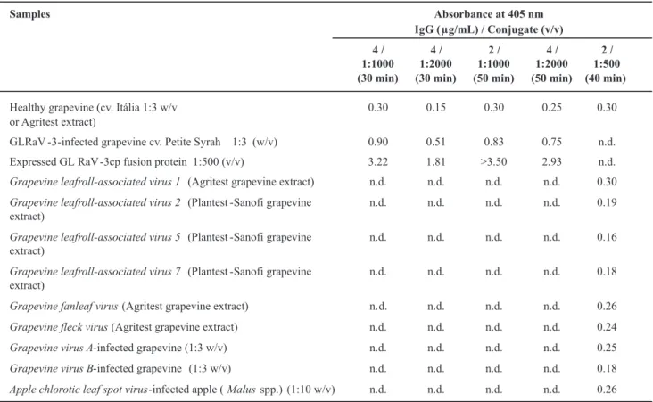
non-TABLE 1 - Results of DAS-ELISA (absorbance values at 405 nm) from grapevine (Vitis spp.) samples using antiserum against GLRaV-3
fusion protein, expressed in Escherichia coli cells
n.d., not determined
serologically related viruses (Table 1). Performance of the antiserum was similar to that obtained with commercial antibodies. Absorbance values from grapevine infected samples suggest that the virus titer was low in evaluated grapevine plants. In this work, DAS-ELISA was chosen to check the antiserum because of its low cost, reliability and practicality as a large-scale virus diagnostic method (Zimmermann et al., 1990; Ling et al., 2000).
Although a number of polyclonal antisera has been raised against recombinant viral proteins, only in a few cases were they effective in detecting the virus by ELISA, as previously reported by Ling et al. (2000) to GLRaV-3, using DAS-ELISA and by Ling et al. (2007) to 2, using PTA-ELISA. In this work, the complete GLRaV-3 capsid protein gene was expressed, while the GLRaV-GLRaV-3 coat protein clone expressed by Ling et al. (2000) covered 96% of the coat protein gene, lacking 13 amino acid residues of the C-terminus. Antibodies raised against recombinant viral proteins are more frequently useful for virus detection through other means, including Western blot, immuno-electron microscopy (ISEM), immunocapture RT-PCR or indirect ELISA (Ling et al., 2007).
Antibodies produced against recombinant antigens may not be functional when used in non-denaturing procedures such as ELISA (Minafra et al., 2000), being more
probably functional when used in denaturing assays such as Western blots (Nickel et al., 2004). Changes in the antigenic structure of the viral CP expressed in bacteria could be an explanation for the weak recognition of the native CP in infected plant samples (Minafra et al., 2000). In the present study the antibodies produced against the in vitro-expressed CP recognized the GLRaV-3cp sensitively and specifically in both systems.
The antibodies produced against the recombinant antigen GLRaV-3cp isolate Pet-4 were not tested against GLRaV-3 isolates Pet-1 to Pet-3, all collected in Northeastern Brazil, from which Pet-4 was shown to differ by only 11 amino acids (Fajardo et al., 2007). However, these antibodies were tested and recognized GLRaV-3 in other infected samples of different origins, such as distinct accessions of Cabernet Sauvignon (A405nm = 1.02), Centennial (1.04), Chardonnay (1.07) and Clara (0.94) cultivars (healthy grapevine A405nm = 0.26).
Barbieri et al. (2004) concluded, based on observations with Watermelon mosaic virus (WMV), family
Potyviridae, genus Potyvirus, that antibodies produced against expressed CPs in E. coli tend to be more specific, reducing the occurrence of unexpected heterologous reactions. This property was shown in the results of Table 1, as expected, the nine non-serologically GLRaV-3-related
Absorbance at 405 nm
IgG ( g/mL) / Conjugate (v/v) Samples
4 /
1:1000
(30 min) 4 /
1:2000
(30 min) 2 /
1:1000
(50 min) 4 /
1:2000
(50 min) 2 /
1:500
(40 min)
Healthy grapevine (cv. Itália 1:3 w/v
or Agritest extract)
0.30 0.15 0.30 0.25 0.30
GLRaV -3-infected grapevine cv. Petite Syrah 1:3 (w/v) 0.90 0.51 0.83 0.75 n.d.
Expressed GL RaV -3cp fusion protein 1:500 (v/v) 3.22 1.81 >3.50 2.93 n.d.
Grapevine leafroll-associated virus 1 (Agritest grapevine extract) n.d. n.d. n.d. n.d. 0.30
Grapevine leafroll-associated virus 2 (Plantest -Sanofi grapevine extract)
n.d. n.d. n.d. n.d. 0.19
Grapevine leafroll-associated virus 5 (Plantest -Sanofi grapevine extract)
n.d. n.d. n.d. n.d. 0.16
Grapevine leafroll-associated virus 7 (Plantest -Sanofi grapevine extract)
n.d. n.d. n.d. n.d. 0.18
Grapevine fanleaf virus(Agritest grapevine extract) n. d. n.d. n.d. n.d. 0.26
Grapevine fleck virus(Agritest grapevine extract) n.d. n.d. n.d. n.d. 0.24
Grapevine virus A-infected grapevine (1:3 w/v) n.d. n.d. n.d. n.d. 0.25
Grapevine virus B-infected grapevine (1:3 w/v) n.d. n.d. n.d. n.d. 0.18
REFERENCES
BARBIERI, M.R., CARVALHO, M.G., ZAMBOLIM, E.M. & ZERBINI, F.M. Expressão em Escherichia coli da proteína capsidial do Watermelon mosaic virus e produção de anti-soro. Fitopatologia Brasileira 29:215-219. 2004.
CLAR�, M.F. & ADAMS, A.N. Characteristics of the microplate method of enzyme linked immunosorbent assay for the detection of plant viruses. Journal of General Virology 34:475-483. 1977.
FAJARDO, T.V.M., �UHN, G.B. & NIC�EL, O. Doenças virais. In: Fajardo, T.V.M. (Ed.). Uva para processamento - Fitossanidade.Uva para processamento - Fitossanidade. Série Frutas do Brasil 35. Brasília DF. Embrapa Informação Tecnológica. 2003. pp. 45-62.
FAJARDO, T.V.M., DIANESE, E.C., EIRAS, M., CERQUEIRA, D.M., LOPES, D.B., FERREIRA, M.A.S.V. & MARTINS, C.R.F. Variability of the coat protein gene of Grapevine leafroll-associated virus 3 in Brazil. Fitopatologia Brasileira 32:335-340. 2007.Fitopatologia Brasileira 32:335-340. 2007.
LING, �.S., ZHU, H.�., ALVIZO, H., HU, J.S., DRONG, R.F., SLIGHTOM, J.L. & GONSALVES, D. The coat protein gene of grapevine leafroll associated closterovirus-3: cloning, nucleotide sequencing and expression in transgenic plants. Archives of
Virology 142:1101-1116. 1997.
LING, �.S., ZHU, H.�., JIANG, Z.� & GONSALVES, D. Effective application of DAS-ELISA for detection of grapevine leafroll associated closterovirus-3 using a polyclonal antiserum developed from recombinant coat protein. European Journal of Plant Pathology 106:301-309. 2000.
LING, �.S., ZHU, H.�., PETROVIC, N. & GONSALVES, D. Serological detection of Grapevine leafroll virus 2 using an antiserum developed against the recombinant coat protein. Journal of Phytopathology 155:65-69. 2007.
MARTELLI, G.P., AGRANOVS��, A.A., BAR-JOSEPH, M., BOSCIA, D., CANDRESSE, T., COUTTS, R.H.A., DOLJA, V.V., FAL�, B.W., GONSALVES, D., JEL�MANN, W., �ARASEV, A.V., MINAFRA, A., NAMBA, S., VETTEN, H.J., WISLER, G.C. & �OSHI�AWA, N. The family Closteroviridae revised. Archives of Virology 147:2039-2044. 2002.
MINAFRA, A., CASATI, P., ELICIO, V., ROWHANI, A., SALDARELLI, P., SAVINO, V. & MARTELLI, G.P. Serological detection of Grapevine rupestris stem pitting-associated virus (GRSPaV) by a polyclonal antiserum to recombinant virus coat protein. Vitis 39:115-118. 2000.
NIC�EL, O., TARGON, M.L.N.P., FAJARDO, T.V.M., MACHADO, M.A. & TRIVILIN, A.P. Polyclonal antibodies to the coat protein of Apple stem grooving virus expressed in Escherichia coli: production and use in immunodiagnosis. Fitopatologia Brasileira 29:558-562. 2004.
NOUEIR�, A.O., LUCAS, W.J. & GILBERTSON, R.L. Two proteins of a plant DNA virus coordinate nuclear and plasmodesmal transport. Cell 76:925-932. 1994.
RIGOTTI, S., BITTERLIN, W. & GUGERLI, P. Production of monoclonal antibodies to Grapevine leafroll-associated virus 7 (GLRaV-7). Extended Abstracts, 15th Meeting of the International
Council for the Study of Virus and Virus-like Diseases of the Grapevine, Stellenbosch, South Africa. 2006. pp. 200-202.
SAMBROO�, J. & RUSSEL, D. Molecular Cloning: A Laboratory Manual. Third edition. New �ork N�. Cold Spring Harbor Laboratory Press. 2001.
TARGON, M.L.P.N., NI�OLAEVA, O., MANJUNATH, �.L., LEE, R.F., MULLER, G.W. & MACHADO, M.A. Coat protein gene of a Brazilian isolate of the Citrus tristeza virus: cloning, expression in E. coli and production of polyclonal antiserum. Fitopatologia Brasileira 22:99-102. 1997.
ZIMMERMANN, D., BASS, P., LEGIN, R. & WALTER, B. Characterization and serological detection of four closterovirus-like particles associated with leafroll disease on grapevine. Journal of Phytopathology 130:205-218. 1990.
Received 7 August 2007 – Accepted 11 December 2007 – FB 7084
viruses (GLRaV-1, GLRaV-2, GLRaV-5, GLRaV-7, GFLV, GFkV, GVA, GVB and ACLSV) (Ling et al., 2007) did not react with the produced GLRaV-3 antisera.
Multiple infections in grapevines, found frequently under field conditions, make virus diagnosis based on symptoms difficult and misleading. Multiple grapevine virus infections also make conventional purification of
Closteroviridae viruses for antisera production impracticable, especially when individual virus species diagnosis is required. The availability of antisera of high sensitivity and specificity, such as those obtained in this study from recombinant antigens, will allow reliable virus diagnosis in these woody hosts.
ACKNOWLEDGEMENTS