HFE gene mutation and oxidative
damage biomarkers in patients with
myelodysplastic syndromes and its
relation to transfusional iron overload:
an observational cross-sectional study
Geane Felix De Souza,1,2Howard Lopes Ribeiro Jr,1,2Juliana Cordeiro De Sousa,1,2 Fabíola Fernandes Heredia,2Rivelilson Mendes De Freitas,3
Manoel Ricardo Alves Martins,4Romélia Pinheiro Gonçalves,5 Ronald Feitosa Pinheiro,2,4Silvia Maria Meira Magalhães2,4
To cite:De Souza GF, Ribeiro HL, De Sousa JC, et al. HFE gene mutation and oxidative damage biomarkers in patients with
myelodysplastic syndromes and its relation to transfusional iron overload: an observational cross-sectional study.BMJ Open 2015;5:e006048.
doi:10.1136/bmjopen-2014-006048
▸ Prepublication history for this paper is available online. To view these files please visit the journal online (http://dx.doi.org/10.1136/ bmjopen-2014-006048). Received 4 July 2014 Revised 10 December 2014 Accepted 11 December 2014
For numbered affiliations see end of article.
Correspondence to Dr Silvia Maria Meira Magalhães; silviamm@ufc.br
ABSTRACT
Objective:A relation between transfusional IOL (iron overload), HFE status and oxidative damage was evaluated.
Design, setting and participants:An observational cross-sectional study involving 87 healthy individuals and 78 patients with myelodysplastic syndromes (MDS) with and without IOL, seen at University Hospital of the Federal University of Ceará, Brazil, between May 2010 and September 2011.
Methods:IOL was defined using repeated measures of serum ferritin≥1000 ng/mL. Variations in the HFE gene were investigated using PCR/restriction fragment length polymorphism (RFLP). The biomarkers of oxidative stress ( plasmatic malonaldehyde (MDA), glutathione peroxidase (GPx) and superoxide dismutase (SOD)) were determined by spectrophotometry.
Results:The HFE gene variations were identified in 24 patients (30.77%) and 5 volunteers (5.74%). The H63D variant was observed in 35% and the C282Y variant as heterozygous in 5% of patients with MDS with IOL. One patient showed double heterozygous variant (C282Y/ H63D) and serum ferritin of 11 649 ng/mL. In patients without IOL, the H63D variant was detected in 29.34%. Serum MDA levels were highest in patients with MDS with IOL, with a significant difference when compared with patients without IOL and healthy volunteers, pointing to the relationship between IOL and oxidative stress. The GPx and SOD were also significantly higher in these patients, indicating that lipid peroxidation increase was followed by an increase in antioxidant capacity. Higher ferritin levels were observed in patients with HFE gene variation. 95.7% of patients with MDS with the presence of HFE gene variations had received more of 20 transfusions.
Conclusions:We observed a significant increase in MDA levels in patients with MDS and IOL, suggesting an increased lipid peroxidation in these patients. The accumulation of MDA alters the organisation of membrane phospholipids, contributing to the process of cellular degeneration. Results show that excess iron
intensifies the process of cell damage through oxidative stress.
Trial registration number:Local Ethics Committee (licence 150/2009).
Strengths and limitations of this study
▪ Oxidative stress and its effects on cellular
biology, DNA damage and carcinogenesis have become a hot topic in MDS research. The para-meters evaluated in this study are important indi-cators the oxidative status and pointed to the relationship between IOL and oxidative stress in patients with MDS.
▪ Our study found iron overload, a significant
prog-nostic factor for overall survival in lower risk MDS patients. Worse clinical outcomes, including cardiac, hepatic and endocrine dysfunction, leu-kemic progression and infectious complications have also been associated with iron overload.
▪ Despite its limitation as an acute phase reactant,
serum ferritin remains a valuable tool for diagno-sis and monitoring iron overload, given its wide availability, low cost and well standardized mea-sures. More prolonged follow-up can confirm our findings, how the ferritin levels higher in all groups of patients and healthy volunteers with gene HFE mutation.
▪ The results of this study suggested that the
pres-ence of HFE gene variations, can be directly corre-lated with the need for blood transfusion, which can induce to iron overload. There was a direct relationship between oxidative stress markers and iron overload, but no was observed significant effect with the HFE gene variation and the oxida-tive stress markers in patients with MDS.
▪ The number of cases evaluated herein may not
INTRODUCTION
Myelodysplastic syndromes (MDS) comprise a heteroge-neous range of haematopoietic diseases characterised by bone marrow failure and a variable propensity to evolve into acute myeloid leukaemia. Most patients have anaemia at some point in the course of the disease and many develop transfusion dependence and consequent iron overload (IOL), considered to be a negative inde-pendent prognostic factor associated with a higher risk of leukaemic transformation and shorter survival.1 The pathogenesis of MDS is complex and involves the haem-atopoietic stem cells, bone marrow microenvironment and the interaction between them. Recently, attention has been focused on oxidative stress and its negative effect on self-renewal and the number of haematopoietic stem cells. Nuclear and mitochondrial DNA can be dir-ectly damaged by hydroxyl radicals resulting in genomic instability contributing to disease progression.2 3 Excess of iron due to multiple transfusions is toxic through the production of free radicals derived from activated oxygen species, which eventually form hydroxyl radicals from superoxide or hydrogen peroxide resulting in organ dysfunction.4Hereditary haemochromatosis (HH) is an autosomal recessive disorder characterised by the enhanced intestinal absorption of dietary iron associated with the presence of variations in the HFE gene located in the short arm of chromosome 6.5The gene variations result in a tyrosine substitution by cysteine residue at position 282 (C282Y allele), exchange of histidine to aspartic acid at amino acid position 63 (H63D allele) and substitution of cysteine for serine at amino acid pos-ition 65 (S65C allele). Patients with HH are most fre-quently either homozygous for C282Y or compound heterozygous for C282Y/H63D.6HFE gene variants cor-relate with body iron levels and have shown association with cancer risk including childhood acute lympho-blastic leukaemia (ALL).7 Increased IOL and iron-mediated oxidative stress may be directly involved in the pathogenesis of MDS. We hypothesise if the presence of HH gene variations in homozygous as well as heterozy-gous forms could contribute to a significantly higher rate of iron accumulation in the context of transfusion dependence.8 The aim of this study was to evaluate the presence of variations in the HFE gene and the oxida-tive status in patients with MDS with IOL and compare these findings with those of patients without IOL and healthy individuals.
MATERIALS AND METHODS
This observational cross-sectional study included 78 sequential adult patients with MDS, 58 without and 20 with IOL, seen at University Hospital of the Federal University of Ceará, Brazil, between May 2010 and September 2011. The disease was classified according to the French American British (FAB) and/or WHO cri-teria9 and the International Prognostic Scoring System (IPSS).10 IOL was defined using repeated measures of
serum ferritin ≥1000 ng/mL. The control group was composed of 87 healthy volunteers. All patients were included at diagnosis and all samples were collected prior to any treatment or chelation therapy. Those with some conditions known to influence oxidative stress bio-markers were excluded ( pregnancy, alcoholism, smoking, alcoholism, use of vitamins, chronic renal failure and hepatitis).
DNA EXTRACT, GENOTYPING AND PCR-RFLP ANALYSIS
Genomic DNA was extracted from peripheral blood using the method described by Biometrix Diagnostica/ DNA Biopur. HFE genotyping for the C282Y, H63D and S65C mutations was performed using the PCR/restriction fragment length polymorphism (PCR-RFLP). The proto-col was the same as that described by Feder11 and Simonsen.12 Fragments of 296bp and 145bp for the wild alleles (282CC), of 296bp, 116bp and 29bp for the homo-zygous allele (282YY) and of 296bp, 145bp, 116bp and 29bp for the heterozygous allele (282CY) were used for identification. Only one fragment of 496bp was used for the homozygous allele (63DD) and 496bp, 352bp and 144bp were used for the heterozygous allele (63HD). The fragments 352bp and 144bp corresponded to the wild allele (63HH). The fragments of 274bp, 147bp, 69bp and 6bp were used to detect the wild allele (65SS), the fragments 274bp, 216bp, 147bp, 69bp and 6bp for the heterozygous allele (65SC) and the fragments 274bp, 216bp and 6bp for the allele mutant (65CC) (figure 1).
HAEMATOLOGICAL PARAMETERS, IRON AND OXIDATIVE STATUS
All samples were obtained at least 8 days following the latest transfusion. Peripheral blood samples collected with EDTA were used for haematological analysis and evaluation of glutathione peroxidase (GPx) and super-oxide dismutase (SOD), or collected with heparin for malonaldehyde (MDA) evaluation. Serum iron and transferrin saturation were assayed using standard techniques (Olympus AU 400e—Siemens) and serum ferritin was measured by enzyme immunoassay (Abbott Laboratory). The MDA analysis was based on its reaction with thiobarbituric acid (TBARS) at a temperature of 100°C.13 Antioxidant activity was evaluated using the kit Ransel for GPx and kit Ransod for SOD.
STATISTICAL ANALYSIS
test or by Kruskal-Wallis test followed by Dunns test as appropriate. Significant differences between the patients with and without IOL were tested by two-way test t-Student and Mann-Whitney. Statistical analysis was per-formed using the GraphPrism program (V.5.01). The level of significance was set at p<0.05.
RESULTS
Median age of the control group (group 1), patients with MDS without IOL (group 2) and patients with IOL (group 3) were 75, 63.8 and 68.9 years, respectively. Patients with IOL received an average of 42.8 transfusions and had a mean serum ferritin level of 2880 ng/mL. Patient characteristics are shown intable 1.
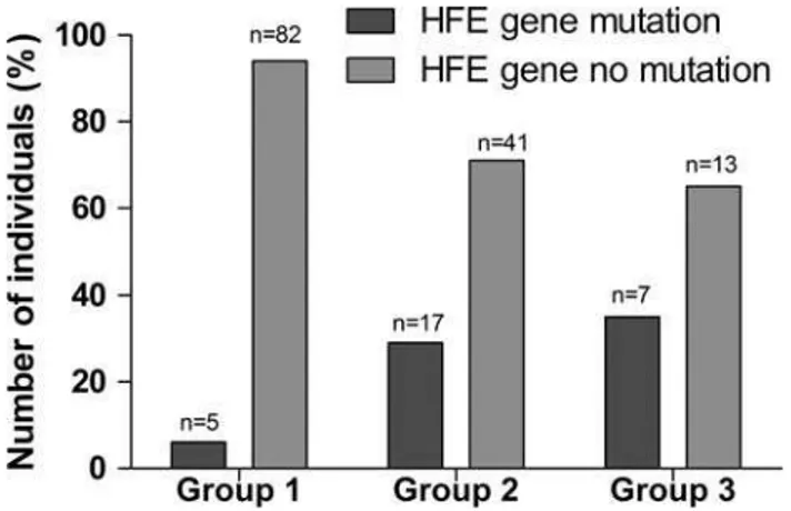
Of the 78 patients, 24 (30.77%) were identified as having at least one variation in the HFE gene, being 14 (58.33%) in male patients. Only 5 volunteers (5.75%) presented with gene variation. In the patients with MDS with IOL, the H63D variant was observed as homozygous in 5% (1/20), as heterozygous in 30% (6/20), and the C282Y variant was detected as heterozygous in 5% (1/20). In one patient, a double heterozygous variant was detected (C282Y/H63D). In the group of patients without IOL, the H63D variant was detected as homozy-gous in 5.2% (3/58) and as heterozyhomozy-gous in 24.14% (14/58). No S65C mutation was detected. The distribu-tion of the genotypes according to variadistribu-tion type is shown intable 2andfigure 2.
The allele frequency for the H63D allele was 3.05%, 14.66% and 17.50% in the control group, patients without IOL and patients with IOL, respectively. For the C282Y allele, the frequency was 2.5%, detected only in patients with IOL.
The genotype frequencies of the HFE variant in con-trols and in patients with and without IOL were signifi -cantly different ( p=0.0013 and 0.0002, respectively), higher in patients with and without IOL. However, no significant difference was observed between these two groups of patients ( p=0.6345).
Seven patients with IOL (35%) were identified with mutation in the HFE gene. Of three mutations in the gene HFE (C282Y; H63D; S65C) evaluated, one of our cases presented two variants (C282Y; H63D) and the highest level of serum ferritin (11 649 ng/mL). Double variant has a higher risk of iron accumulation. Initial investigation showed haemoglobin of 5 g/dL, white cell count 0.641×10⁹/L and platelet count 2×10⁹/L. Cytogenetic analysis demonstrated a complex karyotype (42XY, -18, -19, -21, -22/46, XY). At follow-up, the patient presented with remarkable pancytopenia with transfusion dependence. After transfusion of 24 units of packed red blood cells (RBCs), serum ferritin was as high as 11 649 ng/mL, afigure confirmed later on. The patient was treated with decitabine 20 mg/m2/day for 5 days and died soon afterfinishing thefirst cycle.
The ferritin levels were higher in all groups of patients with HFE gene variation (table 3). Healthy volunteers with HFE gene variation also showed a significant increase in ferritin levels ( p=0.0054).
The majority of the patients with MDS (87.5%) who had the non-variant HFE gene received less than 10 transfusion units. However, the number of patients with MDS with mutations in the HFE gene (95.7%) received more than 20 transfusions. The presence of HFE gene variations in homozygous as well as heterozygous forms was correlated with the need for blood transfusion (χ2 test: 42.92, 9; p<0.0001).
Patients with IOL showed a significant increase in plasma MDA when compared with all groups ( p<0.0001). Levels of MDA were higher in the IOL group compared with the group without IOL. When MDA levels were compared according to the presence of
the HFE gene variant for patients without IOL, no statis-tically significant difference was observed, although there was a trend of increase in the concentration of MDA in the group with HFE variant ( p=0.2789;table 3). The antioxidant enzymes, SOD and GPx, were signifi -cantly higher in patients with IOL when compared with patients without IOL ( p=0.0289 and 0.0267, respectively). GPx activity was also significantly higher in patients with HFE gene variation. However, in the group of patients with no IOL, no significant difference was observed between mutated and non-mutated cases (table 3).
DISCUSSION
Anaemia is the most frequently observed cytopenia in patients with MDS. As elderly patients are especially
vulnerable to anaemia-related comorbidities, most patients require RBC transfusions as a supportive care. Excess iron, due to multiple transfusions (>20 units of packed red cells), is toxic through the production of free radicals derived from activated oxygen species, resulting in organ dysfunction and oxidative stress.14 In the IOL group, we detected an average of over 40 blood transfusions. In Brazil, patients with transfusional iron overload must wait for up to 1 year for an interview by a haematologist. This may explain the high number of patients with increased serum ferritin at diagnosis.
A few studies have reported a positive correlation between IOL and increased production of reactive oxygen species in MDS,15 16 including a recent report by our group in which the preliminary results of some patients have been mentioned.17 Recently, improvement of
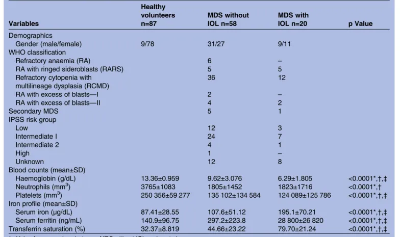
iron-Table 1 Clinical and laboratory characteristics of patients with myelodysplastic syndromes (MDS) with and without iron overload (IOL) and healthy volunteers
Variables
Healthy volunteers n=87
MDS without IOL n=58
MDS with
IOL n=20 p Value
Demographics
Gender (male/female) 9/78 31/27 9/11
WHO classification
Refractory anaemia (RA) 6 –
RA with ringed sideroblasts (RARS) 5 5 Refractory cytopenia with
multilineage dysplasia (RCMD)
36 12
RA with excess of blasts—I 2 –
RA with excess of blasts—II 4 2
Secondary MDS 5 1
IPSS risk group
Low 12 3
Intermediate I 24 7
Intermediate 2 4 1
High 1 –
Unknown 12 8
Blood counts (mean±SD)
Haemoglobin (g/dL) 13.36±0.959 9.62±3.076 6.29±1.805 <0.0001*,†,‡
Neutrophils (mm3) 3765±1083 1805±1452 1823±1716 <0.0001*,†
Platelets (mm3) 250 356±59 277 135 102±134 584 124 089±125 786 <0.0001*,†,‡
Iron profile (mean±SD)
Serum iron (µg/dL) 87.41±28.55 107.6±51.12 195.1±70.21 <0.0001*,†,‡
Serum ferritin (ng/mL) 140.9±96.75 297.2±223.8 28 800±26 820 <0.0001*,†,‡
Transferrin saturation (%) 32.37±8.819 44.66±23.22 79.70±21.24 <0.0001*,†,‡
*p Value for comparison between MDS without IOL and control group. †p Value for comparison between MDS with IOL and control group. ‡p Value for comparison between MDS with IOL and MDS without IOL.
MDS, myelodysplastic syndromes; IOL, iron overload; IPSS, International Prognostic Scoring System; WHO, World Health Organization.
Table 2 HFE genotype frequency distribution according to polymorphism type
Genotype (n=165) WT/C282Y, n (%) H63D/H63D, n (%) H63D/WT, n (%) C282Y/H63D, n (%) WT/WT, n (%)
Healthy volunteers (87) 5 (5.75) 82 (94.25)
MDS without IOL (58) 3 (5.2) 14 (24.1) 41 (70.7)
MDS with IOL (20) 1 (5.0)* 1 (5.0) 5 (25.0) 1 (5.0) 13 (65.0)
*This patient had double heterozygous variant (C282Y/H63D).
mediated oxidative damage after treatment with defera-sirox has been demonstrated by several authors.18–20
In this study, MDA values were significantly higher in patients with IOL when compared with patients without IOL and this was supposed to be independent of the polymorphism in the HFE gene, once the presence of the HFE variant did not show the same effect in patients without IOL. The presence of mutation in the group without IOL did not directly affect the parameters of lipid peroxidation. The antioxidant enzymes were also significantly higher in patients IOL, indicating that lipid peroxidation increase was followed by an increase in anti-oxidant capacity. Although current understanding of these effects is still waiting for more evidence, it is consensual that IOL should be monitored and managed in a selected group of patients based on risk stratification, life expectancy, iron burden and ongoing transfusion need.
According to most published reports worldwide, the C282Y variant corresponds to 80% of cases with clinical manifestations of HH and the remaining patients are mostly compound heterozygous (C282Y/H63D) for the
HFE gene.21 22In the present study, the C282Y allele fre-quency, observed only in patients with IOL, was similar to that previously reported in other haematological dis-eases and IOL.
The genotypic frequency observed for the C282Y/ H63D compound heterozygote was in agreement with the data reported for the American population.23 Double heterozygote has a higher risk of iron accumula-tion as was presented in one of our cases.
Our findings regarding the frequency of HFE gene variants were different from those reported by Nearman et al that compared patients with MDS with refractory anaemia and healthy controls but were similar to the results obtained by Varkonyi et alwho reported a signifi -cant increase in patients with MDS compared with the control group.24 25 List et al analysed 94 patients with MDS for HFE mutations and detected 25.5% of patients heterozygous for H63D,26 afigure similar to the overall frequency observed in this study. However, the frequency of the H63D allele (3.05%) in the control group was lower than that reported for Brazilian blood donors: 2.1% and 13.4% for C282Y and H63D, respectively.27 28
It would be reasonable to hypothesise that the pres-ence of HFE gene variation contributes to increased serum MDA values. However, Parkkila et al29 demon-strated that HFE gene variation did not account for add-itional harm in patients with transfusional IOL with acute myeloid leukaemia. In the present report, statis-tical analysis showed no significant difference when patients with and without gene variation were compared. In the specific group of patients with MDS, the increased absorption of iron secondary to a decrease in the hepcidin level and the transfusional iron loading were the main factors of IOL, probably overcoming the effect of the presence of the HFE variant.
The results of this study suggested that the presence of HFE gene variations in homozygous and heterozygous forms can be directly correlated with the need for blood transfusion, which can induce IOL. Increased MDA
Figure 2 Number of individuals analysed and percentage of HFE gene mutations (C282Y; H63D) in the groups studied.
Table 3 Values of ferritin, MDA, SOD and GPx in the groups according to the presence of mutation HFE gene
Group
Parameters Mean±SD
HFE gene no mutation n=41 (70.7%)
HFE gene mutation
n=17 (29.3%) p Value
MDS without IOL (n=58) FRT (ng/dL) 205.3±139.7 518.6±236.4 <0.0001*
MDA (µM) 9.592±3.073 10.91±4.651 0.2789
SOD (Ug/Hb) 3497±603.8 3590±563.1 0.6376 GPx (Ug/Hb) 76.46±17.73 91.16±7.489 0.0046*
Group
Parameters Mean±SD
HFE gene no mutation n=13 (65%)
HFE gene mutation
n=7 (35%) p Value
MDS with IOL (n=20) FRT (ng/dL) 1883±1236 4731±3684 0.0189†
MDA (µM) 13.30±1.227 14.85±1.195 0.0273†
SOD (Ug/Hb) 4850±420.60 5334±310.10 0.0289†
GPx (Ug/Hb) 144.50±16.95 165.40±15.27 0.0267†
*p Value for comparison between MDS without IOL and with mutation versus MDS without IOL and HFE gene no mutation. †p Value for comparison between MDS with IOL and with mutation versus MDS with IOL and HFE gene no mutation.
levels in patients with MDS and IOL suggests elevated lipid peroxidation and that excess iron intensifies the process of cell damage through oxidative stress. However, the number of cases evaluated may not be rep-resentative and larger studies are necessary to confirm these data.
Author affiliations
1Post-Graduate Program in Medical Science, Department of Clinical Medicine,
Federal University of Ceara, Fortaleza, Ceara, Brazil
2Cancer Cytogenomic Laboratory, Federal University of Ceara, Fortaleza, Ceara,
Brazil
3Department of Pharmacia, Federal University of Piauí, Teresina, Piauí, Brazil 4Department of Clinical Medicine, Federal University of Ceara, Fortaleza, Ceara,
Brazil
5Department of Clinical and Toxicological Analysis, Federal University of
Ceara, Fortaleza, Ceara, Brazil
Twitter Follow Geane de Souza at @geanefelix@ig.com.br
AcknowledgementsThis work was supported by CNPq (Conselho Nacional de Desenvolvimento Científico e Tecnológico) e FUNCAP (Fundação de Amparo à Pesquisa do Estado do Ceará).
Contributors GFdS and SMMM contributed to conception and design. GFdS, SMMM, RFP, RPG and RMdF provided the study materials or patients. HLRJ, JCdS and FFH were involved in collection and assembly of data. GFdS, SMMM and MRAM were involved in data analysis and interpretation. GFdS and SMMM were involved in manuscript writing. All authors approved the final manuscript.
Competing interests None. Patient consent Obtained.
Ethics approval The project received approval from the local ethic committee (0150.12.09).
Provenance and peer review Not commissioned; externally peer reviewed. Data sharing statement No additional data are available.
Open Access This is an Open Access article distributed in accordance with the Creative Commons Attribution Non Commercial (CC BY-NC 4.0) license, which permits others to distribute, remix, adapt, build upon this work non-commercially, and license their derivative works on different terms, provided the original work is properly cited and the use is non-commercial. See: http:// creativecommons.org/licenses/by-nc/4.0/
REFERENCES
1. Malcovati L, Della Porta MG, Pascutto C,et al. Prognostic factors and life expectancy in myelodysplastic syndromes classified according to WHO criteria: a basis for clinical decision making.
J Clin Oncol2005;23:7594–603.
2. Ghoti H, Amer J, Winder A,et al. Oxidative stress in red blood cells, platelets and polymorphonuclear leukocytes from patients with myelodysplastic syndromes.Eur J Haematol2007;79:463–7. 3. Rassool F, Gaymes TJ, Omidvar N,et al. Reactive oxygen
species, DNA damage, and error-prone repair: a model for genomic instability with progression in myeloid leukemia?Cancer Res
2007;67:8762–71.
4. Kohgo Y, Ikuta K, Ohtake T,et al. Body iron metabolism and pathophysiology of iron overload.Int J Haematol2008;88:7–15. 5. Guerreiro RJ, Bras JM, Santana I,et al. Association of HFE common
polymorphisms with Parkinson’s disease, Alzheimer’s disease and mild cognitive impairment in a Portuguese cohort.BMC Neurol
2006;6:24.
6. Beutler E. Hemochromatosis: genetics and pathophysiology.Annu Rev Med2006;57:331–47.
7. Kennedy AE, Kamdar KY, Lupo PJ,et al. Examination of HFE association with childhood leukemia risk and extension to other iron regulatory genes.Leuk Res2014;38:1055–60.
8. Nearman ZP, Szpurka H, Serio B,et al. Hemochromatosis-associated gene mutations in patients with myelodysplastic syndromes with refractory anemia with ringed sideroblasts.Am J Hematol2007;82:1076–9.
9. Vardiman JW, Harris NL, Brunning RD. The World Health (WHO) classification of the myeloid neoplasms.Blood2002;100:2292–302. 10. Greenberg P, Cox C, Lebeau MM,et al. International scoring system
for evaluating prognosis in myelodysplastic syndromes.Blood
1997;89:2079–88.
11. Feder JN, Gnirke A, Thomas W,et al. A novel MHC class I-like gene is mutated in patients with hereditary haemochromatosis.Nat Genet
1996;13:399–408.
12. Simonsen K, Dissing J, Rudbeck L,et al. Rapid and simple determination hereditary haemochromatosis mutation by multiplex PCR-SSCP: detection of a new polymorphic mutation.Ann Hum Genet1999;63:193–7.
13. Draper HH, Hadley M. Malonaldehyde determination as index of lipid peroxidation.Methods Enzymol1990;186:421–31.
14. Gattermann N, Rachmilewitz EA. Iron overload in MDS pathophysiology, diagnosis and complications.Ann Hematol
2011;90:1–10.
15. Saigo K, Takenokuchi M, Hiramatsu Y,et al. Oxidative stress levels in myelodysplastic syndrome patients: their relationship to serum ferritin and haemoglobin values.J Int Med Res2011;39:1941–5. 16. Farquhar MJ, Bowen DT. Oxidative stress and the myelodysplastic
syndromes.Int J Hematol2003;77:342–50.
17. De Souza GF, Barbosa MC, Santos TE,et al. Increased parameters of oxidative stress and its relation to transfusion iron overload in patients with myelodysplastic syndromes.J Clin Pathol
2013;66:996–8.
18. Kikuchi S, Kobune M, Iyama S,et al. Improvement of iron-mediated oxidative DNA damage in patients with transfusion-dependent myelodysplastic syndrome by treatment with deferasirox.Free Radic Biol Med2012;53:643–8.
19. Ghoti H, Fibach E, Merkel D,et al. Changes in parameters of oxidative stress and free iron biomarkers during treatment with deferasirox in iron-overloaded patients with myelodysplastic syndromes.Haematologica2010;95:1433–4.
20. Saigo K, Kono M, Takagi Y,et al. Deferasirox reduces oxidative stress in patients with transfusion dependency.J Clin Med Res
2013;5:57–60.
21. Santos PCJL, Krieger JE, Pereira AC. Molecular diagnostic and pathogenesis of hereditary hemochromatosis.Int J Mol Sci
2012;13:1497–511.
22. Beutler E. Iron absorption in carriers of the C282Y hemochromatosis mutation.Am J Clin Nutr2004;80:799–800.
23. Pietrangelo A. Hereditary hemochromatosis: pathogenesis, diagnosis, and treatment.Gastroenterology2010;139:393–408. 24. Varkonyi J, Demeter J, Tordai A,et al. The significance of the
hemochromatosis genetic variants in multiple myeloma in comparison to that of myelodysplastic syndrome.Ann Hematol
2006;85:869–71.
25. Varkonyi J, Tarkovacs G, Karadi I,et al. High incidence of hemochromatosis gene mutations in the myelodysplastic syndrome: the Budapest Study on 50 patients.Acta Haematol
2003;109:64–7.
26. List AF, Baer MR, Steensma DP,et al. Deferasirox reduces serum ferritin and labile plasma iron in RBC transfusion-dependent patients with myelodysplastic syndrome.J Clin Oncol2012;30:2134–9. 27. Terada CT, Santos PC, Cançado RD,et al. Iron deficiency and
frequency of HFE C282Y gene mutation in Brazilian blood donors.
Transfus Med2009;19:245–51.
28. Viana-Baracioli LM, Tukamoto Junior NC, Ricci Junior O,et al. Comparison of oxidative stress and the frequency of polymorphisms in the HFE gene between hemoglobin S trait blood donors and sickle cell disease patients.Genet Mol Res2011;10:3446–54. 29. Parkkila S, Niemelä O, Britton RS,et al. Molecular aspects of iron
absorption and HFE expression.Gastroenterology
cross-sectional study
iron overload: an observational
syndromes and its relation to transfusional
biomarkers in patients with myelodysplastic
HFE gene mutation and oxidative damage
Feitosa Pinheiro and Silvia Maria Meira Magalhães
Manoel Ricardo Alves Martins, Romélia Pinheiro Gonçalves, Ronald Sousa, Fabíola Fernandes Heredia, Rivelilson Mendes De Freitas, Geane Felix De Souza, Howard Lopes Ribeiro, Jr, Juliana Cordeiro De
doi: 10.1136/bmjopen-2014-006048
2015 5:
BMJ Open
http://bmjopen.bmj.com/content/5/4/e006048
Updated information and services can be found at:
These include:
References
#BIBL http://bmjopen.bmj.com/content/5/4/e006048
This article cites 29 articles, 8 of which you can access for free at:
Open Access
http://creativecommons.org/licenses/by-nc/4.0/
non-commercial. See:
provided the original work is properly cited and the use is
non-commercially, and license their derivative works on different terms, permits others to distribute, remix, adapt, build upon this work
Commons Attribution Non Commercial (CC BY-NC 4.0) license, which This is an Open Access article distributed in accordance with the Creative
service
Email alerting
box at the top right corner of the online article.
Receive free email alerts when new articles cite this article. Sign up in the
Collections
Topic
Articles on similar topics can be found in the following collections (37)Haematology (incl blood transfusion)
Notes
http://group.bmj.com/group/rights-licensing/permissions
To request permissions go to:
http://journals.bmj.com/cgi/reprintform
To order reprints go to:
http://group.bmj.com/subscribe/