H istopathological Study of Experimental and Natural
Infections by
Trypanosoma cruzi
in
D idelphis marsupialis
João Carlos Araujo Carreira/+, Ana M aria Jansen, M aria P D eaneUU, H enrique Leonel Lenzi*
Laboratório de Biologia de Tripanosomatídeos, Departamento de Protozoologia *Departamento de Patologia, Instituto Oswaldo Cruz, Av. Brasil 4365, 21045-900 Rio de Janeiro, RJ, Brasil
Didelphis marsupialis, the most important sylvatic reservoir of Trypanosoma cruzi, can also main-tain in their anal scent glands the multiplicative forms only described in the intestinal tract of triatomine bugs. A study of 21 experimentally and 10 naturally infected opossums with T. cruzi was undertaken in order to establish the histopathological pattern under different conditions.
Our results showed that the inflammation was predominantly lymphomacrophagic and more severe in the naturally infected animals but never as intense as those described in Chagas’ disease or in other animal models.
The parasitism in both groups was always mild with very scarce amastigote nests in the tissues. In the experimentally infected animals, the inflammation was directly related to the presence of amastigotes nests.
Four 24 days-old animals, still in embryonic stage, showed multiple amastigotes nests and moderate inflammatory reactions, but even so they survived longer and presented less severe lesions than experi-mentally infected adult mice.
Parasites were found in smooth, cardiac and/or predominantly striated muscles, as well as in nerve cells. Differing from the experimentally infected opossums parasitism in the naturally infected animals predominated in the heart, esophagus and stomach. Parasitism of the scent glands did not affect the histopathological pattern observed in extraglandular tissues.
Key words: Trypanosoma cruzi - Didelphis marsupialis - opossum - histopathology
Marsupials of the genus Didelphis are consid-ered the most important natural reservoir of
Trypanosoma cruzi. The first case of natural in-fection by T. cruzi in marsupials was reported by Robertson (1929) in D. marsupialis. Further stud-ies have confirmed the importance of this reser-voir showing levels of natural infection ranging from 17 to 100% (Rodrigues & Mello 1942, Guimarães & Jansen 1943, McKeever & Gorman 1958, Herrera & Urdaneta-Morales 1992, Travi et al. 1994).
Studying natural infections, Zeledon et al. (l970) found only 19 infected out of 100 opos-sums, detecting few but typical amastigotes in sec-tions of the heart, esophagus, small and large in-testine and skeletal muscle. Petana (1969) observed only very few and small amastigote nests in the heart muscle of one naturally infected opossum. Brito and Deane (1966) examined 8 opossums, and although only 1 had amastigotes in the stomach,
they observed interstitial inflammatory reactions in the heart, stomach and intestine in all of them. Barr et al. (1991) examining 48 opossums, ob-served myocarditis in 22, although only 6 exhib-ited amastigotes nests in the heart and in the tongue. These authors did not mention inflammatory reac-tions related to T. cruzi in other sites in the animal. More recently Herrera and Urdaneta-Morales (1992) observed only few parasites in the heart of 2 naturally infected opossums.
Parasitological studies (Deane et al.1984a, Jansen et al. 1985, Jansen et al. 1991) of opossums experimentally infected with T. cruzi showed that since very young, opossums resist to experimental infections with high inocula and that two types of infection could occur: (1) apparently self-cured, where the results of sub-inoculation (in mice), xe-nodiagnosis and hemoculture became negative within a few weeks after inoculation, and the im-munofluorescent antibody test (IFAT) was always positive at low titers and no invasion of the scent glands could be observed; (2) infection where the parasites were often intermittently recovered through xenodiagnosis and/or hemoculture, after many months; IFAT was persistently positive at high titers and most of the animals presented
para-+Corresp.author.: E. mail: carreira@dcc001.cict.fiocruz.br
UIn memoriam, deceased 13 August 1995
sites in the scent glands. These differences were considered by Deane et al. (1984a,b) as related to the T. cruzi-strain, since: Y strain inoculations re-sulted in the first and F or sylvan-isolates inocula-tions resulted in the second pattern of infec-tion. An important aspect of the T. cruzi -opos-sum interaction is the cycle of the parasite in the opossum’s scent glands where, T. cruzi shows simi-lar evolutional forms as in the triatomid bugs (Deane et al. 1984a, Lenzi et al. 1984, Deane et al. 1986). Several authors have confirmed the occur-rence of epimastigotes and trypomastigotes in the scent glands of naturally infected opossums (Naiff et al. 1986, Steindel et al.1987, Fernandes et al. 1989). The passage of T. cruzi antigens through the glandular epithelium to blood circulation, stimulating antibody production, was observed by Jansen et al. (1988), however the histopathological consequences of this event in extraglandular tis-sues was still unknown. Despite several works de-scribing natural infection of opossums there are only a few focusing on the histopathological as-pects. We undertook our study at this point in or-der to describe histopathological findings of natu-rally and experimentally infected opossums with laboratory appraised T. cruzi-strains under differ-ent conditions.
MATERIALS AND METHODS
The host - Opossums, identified as D. marsupialis, were: (a) specimens conceived, born and reared in captivity and, (b) specimens trapped in Jaguanum Island, State of Rio de Janeiro, south-east Brazil.
The parasite - T. cruzi strains used for experi-mental infections were: (a) Y, isolated, from a hu-man patient (Silva & Nussenzweig 1953) and (b) opossums strains G-N and G-49, isolated from naturally infected D. marsupialis in the Amazon region (Yoshida 1983) and in Rio de Janeiro (iso-lated in our laboratory 1985), respectively.
Among opossums trapped in the field, 10 were infected with T. cruzi, 3 with T. (Mega-trypanum) freitasi and 3 were free of trypanosome infection.
Inoculations - The animals were subcutane-ously (SC) inoculated with bloodstream trypomastigotes (BST) from maintenance mice, or with metacyclic trypomastigotes from triatomine bugs. Inocula were adjusted on the basis of num-ber of 5 x 10 3 parasites per gram of body-weight for 24 and 45 days-old opossums and 2 x 10 3 para-sites per gram of body-weight for the others.
Follow-up - Experimentally or naturally in-fected opossums have been followed with parasi-tological and/or serological tests made at monthly or bi-monthly intervals for periods which covered
more than one year for some naturally infected animals.
Parasitological diagnosis - It was based on: (a) direct blood examination of fresh preparation dur-ing the acute phase; (b) hemoculture in NNN me-dium examined through a period of one to three months.
Serology was done on blood samples obtained by vein (femoral or caudal) or heart puncture and the sera were tested by immunoflourescent anti-body test (IFAT) described by Jansen et al. (1985).
Necropsy - The animals were killed by intra-muscular injection of Ketalar and chloroform in-halation. Tissues samples were fixed in formalin-Millonig (Carson et al. 1973) and paraffin sections stained with hematoxilin and eosin (H&E). At least one section of the following tissues was examined from all the animals: tongue, tonsils, thyroid, lymph nodes, lung, heart, esophagus, stomach, liver, spleen, diaphragm muscle, small and large intes-tines, skeletal muscle, bone marrow of different places, pancreas, eyes, brain, cerebellum, skin, ears, gallbladder, bladder, marsupial pouch, epiploon, mesentery, bulbourethral glands, hypophysis, spi-nal chord, kidney, genital tracts and scent glands. The opossums were killed very young, when still attached to their mother teats, had the whole body processed.
RESULTS
Most of the examined animals presented few amastigotes in small, rare and dispersed nests lo-calized mainly in skeletal muscle. The inflamma-tory reactions were constituted predominantly of lymphocytes and macrophages. In experimentally infected animals the parasites were randomly dis-tributed with mild inflammatory reactions. On the contrary, in the naturally infected opossums the nests of amastigotes were preferentially located in the digestive tract and heart with more intense in-flammation and occasional tissue destruction.
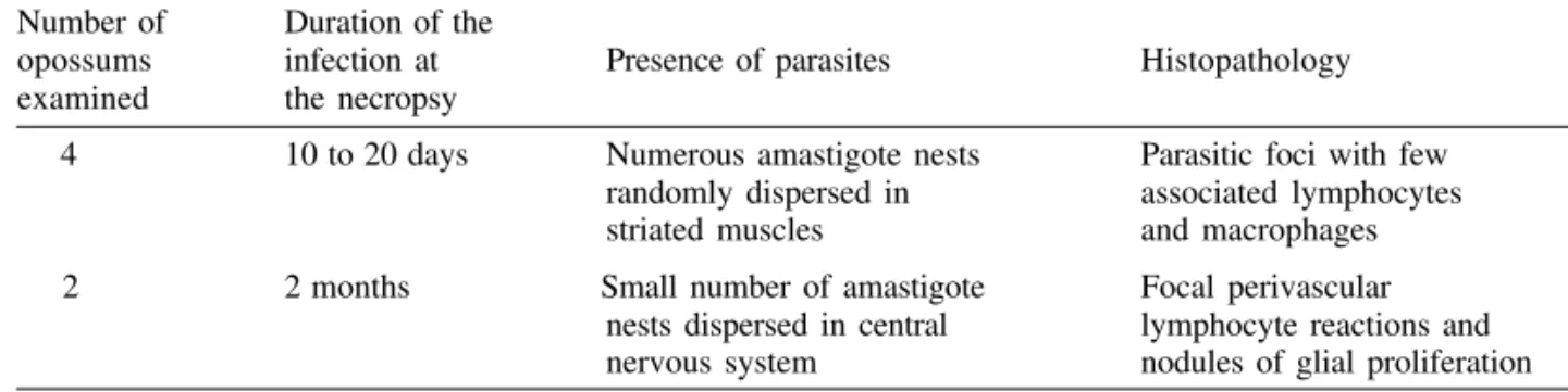
Table I shows the histopathological lesions in newborn opossums: in the 45 days old animals inoculated with Y strain, the parasites were lim-ited to the central nervous system associated with a mild inflammatory reaction (Fig. 1), differing from 24 days old opossums in which the parasites were randomly dispersed in striated muscles with lymphocytes and macrophages only rarely associ-ated (Fig. 2).
for 3-4 weeks, after which also the hemocultures became negative. No invasion of the scent glands were detected in fresh preparations of the scent glands content. The total immunoglobulins titers oscillated between 1:40-1:80.
A different picture could be observed in ani-mals inoculated with the opossum G-N strain at the same age in which we found small amastigote nests distributed in striated muscle fibers and scent glands.
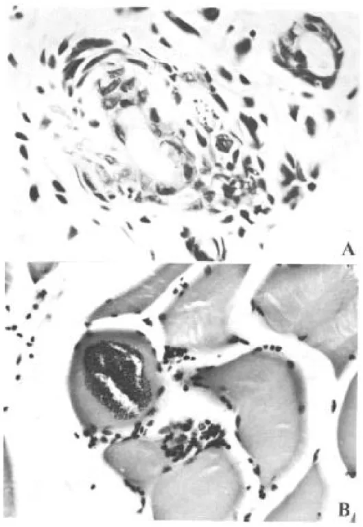
Although only one out of the 10 opossums in-oculated with G-49 strain presented parasites in the tissues (Fig. 3), the duration of the chronic infec-tion did not determine the severity of the lesions.
Indeed, opossums inoculated with this strain at 80 days old and killed between 5 and 12 months after inoculation exhibited almost the same histopathological pattern (Table II).
Two opossums experimentally infected with the G-49 strain and killed in the acute phase (one month) displayed a large number of parasite nests dispersed throughout several tissues (data not shown). Both animals presented patent parasitemia - up to 1 x10⁵ flagellates/ml and parasites in the scent glands.
The histopathological findings were similar in the animals with or without parasites in the scent glands (Table II).
TABLE II
Histopathologic follow-up of 10 pouch young opossums (≅ 80 days) inoculated subcutaneously with a
Trypanosoma cruzi isolate (G-49) from a naturally infected opossum Didelphis marsupialis - 2 x 10³ metacyclic forms per gram of body-weight
Opossum Duration of infection Presence of parasites Histopathology no. at necropsy (months)
1 5 Abdominal wall, tongue, Mild interstitial inflammatory heart, cranial muscle reactions in heart and tongue
2 6 - Scarse areas of inflammatory
reaction in heart and brain
3 7 Scent glands Scarse inflammatory
reaction in skeletal muscles
4 10 Scent glands
-5 10 -
-6 10 Scent glands
-7 11 Scent glands Scarse inflammatory
reaction in heart
8 11 Scent glands
-9 12 - Scarse inflammatory
reaction in skeletal muscle
10 12 Scent glands
-(-) No parasites observed or no significant histopathologic lesions. TABLE I
Histopathologic lesions related to age in four and two (24 and 45 days old respectively) opossums inoculated subcutaneously with Y strain of Trypanosoma cruzi - 5 x 10³ blood forms per gram of body weight Number of Duration of the
opossums infection at Presence of parasites Histopathology examined the necropsy
4 10 to 20 days Numerous amastigote nests Parasitic foci with few randomly dispersed in associated lymphocytes striated muscles and macrophages
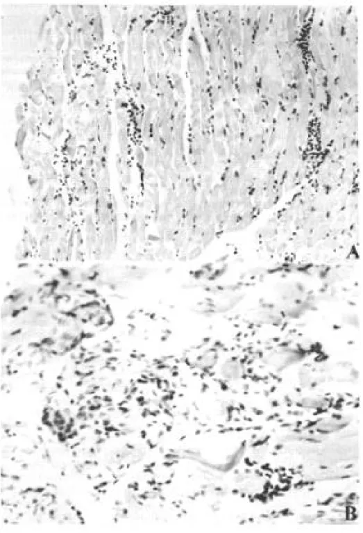
Only 4 out of 10 naturally infected opossums showed amastigotes in the tissues. The heart, stom-ach and esophagus were involved in 2 of them; bladder and ureter in 1. Inflammatory reaction was focal, without fibrosis and directly related to the intensity of tissue parasitism which was usually light and more concentrated in the heart and di-gestive tract (Fig. 4, Table III).
In the heart there was focal and interstitial myocarditis, mainly in atria involving the subendothelial layer. In a single case there was in-flammation in intramural ganglion. In the stom-ach the infiltrate invaded the Auerbstom-ach plexus and
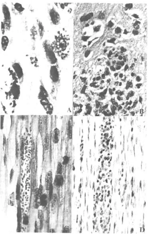
Fig. 2:nests of amastigotes in opossums experimentally infected with G-N strain at 80 days old. A very little nest in perifolicular cell of the skin (A) and a large one in the diaphragm muscle with little inflammatory reaction (B) (H&E - A: x 640 and B: x 310).
muscular layers.
A similar inflammatory reaction pattern lower in intensity was observed in the six T. cruzi in-fected animals with no detectable tissue parasit-ism (Fig. 4).
Besides the natural infection by T. cruzi, all the opossums harbored different species of helmints as well as protozoa, bacteria and fungi.
Opossums naturally infected with T. freitasi as well as the uninfected ones displayed some non-specific lesions - probably due to the presence of other parasites such as helminths and bacteria that were observed in most of animals.
DISCUSSION
Opossum probably starts to resist to infections with Y strain from the 45th dayof life on since all 24 days old inoculated animals died in contrast to the survival of the older ones (Deane et al. 1984c).
Fig. 4:histopathologic lesions of opossums naturally infected. Diffuse inflammatory reaction in the muscle of the leg (A) and in the smooth muscle of the gastric wall with involvement of Auerbach plexus (B) (H&E - A: x 200 and B: x 350).
sue lesions were not detected. Only few parasites and a mild inflammatory reaction in the central nervous system was observed in the 45 days old infected opossums necropsed 2 months after the inoculation with Y strain trypomastigote (Table I). The parasitological follow-up correlated en-tirely with the histopathological findings depend-ing also on the inoculated strain. Opossums ex-perimentally infected with Y strain displayed a short period of patent parasitemia with rare posi-tive fresh blood examinations and no scent glands parasitism. On the contrary, the infections with
G-N strain resulted in high parasitemia up to 1x10⁷ parasites/ml and heavily parasitized scent glands. The monthly hemocultures done during the subpatent period were consistently positive in the G-N inoculated animals, contrasting to those in-oculated with the Y strain (data not shown).
not cause an increase in the inflammatory reactions as it could be expected. Until now, it is unknown if the glandular T. cruzi derived antigens exert stimu-lating or suppressive effects on the immune sys-tem, interfering on the host responses.
The opossums naturally infected with another trypanosomatid - T. freitasi - presented no lesions at all.
Comparing opossums naturally infected by T. cruzi with the experimentally infected ones, the former exhibited more intense inflammatory reac-tions in tissues and a tendency for parasitism to occur in the heart and digestive tract. The pattern of inflammatory reactions in both groups was quali-tatively similar, composed of macrophages and lymphocytes with occasional small numbers of eosinophils and/or plasma cells.
Parasitism in the digestive system by T. cruzi
in natural infections of opossums was already ob-served (Brito & Deane 1966, Zeledon 1970, Barr et al. 1991). Brito and Deane (1966) suggested that it probably could occur through oral infections. Although this hypothesis can be true, T. cruzi
para-sites in the digestive system can also occur follow-ing other infection route as it could be observed in some of the subcutaneously inoculated animals.
In our understanding, several factors contrib-ute to explain the more intense inflammatory reac-tions in natural infecreac-tions, such as the characteris-tics of the strain, schedule of infection and/or the occurrence of polyparasitism. This may possibly play a very important role in the modulation of the inflammatory reactions. The frequent presence of other parasites (like helminths and bacteria) in the organism of the animals could cause a more se-vere immune response due to superimposition of immunological reactions stimulated by different agents. Favoring this hypothesis is the fact that all the animals, caught in the wild and heavily infected by helminths and other parasites, presented more pronounced histopathologic lesions than those ani-mals born in the laboratory.
It was reported that the opossum is able to set up a humoral response to T. cruzi antigens and that the levels of serum specific antibodies is depen-dent on the strain of parasite (Jansen et al. 1985).
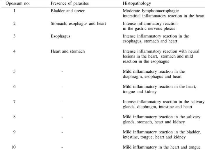
TABLE III
Histopathologic lesions of 10 opossums (Didelphis marsupialis) naturally infected with Trypanosoma cruzi
Opossum no. Presence of parasites Histopathology
1 Bladder and ureter Moderate lymphomacrophagic
interstitial inflammatory reaction in the heart
2 Stomach, esophagus and heart Intense inflammatory reaction in the gastric nervous plexus
3 Esophagus Intense inflammatory reaction in the esophagus, stomach and heart
4 Heart and stomach Intense inflammatory reaction with neural lesions in the heart, stomach and mild reaction in the esophagus
5 - Mild inflammatory reaction in the diaphragm, esophagus and heart
6 - Mild inflammatory reaction in the heart, tongue and kidney
7 - Intense inflammatory reaction in the salivary glands, diaphragm, intestine and heart
8 - Mild inflammatory reaction in the salivary glands, stomach, heart and kidney
9 - Mild inflammatory reaction in the bladder, intestine, tongue, heart and kidney
10 - Mild inflammatory in the heart and tongue
It was also reported by Jansen et al. (1991) that the G-49 strain caused higher levels of immunoglobu-lins and parasitemia compared to G-327, both be-ing of sylvatic origin and isolated from naturally infected opossums.
The Y strain, on the other hand, is known to present low levels of infection and immunoglobu-lins in the opossums (Jansen et al. 1985), and in some cases the animals rid themselves of the para-sites (Thomaz et al. 1984). We observed that the Y strain displays correlated histopathological behav-ior in agreement with parasitological conditions. For instance, when the opossums were close to 80 days-old and the immune system had become ma-ture, neither inflammatory reactions nor parasites in tissues were detected. Otherwise, opossums in-oculated with G-N strain in the same age group presented several amastigotes nests and light tis-sue inflammation.
When we studied animals inoculated with Y strain at 45 and 24 days, we observed several amastigotes nests in tissues and scattered inflam-matory focuses. Even so, the opossums had a longer survival period compared to mice, as observed by Deane et al. (1984c). These results indicate that young opossums, even when they were immuno-logically immature, they were capable of setting up inflammatory responses sufficient to control the infection. When the animal reached immunologi-cal maturity close to 80 days of age, they acquired capability to control the infection. Tissue parasit-ism decreased and amastigotes nests were no longer observed.
The results obtained with the G-49 strain showed that only the animal killed 5 months after inoculation presented tissue parasitism. This data, together with that of natural infection (where amastigotes were found only in 4 of the 10 exam-ined opossums), suggest that sometimes the opos-sums can control the infection by decreasing the number of parasite foci in tissue and subsequent inflammatory reactions.
On the other hand, due to differences of the strain characteristics and the size of inoculum, the opossums do not always control the colonization of the tissue by the parasite. The parasites can re-main in the tissues for a long time, with very lim-ited inflammatory reactions, as we could observe in animals infected with the G-N strain.
T. cruzi is a very heterogenous species; conse-quently, the infection by this parasite may be very complex, depending on the host and parasite fac-tors.
Our observations on the behavior of T. cruzi in the D. marsupialis indicate that even those natu-rally infected animals showing more severe inflam-matory reactions did not present a pronounced
histopathological changes as observed in Chagas’ disease, and in T. cruzi infected mice and dogs (Andrade & Carvalho 1969, Aluja 1985, Andrade & Sadigursky 1987, Molina & Kierszenbaum 1988).
The size of the inoculum, in some way, may modulate the parasite dispersion in the host body. Jansen et al. (1991), have shown that T. cruzi may or may not reach the opossums’ scent glands de-pending on the levels of parasitemia induced by the size of the inoculum. This fact can explain why our naturally infected opossums did not present anal scent glands infection, though some isolates when obtained from the glands and inoculated in others (Jansen et al.1991), were capable of further glandular colonization. In experimental conditions higher parasitemia was induced by larger inocu-lum than that observed in natural infections.
Deane et al. (1984a), observed that opossums select different sub-populations from T.cruzi. Our study shows that this selection is correlated to histopathological findings.
This paper presents for the first time a detailed histopathological study of D. marsupialis harbor-ing simultaneously the two multiplicative life cycles of T. cruzi: amastigote multiplication in the tissues and triatomine vector-like cycle in the scent glands.
ACKNOWLEDGMENTS
To Luzia Fatima Gonçalves Caputo for technical assistance.
REFERENCES
Aluja AS 1985. Miocarditis por Trypanosoma cruzi en un perro. Veterinaria Mex 16: 41-44.
Andrade SG, Carvalho ML 1969. Efeito da excitação do sistema retículo-endotelial pelo adjuvante de Freud, na doença de Chagas experimental. Rev Inst Med Trop São Paulo11: 229-235.
Andrade SG, Sadigursky M 1987. The conduction sys-tem of the heart in mice chronically infected with
Trypanosoma cruzi: histopathological lesions and electrocardiographic correlations. Mem Inst Oswaldo Cruz 82: 59-66.
Barr SC, Brown CC, Dennis VA, Klei TR 1991. Infec-tion of inbred mice with 3 Trypanosoma cruzi iso-lated from Louisiana (USA) mammals. J Parasitol 76: 918-921.
Block M 1964. VI The blood forming tissues and the blood of the newborn opossum (Didelphis marsupialis). Ergeb Anat Entwicklungsgesh 37: 237- 366.
Brito T, Deane LM 1966. Aspectos anátomo-patológicos da infecção natural de alguns animais silvestres pelo
Trypanosoma cruzi. Rev Inst Med Trop São Paulo 8: 79-82.
re-evalua-tion. Am J Clin Path 59: 365-373.
Deane MP, Jansen AM, Lenzi HL 1984a. Trypanosoma cruzi: vertebrate and invertebrate cycles in the same mammal host, the opossum Didelphis marsupialis. Mem Inst Oswaldo Cruz 79: 513-515.
Deane MP, Jansen AM, Mangia RHR, Gonçalves AM, Morel CM 1984b. Are our laboratory “strain” rep-resentative samples of Trypanosoma cruzi popula-tions that circulate in nature? Mem Inst Oswaldo Cruz (Suppl) 79: 19-24.
Deane MP, Jansen AM, Mangia RHR, Lenzi HL 1984c. A study of experimental infection of the opossum
Didelphis marsupialis. Annals of the XI Annual Meeting of Basic Research on Chagas’ Disease (Ab-stract BI-39).
Deane MP, Lenzi HL, Jansen AM 1986. Double devel-opment cycle of Trypanosoma cruzi in the opossum.
Parasitol Today 2: 146-147.
Fernandes AJ, Diotaiuti L, Pinto Dias JC, Romanha AJ, Chiari E 1989. Infecção natural das glândulas anais de gambas Didelphis marsupialis pelo Trypanosoma cruzi no município de Bambuí - MG. Mem Inst Oswaldo Cruz 84: 87-93.
Guimarães FN, Jansen G 1943. Novo transmissor silvestre do Trypanosoma (Schizotrypanum) cruzi
(Chagas 1909). Mem Inst Oswaldo Cruz 38: 473-441.
Herrera L, Urdaneta-Morales S 1992. Didelphis marsupialis: a primary reservoir of Trypanosoma cruzi in urban areas of Caracas, Venezuela. Ann Trop Med Parasitol 86: 607-612.
Jansen AM, Carreira JCA, Deane MP 1988. Infection of a mammal by monogenetic insect trypanosomatids (Kinetoplastida,Trypanosomatidae). Mem Inst Oswaldo Cruz 83: 271-272.
Jansen AM, Galvão BC, Moriearty PL, Deane MP 1985.
Trypanosoma cruzi in Didelphis marsupialis: an indirect fluorescent antibory test for diagnosis and follow up of natural and experimental infections.
Trans R Soc Trop Med Hyg 79: 474-476.
Jansen AM, Leon L, Machado GM, Da Silva MH, Souza-Leão S, Deane MP 1991 Trypanosoma cruzi in the opossum Didelphis marsupialis: Parasitological and serological follow-up of the acute infection. Exp Parasitol 73: 249-259.
Lenzi HL, Jansen AM, Deane MP 1984.The recent dis-covery of what might be a primordial escape mecha-nism for Trypanosoma cruzi. Mem Inst Oswaldo Cruz (Suppl) 79: 7-12.
McKeever J, Gorman L 1958. Occurrence of a
Trypanosoma cruzi-like organism in some mammals from Southwestern Georgia and Northwestern Florida. J Parasitol 44: 583-587.
Molina HA, Kierszenbaum F 1988. Kinetics of devel-opment of inflammatory lesions in myocardial and skeletal muscle in experimental Trypanosoma cruzi
infection. J Parasitol 74: 370-374.
Naiff RD, Naiff MF, Barret TV, Arias JR 1986.
Trypanosoma cruzi nas glândulas anais do Didelphis marsupialis: primeiro registro de infecções naturais. Annals of the X Meeting of the Brazilian Society of Parasitology (Abstract - 165) .
Petana WB 1969. American trypanosomiasis in British Honduras - VI - A natural infection with
Trypanosoma (Schizotrypanum) cruzi in the opos-sum Didelphis marsupialis (Marsupialia, Didelphoidea) and experimental investigations of different wild-animal species as possible reservoirs for the parasite. Ann Trop Med Parasitol 63: 47-56. Robertson A 1929. Note on trypanosome morphologi-cally similar to Trypanosoma cruzi Chagas, 1909, found in opossum, Didelphis marsupialis captured at Tela, Honduras, Central America. XVIII th Ann
Rep Med Dept Co. 293 - 310.
Rodrigues BA, Mello GB 1942. Contribuição ao estudo da tripanosomíase americana. Mem Inst Oswaldo Cruz 37: 77-94.
Silva LIP, Nussenzweig W 1953. Sobre uma cepa de
Trypanosoma cruzi altamente virulenta para camundongo branco. Folia Clin Biol 20: 191-208. Steindel M, Scholz AF, Toma HK, Schlemper Jr BR
1987. Parasitism of blood and anal glands of natu-rally infected opossum (Didelphis marsupialis) from the Arvoredo Island Santa Catarina by Trypanosoma cruzi. Mem Inst Oswaldo Cruz (Suppl) 82: 66. Thomaz N, Jansen AM, Deane MP 1984. Trypanosoma
cruzi. The complement-mediated lysis (Coml) in experimentally infected opossum Didelphis marsupialis.Annals of the XI Annual Meeting of Basic Research on Chagas’ Disease (Abstract, I-48), p.116.
Travi BL, Jaramilllo C, Montoya J, Segura I, Zea A, Gonçalves A, Velez ID 1994. Didelphis marsupialis,
an important reservoir of Trypanosoma (Schizotrypanum) cruzi and Leishmania (Leishma-nia) chagasi in Colombia. Am J Trop Med Hyg 50:
557-565.
Yoshida N 1983. Surface antigens of metacyclic trypomastigotes of Trypanosoma cruzi. Infect Immun 40: 836-839.
Zeledon R, Solono G, Saenzs G, Swatzwelder JC 1970. Wild reservoir of Trypanosoma (Schizotrypanum) cruzi with special mention of the opossum, Didelphis marsupialis, and its role in the epidemiology of Chagas’ disease in an epidemic area of Costa Rica.