219 219 219 219 219 Mem Inst Oswaldo Cruz, Rio de Janeiro, Vol. 93(2): 219-224, Mar./Apr. 1998
Purification and Partial Characterization of
Trypanosoma
cruzi
Triosephosphate Isomerase
SC Bourguignon/
+, MN Meirelles*, RS Pacheco**, S Giovanni De Simone/*/
++Departamento de Biologia Celular e Molecular, Instituto de Biologia, Universidade Federal Fluminense, Niterói, RJ, Brasil *Departamento de Ultraestrutura Celular **Laboratório de Sistemática Bioquímica ***Laboratório de Microseqüenciamento de Proteínas, Departamento de Bioquímica e Biologia Molecular, Instituto Oswaldo Cruz,
Av. Brasil 4365, 21045-900 Rio de Janeiro, RJ, Brasil
The enzyme triosephosphate isomerase (TPI, EC 5.3.1.1) was purified from extracts of epimastigote forms of Trypanosoma cruzi. The purification steps included: hydrophobic interaction chromatography on phenyl-Sepharose, CM-Sepharose, and high performance liquid gel filtration chromatography. The CM-Sepharose material contained two bands (27 and 25 kDa) with similar isoelectric points (pI 9.3-9.5) which could be separated by gel filtration in high performance liquid chromatography. Polyclonal antibodies raised against the porcine TPI detected one single polypeptide on western blot with a mo-lecular weight (27 kDa) identical to that purified from T. cruzi. These antibodies also recognized only one band of identical molecular weight in western blots of several other trypanosomatids (Blastocrithidia culicis, Crithidia desouzai, Phytomonas serpens, Herpertomonas samuelpessoai). The presence of only one enzymatic form of TPI in T. cruzi epimastigotes was confirmed by agarose gel activity assay and its localization was established by immunocytochemical analysis. The T. cruzi purified TPI (as well as other trypanosomatid’ TPIs) is a dimeric protein, composed of two identical subunits with an approxi-mate mw of 27,000 and it is resolved on two dimensional gel electrophoresis with a pI of 9.3. Sequence analysis of the N-terminal portion of the 27 kDa protein revealed a high homology to Leishmania mexicana
and T. brucei proteins.
Key words: Trypanosoma cruzi - triosephosphate isomerase - purification - sequence
Chagas’ disease is caused by the protozoan hemoflagellate Trypanosoma cruzi. This and other kinetoplastids are distinguished from diverse eu-caryotic cells by the presence of glycosomes, a mi-crobody-like organelle containing glycolytic and glycerol metabolic enzymes (Opperdoes 1987, Michels 1989, Blattner et al. 1992). Correspond-ing glycolytical isoenzymes were also found to be present in the cytoplasm of most protozoa studied so far. Most of these proteins are encoded by dif-ferent genes (Sullivan et al. 1985, Huang et al. 1989) and the glycosomal enzymes are synthesised in the cytoplasm by free ribosomes and imported post-translationally without apparent cleavage or modification. Several of these glycosomal and cy-tosolic isoenzymes have been purified from T. brucei (Misset et al. 1986, 1987) and T. cruzi
(Bourguignon et al. 1997) and were distinguished
structurally from their mammalian counterparts (Michels & Opperdoes 1991). In view of this and based on the observation that the inhibition of some glycosomal enzymes from T. brucei (Durieux et
al. 1991) may result in the death of the parasite (Opperdoes et al. 1990, Willson et al. 1993) these enzymes have suggested as promising targets for drug design.
The T. brucei (Swinkels et al. 1986) and Leish-mania mexicana (Kokl et al. 1994)
triose-phos-phate isomerase (TPI, EC 5.3.1.1) genes have been cloned and characterized. This enzyme was shown to be a dimer composed by identical subunits and it was also shown to catalyze the isomerization of the two products of aldolase, D-glyceraldehyde 3-phosphate and dihydroxyacetone 3-phosphate, in the absence of carbohydrates. Moreover, it presents a key position in the metabolism, generating dihy-droxyacetone phosphate in anaerobic condition, through reverse action of the glycerol kinase and glyceraldehyde-phosphate dehydrogenase. The overall structures of these enzymes are strictly con-served despite substantial variation, and approxi-mately 50% in its amino acid sequence has re-mained identical (Fothergill-Gilmore & Michels 1993). In this work we describe the purification and partial characterization of the TPI enzyme from
Suported in part by CNPq, Finep and Fiocruz.
+Recipient of a CNPq fellowship.
++Corresponding author. Fax: +55-21-590.3495. E-mail:
220 220 220 220
220 T. cruzi TPI SC Bourguignon et al.
T. cruzi. In addition, using an enzymatic assay by agarose gel electrophoresis we show that the T. cruzi Dm28c clone expresses only one form of this
enzyme.
MATERIALS AND METHODS
Materials - Phenyl- and CM-Sepharose were
obtained from Pharmacia Fine Chemicals (Uppsala, Sweden); ethylenediamino tetratacetic acid (EDTA), dithiotreitol (DTT), ammonium sulfate, rabbit GAPDH, agarose, trietanolamine, porcine TPI, nonidet P40 (NP40), (3-[4,5-dimethylthiazol-2yl]5-2,5 diphenyltetrazolium bromide) (MTT), phenazine metnosulfate (PMS), glutaraldehyde, paraformaldehyde, cacodilate, chromatographic weight standard proteins and goat anti-rabbit per-oxidase conjugated from Sigma Chemical Co. (St. Louis, Mo, U.S.A.); ampholines, silver staining kit, SDS-molecular weight standard proteins, poly (vinylidenedifluoride) (PVDF) and nitrocellulose membrane from Bio-Rad (Richmond, CA, U.S.A.). The Shinpack Diol-150 HPLC column and the se-quencing reagents were from Wako Pure Chemi-cals (Richmond, CA, U.S.A.) while the electro-phoresis reagents were obtained from Serva (Heidelberg, Germany). Freund’s adjuvants and P10 filters were from Gibco and Amicon (Beverly, MA, U.S.A.) respectively. Liver infusion tryptose (LIT) and brain heart infusion-tryptose (BHI) me-dium were obtained from Difco (Detroit, Michi-gan, U.S.A.). All other reagents and chemicals were from Merck (Darmstadt, Germany).
Parasites - Epimastigote forms from Dm 28c
clone were maintained in liver infusion tryptose (LIT) medium (Contreras et al. 1985) while amastigotes from cellular culture were maintained according to Giovanni De Simone et al. (1987).
Blastocrithidia culicis, Crithidia desouzai,
Phytomonas serpens, L. guianensis and Her-pertomonas samuelpessoai were cultivated in BHI medium (Giovanni De Simone et al. 1987). All parasites were harvested at log phase and washed three times by centrifugation (3,000 g, 10 min) in PBS.
Extraction of the enzyme and binding to phe-nyl-Sepharose column - After washes with PBS,
T. cruzi epimastigote forms were freeze-thawed three times in the same buffer and the insoluble material was pelleted by centrifugation (40,000 g, 30 min, 4oC). The supernatant was designed crude extract and ammonium sulfate was added to a fi-nal concentration of 1M. The extract was applied to a 20 ml phenyl-Sepharose CL-4B column equili-brated with 25mM Tris-HCl pH 7.8, containing 5 mM EDTA, 1mM DTT and 1M (NH4)2SO4 and fractionated using an ammonium sulfate reverse gradient.
Cation-exchange chromatography - After
re-lease from phenyl-Sepharose, the fraction (peak 3) was concentrated on a Centripep P10 filter fol-lowed by replacement of the buffer (0.1M trietanolamine-HCl pH 8.0 containing 1 mM EDTA) and fractionation of the sample on a fast flow CM-Sepharose column. Fractions of 3 ml were collected and concentrated, separately, using Centripep P10 filters and analyzed by sodium dodecyl sulfate polyacrylamide gel electrophore-sis (SDS-PAGE).
Size exclusion HPLC - The CM-Sepharose
pooled peak 2 was concentrated using centricon filters and injected in a Shinpack Diol-150 HPLC column (5 mm; 50 cm x 7.9 mm I.D.), previously equilibrated in 50 mM phosphate buffer, pH 7.2. The proteins were fractioned on an automatic HPLC system (Shimadzu, 6A model) during 28 min at 25oC (Fig. 4). Fractions of 1 ml were col-lected and concentrated as described before. For molecular mass characterization the column was calibrated in the same buffer with the following markers: b-galactosidase (105 kDa), bovine serum
albumin (66 kDa), ovalbumin (45 kDa) and car-bonic anhydrase (29 kDa).
One and two-dimensional PAGE -
One-dimen-sional SDS-PAGE was performed using polyacry-lamide gels in Laemmli buffers (Laemmli 1970) under reduction conditions. Two-dimensional PAGE (2D-PAGE) consisted of isotachophoresis followed by electrophoretic analysis on uniform 15% SDS-PAGE (O’ Farrel et al. 1977). The gels were stained with Coomassie blue R-250 or silver (Bio-Rad silver stain kit). Bovine serum albumin (BSA 66 kDa), hen egg white ovalbumin (45 kDa), bovine carbonic anhydrase (29 kDa), and hen egg white lysozyme (14,4 kDa) were used as standards for characterization of molecular mass.
Agarose gel electrophoresis - This
electro-phoresis was conduced in a Multifor apparatus using a continuous buffer (414 mM Tris and 50 mM citric acid pH 8.1) under 50 V in a water-cooled apparatus for approximately 2 hr ( Harry & Hopkinson 1976). Afterwards the gels were re-moved, washed (0.2 M Tris-HCl, pH 8.0) and in-cubated in the same buffer (50 ml) containing 0.1% (w/v) of sodium pyruvate and sodium arsenate, 20% (w/v) NAD+, 4% (w/v) MTT and 2% (w/v) PMS. The staining was allowed to proceed at 25oC in the dark. Extract from L. guianensis was run simultaneously and the development of the TPI used as internal standard.
Preparation of anti-TPI antibodies - Polyclonal
antibodies were raised in rabbits by subcutaneous injection of 20 mg commercially available porcine
221 221221 221221 Mem Inst Oswaldo Cruz, Rio de Janeiro, Vol. 93(2), Mar./Apr. 1998
adjuvant. The animals were bled seven days after the last booster injection. Antibody title and speci-ficity were evaluated by ELISA and western blot-ting.
Immunocytochemistry -T. cruzi epimastigote
and amastigote forms were processed for Lowicryl K4M at -20oC as previously described (Bendayan et al. 1987). Ultra-thin sections were cut with a RMC MT-7 ultramicrotome and sequentially la-belled with polyclonal antibody anti-TPI (diluted in TBS 0.1 M, BSA 1%, Tween 1%) and (1:10 dilution) of protein A-gold complexes (15 nm). Photographs were taken using a Zeiss EM 10C electron microscope (Fig. 7).
Western-blotting and protein estimation
-Immunoblots were performed according Towbin and Gordon (1979) and protein was estimated by the Lowry’s method (Lowry et al. 1951).
Protein microsequencing - For sequencing
ex-periments, the isolated protein was analyzed di-rectly by the polybrene method or it was run a 15%-acrylamide SDS-PAGE (Laemmli 1970) and ted onto a PVDF membrane using a semidry blot-ting apparatus. NH2-terminal amino acid sequence analysis was performed by automatic Edman deg-radation using a gas-phase protein microsequencer (Model PSQ-1; Shimadzu) coupled to an on line HPLC system (model 6A; Shimadzu) using the conditions described by Giovanni De Simone et al. (1994). The obtained amino acid sequence was compared with the GenBank and Swiss-PROT pro-tein database.
RESULTS AND DISCUSSION
In this work we report the purification, in ap-parent homogeneity, of triose phosphate isomerase
from T. cruzi epimastigote forms by combining phenyl-Sepharose, CM-Sepharose and gel filtra-tion high performance liquid chromatography. The soluble freeze-thawed extract (40,000 g) of T. cruzi
was fractionated by phenyl-Sepharose column us-ing an ammonium sulfate reverse gradient (Fig. 1). When the fraction 3 was subsequently applied to a CM-Sepharose column at pH 8.0, four principal peaks were revealed. The TPI activity was eluted at 45-70 mM of NaCl (Fig. 2). The SDS-PAGE analysis showed that the peak 1 contained proteins with 27 and 15 kDa (Fig. 2, lane A) while the peak 2 represented bands with approximately 27 and 25 kDa (Fig. 2 lane B, insert). These last two proteins presented a similar isoelectric point 9.3-9.5 (data not shown), however by western blot studies us-ing the rabbit sera anti-TPI (Fig. 3), it was shown that only the 27 kDa molecular species was the immuno active protein.
In order to purify homogeneously and to deter-mine the Mr of the TPI component in the native state, a gel filtration-fractionation protocol was performed. The cross reactive component (peak 1) was eluted with an approximate Mr of about 55,000 (Fig. 4) suggesting that the enzyme was a dimeric protein. Subsequent SDS-PAGE analysis con-firmed the purification (data not shown).
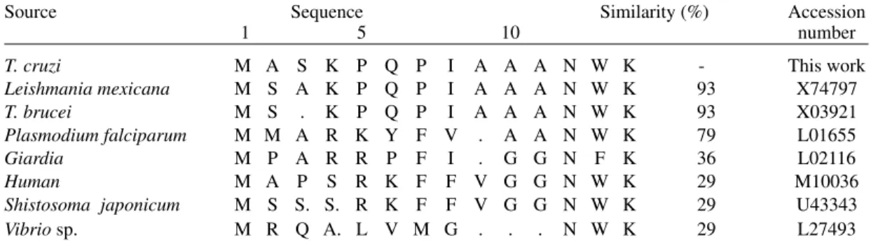
The partial N-terminal sequence data analysis of the first 15 amino acid of the protein revealed 93% and 100% of homology with the TPI from T. brucei and L. mexicana, respectively(Table). This high homology in TPI of different organisms is reinforced by the results of the immunoblot tests. The use of a heterologous serum anti-TPI revealed only one identical migrating band in different para-sites (B. culicis, P. serpens, C. desouzai, H.
samuel-Fig. 1: hydrophobic chromatography on a phenyl-Sepharose column (12.5 x 1.5 cm, I.D.) and SDS-PAGE (12%) of the freeze-thawing epimastigote supernatant. The column was equilibrated in 25 mM Tris-HCl pH 7.8 containing 5 mM EDTA, 1mM DTT, 1 mM NaN3 and 1 M (NH4)2SO4. Fractions of 5 ml were collected from the column at flow rate of 20 ml h-1 using a reverse gradient
of (NH4)2SO4 (100%-0%) in the same buffer. Coomassie stained, SDS-gel electrophoresis of fraction 2 (A) and 3 (B) are shown inside the figure. The molecular weight of the standard proteins are shown in the right side.
222 222 222 222
222 T. cruzi TPI SC Bourguignon et al.
Fig. 2: cation exchange chromatography of the triosephosphate isomerase fraction (peak 3, Fig. 1) and SDS-PAGE (12%) of the main peak 1 (a) and 2 (b) recovered from CM-Sepharose column followed by silver staining. The molecular weight of the standard proteins are shown in the right side. The CM-Sepharose column (6.5 x 1.0 cm, I.D.) was equilibrated with 25 mM Tris-HCl, pH 8.0, buffer and the fractions (1,4 ml) col-lected at a flow rate of 5 ml h-1 using a gradient of NaCl (0-200
mM) in the same buffer.
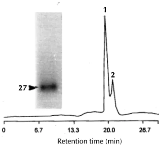
Fig. 4: gel filtration chromatography analysis of the CM-Sepharose triosephosphate isomerase-containing peak 2 using a Shinpack Diol-150 HPLC column (50 cm x 7.9 mm, I.D.) and SDS-PAGE (12%) of the purified TPI (peak 1). About 100 mg of protein was analyzed in the gel filtration column and elution performed using 50 mM phosphate buffer (pH 7.2) at a flow rate of 1ml min-1. Absorbance was measured at 280 nm using
AUFS = 4. Arrow shows the coomassie blue stained protein (peak 1).
Fig 3: western blotting analysis of fractions in the course of triosephosphate isomerase purification incubated with rabbit serum anti-porcine TPI. Lanes a, peak 3 from phenyl-Sepharose column (Fig. 1); b, peak 2 from CM-Sepharose column (Fig. 2); c, porcine TPI. Approximately 50 mg of protein was ap-plied in each slot. Size markers are indicated on the left side proteins.
Fig. 5: immunological characterization of the triose phosphate isomerase from various trypanosomatids using the triose phos-phate isomerase antiserum. A: porcine TPI; B: Blastocrithidia culicis; C: Phytomonas serpens; D: Crithidia desouzai; E:
Herpertomonas samuelpessoai. Approximately 100 mg of pro-tein from the various species of parasite was applied in each slot. At the left side of the figure are indicated the molecular mass, in kDa, of marker proteins.
pessoai) extracts (Fig. 5), suggesting that only one form of enzyme exists in these parasites including
T. cruzi (Fig. 3).
Highly active preparation of TPI has been ob-tained from T. brucei isolated glycosomes (Misset & Opperdoes 1984, Misset et al. 1986), however the number of this organelle in this parasite is four times greater than in T. cruzi (Soares & De Souza 1988, Tetley & Vickerman 1990). These observa-tions may indicate difficulty to obtain the TPI di-rectly from T. cruzi purified organelle. Therefore for purify the TPI we decided to use the superna-tant of the total extract after freeze-thawing frac-tionation. Despite the presence of different puta-tive enzyme forms as demonstrated in different cell types (Tang et al. 1990), only one form of TPI could be detected in T. cruzi epimastigotes by
immunoblotting (Fig. 3) and by agarose electro-phoresis (Fig. 6) analysis. In addition, this last analysis demonstrated that the T. cruzi protein is more positively charged than the L. guinanenis TPI enzyme. This fact is strengthen by the demonstra-tion of the lower isoelectric point (9.0) of L. mexicana mexicana TPI enzyme (Kohl et al. 1994). Immunocytochemical studies showed that gold immunecomplexes were distributed in the cyto-plasm of epimastigotes and amastigote cells and no gold particles were displayed delimiting glycosome organelles. Although a dual localiza-tion of a single TPI form has been described in
Leishmania (Michels 1989), in our studies it is not
27 27 27 27 27
1 11 1 1
2 22 2 2
Retention time (min)
a b c a b ca b c a b ca b c
14 14 14 14 14---29 29 29 29 29---45 45 45 45 45---66 66 66 66
66---14 14 14 14 14 29 29 29 29 29 45 45 45 45 45 66 66 66 66 66
223 223223 223223 Mem Inst Oswaldo Cruz, Rio de Janeiro, Vol. 93(2), Mar./Apr. 1998
clear whether glycosomes were detected by the electron micrography depicted. This fact did not eliminate the possibility of a dual localization of the T. cruzi TPI protein. Indeed the anaerobic
path-way is used by amastigote forms while the aerobic oxidation of substrates, such as amino acids (Engel et al. 1987) and fatty acids (Wood & Shiller 1975) is preferred by epimastigote and trypomastigote, respectively. Therefore, we believe that similar to other parasites T. cruzi presents only one form of
TPI with a possible dual (cytoplasm and glycosomes) localization. This assumption is sup-ported by the western blotting results (Fig. 3) which detected only one reactive band in total T. cruzi
extract, by the agarose electrophoresis activity analysis (Fig. 6) and by the cloning results from Kohl et al. (1994) which detected only one TPI gene for L. mexicana.
TABLE
Comparison of the N-terminal amino acid sequence of the Trypanosoma cruzi triosephosphate isomerase with translated triosephosphate isomerase sequences described in the literature
Source Sequence Similarity (%) Accession
1 5 10 number
T. cruzi M A S K P Q P I A A A N W K - This work Leishmania mexicana M S A K P Q P I A A A N W K 93 X74797 T. brucei M S . K P Q P I A A A N W K 93 X03921 Plasmodium falciparum M M A R K Y F V . A A N W K 79 L01655 Giardia M P A R R P F I . G G N F K 36 L02116 Human M A P S R K F F V G G N W K 29 M10036 Shistosoma japonicum M S S. S. R K F F V G G N W K 29 U43343 Vibrio sp. M R Q A. L V M G . . . N W K 29 L27493
The numbering refers to T. cruzi sequence, and the dots gaps that have been introduced in order to align these sequences.
Fig. 7: thin sections of Lowicryl embedded cells incubated with rabbit anti-triosephosphate isomerase antibodies and protein A-gold complexes. Labelling of the amastigote (A) and epimastigote (B) forms were evident in the cytoplasm (arrow). C: cytoplasm; K: kinetoplast and N: nucleus. (x 30.000). Fig. 6: agarose gel electrophoresis. Electrophoresis and the
triosephosphate isomerase activity were developed as described under “Materials and Methods”. a: Leishmnia guianensis crude extract; b: Trypanosoma cruzi crude extract. Arrows point to the stained bands.
a b + a b + a b + a b + a b +
In summary, we have been able to purify ho-mogeneous T. cruzi TPI using an alternative pro-cedure. The enzyme was localized preferentially in the cytoplasm of both amastigote and epimastigote forms and it showed a native molecu-lar weight of 52 kDa identical to other TPI studied thus far. Moreover, the T. cruzi enzyme differs from
L. guianensis (as shown in this work), T. brucei
(pI 9.8; Willson et al. 1993) and L. mexicana (pI 9.0; Kohl et al. 1994) TPIs in that its isoelectric point is slightly more acid and basic, respectively.
ACKNOWLEDGEMENTS
224 224 224 224
224 T. cruzi TPI SC Bourguignon et al.
REFERENCES
Bendayan M, Nanci A, Kan FWK 1987. Effect of tissue processing on colloidal gold cytochemistry. J Histochem Cytochem 35: 983-996.
Bourguignon SC, Alves CR, Giovanni De Simone S 1997. Detrimental effect of nitric oxide on Trypano-soma cruzi and Leishmanina major like cells. Acta Tropica66: 109-118.
Blattner J, Swinkels B, Dörsam H, Prospero T, Subramani S, Clayton C 1992. Glycosome assem-bly in trypanosomes: variation in the acceptable de-generacy of a COOH-terminal microbody targeting sinal. J Cell Biol19: 1129-1136.
Contreras VT, Salles JM, Thomas N, Morel CM, Goldenberg S 1985. In vitro differentiation of Try-panosoma cruzi under chemically defined condi-tions. Molec Biochem Parasitol16: 315-327. Durieux PO, Schutz P, Brun R, Kohler P 1991.
Alter-ations in Krebs cycle enzyme activities and carbohy-drate catabolism in two strains of Trypanosona brucei during differentiation of their bloodstream to procyclic stages. Molec Biochem Parasitol45: 19-28. Engel JC, Cazzulo BMF, Stoppani AOM, Cannata JJB,
Cazzulo JJ 1987. Aerobic glucose fermentation by Trypanosoma cruzi axenic culture amastigote-like forms during growth and differentiation to epimastigotes. Molec Biochem Parasitol26: 1-10. Fothergill-Gilmore LA, Michels PAM 1993. Evolution
of glycolysis. Prog Biophys Molec Biol 59: 105-235. Giovanni De Simone S, Pontes de Carvalho LC, Oliva OFP, Andrade S, Galvão-Castro B 1987. Trypano-soma cruzi strain specific monoclonal antibodies: Identification of Colombian strain flagellates in the insect vector. Trans R Soc Trop Med Hyg 81: 750-754.
Giovanni De Simone S, Santos R, Araujo MF, Pinho RT 1994. Preparative isolation of the lectin jacalin by anion-exchange high performance liquid chromatog-raphy. J Chromatogrphy A688: 356-362.
Harry H, Hopkinson DA 1976. Handbook of Enzyme Electrophoresis in Human Genetics, Elsevier Pub-lishing Co. Inc., New York, 120 pp.
Huang XY, Barrios LAM, Vonkhorporn P, Honda S, Albertson DG, Hecht RM 1989. Genomic organiza-tion of the glyceraldehyde-3-phosphate dehydroge-nase gene family of Caenorhabditis elegans. J Mol Biol 206: 411-424.
Kohl L, Callens M, Wierenga RK, Opperdoes FR, Michels AM 1994. Triosephosphate isomerase of Leishmania mexicana mexicana. Cloning and char-acterization of the gene, overexpression in Escheri-chia coli and analysis of the protein. Eur J Biochem 220: 331-338.
Laemmli UK 1970. Cleavage of structural proteins dur-ing the assembly of the head of bacteriophage T4. Nature 227: 680-685.
Lowry OH, Rosebrough NJ, Farr AL, Randall RJ 1951. Protein measurement with the folin phenol reagent. J Biol Chem193: 265-275.
Michels PAM 1989. The glycosome of trypanosomes: Properties and biogenesis of a microbody. Exp
Parasitol69: 310-315.
Michels PAM, Opperdoes FR 1991. The evolutionary origin of glycosomes. Parasitol Today 7: 105-109. Misset O, Opperdoes FR 1984. Simultaneous
purifica-tion of hexokinase, class-I, frutose-biphosphate al-dolase, triosephosphate isomerase and phosphoglyc-erate kinase from Trypanosoma brucei. Eur J Biochem144: 475-483.
Misset O, Boss OJ.M, Opperdoes FR 1986. Glycolytic enzymes of Trypanosoma brucei. Simultaneous pu-rification, intraglycosomal concentrations and physi-cal properties. Eur J Biochem157: 441-453. Misset OE, Van Beeumen J, Lambier AM, Van Der Meer
RE, Opperdoes FR 1987. Glyceraldehyde 3-phos-phate dehydrogenase from Trypanosoma brucei. Comparison of the glycosomal and cytosolic isoen-zymes. Eur J Biochem 162: 501-507.
O’Farrel PZ, Goodman HM, O’Farrel PH 1977. High resolution two-dimensional electrophoresis of basic as well as acidic proteins. Cell12: 1133-1142. Opperdoes FR 1987. Compartimentation of carbohydrate
metabolism in trypanosomes. Ann Rev Microbiol41: 127-151.
Opperdoes FR, Wierenga RK, Noble MEM, Hol WGJ, Willson M, Kuntz DA, Callens M, Perié J 1990. Unique properties of glycosomal enzymes, p. 233-246. In N Agabian, A Cerami (eds), Parasites, Mo-lecular Biology, Drugs and Vaccine Design, Wiley-Liss, New York.
Soares MJ, De Souza W 1988. Cytoplasmic organelles of trypanosomatids: A cytochemical and stereologi-cal study. J Submicroscop Cytol Pathol20: 349-361. Sullivan DT, Carroll WT, Kanik-Ennulat CL, Hitti YS, Lovett JA, Von Kalm L 1985. Glyceraldehyde-3-phosphate dehydrogenase from Drosophila melanogaster. Biol Chem 260: 4345-4350. Swinkels BW, Gibson WC, Osinga KA, Kramer R,
Veeneman GH, Van Boom HJ, Borst P 1986. Char-acterization of the gene for the microbody (glycosomal) triosephosphate isomerase of Trypa-nosoma brucei. EMBO J 5: 1291-1298.
Tang CT, Yuksel KÜ, Jacobson TM, Gracy RW 1990. Isoforms of chicken triosephosphate isomerase are due to specific oxidation of cysteine. Arch Biochem Biophys15: 12-19.
Tetley L, Vickerman K 1990. The glycosoma of trypa-nosomes: Number and distribution as revealed by electron spectroscopic imaging and 3-d reconstruc-tion. R Microscop Soc26: 83-90.
Towbin T, Gordon J 1979. Electrophoretic transfer of proteins from polyacrylamide gels to nitrocellulose sheets: procedures and some applications. Proc Natl Acad Sci76: 4350-4354.
Willson M, Callens M, Kuntz DA, Perié J, Opperdoes FR 1993. Synthesis and activity of inhibitors highly specific for the glycolytic enzymes from Trypano-soma brucei.Mol Biochem Parasitol59: 201-210. Wood DE, Shiller EL 1975. Trypanosoma cruzi: