online | memorias.ioc.fiocruz.br
Purification and biochemical characterization of four iron superoxide
dismutases in
Trypanosoma cruzi
Héctor Mateo, Clotilde Marín, Gregorio Pérez-Cordón, Manuel Sánchez-Moreno/+
Departamento de Parasitología, Instituto de Biotecnologia, Universidad de Granada, Severo Ochoa s/n, E-18071 Granada, España
Four superoxide dismutase (SOD) activities (SOD I, II, III, and IV) have been characterized in the epimastigote form of Trypanosoma cruzi. The total extract was subjected to two successive ammonium sulphate additions between 35 and 85%, and the resulting fraction was purified using two continuous chromatography processes (ion exchange and filtration). Enzymes were insensitive to cyanide but sensitive to hydrogen peroxide, properties characteristic of iron-containing SODs. The molecular masses of the different SODs were 20 kDa (SOD I), 60 kDa (SOD II), 50 kDa (SOD III) and 25 kDa (SOD IV), whereas the isoelectric points were 6.9, 6.8, 5.2 and 3.8, respectively. Subcellular location and digitonin experiments have shown that these SODs are mainly cytosolic, with small amounts in the low-mass organelles (SOD II and SOD I) and the mitochondrion (SOD III), where these enzymes play an important role in minimizing oxidative damage.
Key words: superoxide dismutase - T. cruzi - chromatography - subcellular localization
Trypanosoma cruzi is a blood-borne flagellated protozoan responsible for Chagas disease, a vectorially transmitted disease that, in its chronic form, affects more than 16 million people, especially in Latin America, with high morbidity and a rate of more than half a mil-lion new cases every year, most of them children (WHO 2002). The lack of fast and reliable diagnostic methods together with the strain biodiversity and therapeutic dif-ficulties make it a major health problem in which only prophylactic measures are effective(Binder et al. 1999, Urbina & Docampo 2004).
The presence of the parasite in a new host triggers a series of mechanisms intended to eliminate the foreign agents. Beyond all doubt, one of the most important host strategies to combat infection consists of the production of free radicals (highly reactive chemical species able to denaturalize the parasitic membranes), such as the super-oxide anion (O2-), hydrogen peroxide (H
2O2), peroxyni-trite (ONOO-) and hydroxyl radical (HO-) (Piñeyro et al. 2005). It has been reported that trypanosomatids are de-void of or extremely deficient in catalase and selenium-dependent glutathione peroxidase, the most common an-tioxidant defences in eukaryotes. Instead, they possess an array of enzyme-mediated detoxifying mechanisms, such as superoxide dismutase (SOD), three trypanothi-one-dependent peroxidases, a glutathione-spermidine-dependent peroxidase, a tryparedoxin peroxidase, two non-selenium-glutathione peroxidases and the ascor-bate-dependent hemoperoxidase, meant to maintain the
Financial support: CGL2006-27889-E/BOS, Ministerio de Ciencia y Tecnología
+ Corresponding author: msanchem@ugr.es Received 28 December 2007
Accepted 7 May 2008
intracellular reducing environment and, thereby, toallow the parasite to evade the immune response (Wilkinson & Kelly 2003, Turrens 2004, Hernández et al. 2006).
SODs are a group of antioxidant metalloenzymes that eliminate superoxide radicals by dismutation into H2O2 and molecular oxygen. SODs have strong antioxi-dant properties and have been shown to protect normal cells as well as a number of pathogens from reactive oxygen species (ROS). Classically, SODs have been clas-sified into three types according to the metal cofactor bound at the active site: copper-zinc (Cu-Zn-SOD) and manganese SODs (Mn-SOD), (both of which are present in most prokaryotes and eukaryotes, including humans), and iron SODs (Fe-SOD), detected in some prokaryotes, protozoans and chloroplasts of plants and algae. In recent years, a nickel SOD activity has also been described in
Streptomyces spp. (Youn et al. 1996).
To date, the presence of an iron-containing SOD activ-ity has been reported in numerous trypanosomatids, in-cluding T. cruzi (Ismail et al. 1997, Temperton et al. 1997, Villagrán et al. 2005), Trypanosoma brucei (Dufernez et al. 2006, Wilkinson et al.2006), different species of the genus Leishmania (Leishmania aethiopica, Genetu et al. 2006, Leishmania chagasi, Paramchuk et al. 1997,and
Leishmania donovani, Bogdan et al. 1990), Crithidia fas-ciculata (Le Trant et al. 1983) and Phytomonas spp. (plant trypanosomatid) (Marín et al. 2004), playing an impor-tant role in the survival and growth of the parasites.
In a previous study, we detected four SOD activities in epimatigote forms of T. cruzi, for which different isoelec-tric points (pI) are known (Villagrán et al. 2005), but these have been neither purified nor biochemically character-ized. Thus, this is the first study to focus on the purifica-tion and biochemical characterizapurifica-tion of SODs in T. cruzi.
MATERIALS AND METHODS
Organisms and media - Epimastigotes of T. cruzi
supple-mented with 10% heat-inactivated fetal bovine serum at 28ºC in tissue-culture flasks until the cultures reached a density of approximately 1 x 10⁷ parasites/ml. Cells were collected at the logarithmic growth phase by centrifuga-tion (1500 g for 10 min at room temperature).
Extraction and purification of the SODs - The pellets of cells (0.5-0.6 g mw/ml) were washed twice and re-suspended in 3 ml ice-cold STE buffer (0.25 M sucrose, 25 mM Tris-HCl, 1 mM EDTA, pH 7.8; buffer 1). The cells were disrupted by three consecutive cold cycles of sonic disintegration (30 s each at 60 volts, 1 min between cycles), and inspection by light microscopy was used to verify the complete cellular lysis; after this, the sample was centrifuged at 2500 g for 5 min at 4ºC. The pellet was washed twice with buffer 1 and then centrifuged (2500 g for 10 min at 4ºC) to obtain a total supernatant fraction of approximately 10 ml (Fraction H). Degradation due to protease activities present in the supernatant was
mini-mized by the addition of 25 μl of protease inhibitor (Pro -tease Inhibitor Complete Mini, Roche®). This protein fraction was subjected to two ice-cold consecutive am-monium sulphate additions between 35 and 85% (w/v) salt and centrifuged (9000 g for 25 min at 4ºC) to obtain a precipitate that was finally redissolved in 2.5 ml of 20 mM potassium phosphate buffer, pH 7.8, containing 1 mM EDTA (buffer 2), and desalted in a Sephadex G-25 column (Pharmacia, PD 10) previously equilibrated with 25 ml buffer 2, bringing it to a final volume of 3.5 ml (Fraction P85) (Marín et al. 2004).
The P85 fraction was applied to a QAE-Sephadex
A-50 column (30 x 2Ф cm) equilibrated with buffer 2.
The column was extensively washed with the same buf-fer, and the adsorbed proteins were eluted with a linear gradient of KCl (0-0.6 M). Fractions exhibiting SOD activity (peaks Q1, Q2 and Q3) were pooled and con-centrated by ultrafiltration in Microcon® filter tubes (Amicon®) at 11200 g for 30 min. The peaks were applied separately to a G-75 Sephadex molecular sieve
chroma-tography column (75 x 1.6Ф cm) equilibrated with buffer
2. The enzyme was eluted with 200 ml of the same buffer solution. The fractions with the highest specific SOD ac-tivity (SOD I, SOD II, SOD III and SOD IV) were again collected, concentrated (to a concentration of 2 mg/ml), and used for the experiments described below.
Subcellular location of SODs - A homogenate of the Maracay strain of T. cruzi was prepared by grinding the cell paste with silicon carbide abrasive (Carborundum®), as described by Steiger et al. (1980). Protein concentra-tions and SOD activities were measured in fracconcentra-tions recovered by differential centrifugation following the method of Marín et al. (2004). Fractions H (homoge-nate), N (nuclear), L(high-mass organelles), S (low-mass organelles), M (microsomal) and C (cytosolic) were re-covered. Recovery (based on the sum of fractions) and relative specific activity (RSA) were calculated. For the correct identification of the subcellular fractions, we checked specific marker enzymes according to Uttaro and Opperdoes (1997), i.e., pyruvate kinase for the cy-tosol, hexokinase for the glycosome and alanine trans-ferase for the mitochondria.
Digitonin treatment of intact cells - The cell suspen-sions were diluted to a density corresponding to 2 mg protein/ml in buffer 1 containing increasing concentra-tions of digitonin. After incubation for 5 min at 4ºC, sus-pensions were centrifuged. Aliquots of the supernatants were used to assay the various enzymatic activities ac-cording to Uttaro and Opperdoes (1997).
Protein determination - The protein content of frac-tions H andP85, the peaks and all elution fractions result-ing from the two columns were quantified usresult-ing the Bio-Rad® Bradford test, which uses bovine serum albumin (BSA) as a standard.
Enzyme assay - SOD activity was performed on frac-tions H, P85 and elufrac-tions by spectrometric measurement of NBT-UV light oxidation according to the method de-scribed by Beyer and Fridovich (1987). Buffer 3(50 mM potassium phosphate, pH 7.8) was chosen for these de-terminations. Pyruvate kinase, hexokinase and alanine aminotransferase activities were determined according to Uttaro and Opperdoes (1997).
Determination of molecular weight - Apparent molec-ular weights of the purified SODs (peaks Q1, Q2 and Q3) were calculated by native gel electrophoresis in PhastGel Homogeneous 12.5% gels. The standard mixture of pro-teins used as markers (LMW Calibration Kit, Pharmacia®) contained phosphorylase B (94 kDa), BSA (67 kDa), oval-bumin (43 kDa), carbonic anhydrase (30 kDa), soybean
trypsin inhibitor (20 kDa) and α-lactalbumin (14.4 kDa).
The gels were stained for protein with the silver staining solution as described in the PhastSystem handbook, and the SOD activity was revealed with the method described previously.
Determination of the pI - Isoelectric focusing (IEF) in polyacrylamide PhastGel IEF 3-9 gels were used to determine the pI of components of fraction P85 as de-scribed by Bécuwe et al. (1996). The markers used were trypsinogen (9.3), lentil-lectin acidic band (8.15), horse heart myoglobin (6.8), carbonic anhydrase II (5.9),
α-lactoglobulin A (5.1), soybean trypsin inhibitor (4.6)
and amyloglucosidase from Aspergillus niger (3.6) (Pharmacia®). The markers were stained for protein, and the activity was revealed as described previously.
Inhibition studies - Specific inhibitors with known ef-fects on the different SOD metal cofactors (cyanide and H2O2) were employed. For the cyanide assays, samples were applied to an IEF 3-9 polyacrylamide gel, and then the SOD activity was revealed by adding 20 mM sodium cyanide to the revealing solution. For the H2O2 inhibition test, H2O2 was added to the gel and incubated at room tem-perature for 15 min. Then, the gel was stained for SOD activity as described above.
RESULTS
The epimastigote lysate (FractionH) from T. cruzi
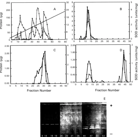
gel electrophoresis of the fractions eluted from the QAE-Sephadex A-50 column was also performed to visual-ize the SOD activities (Fig. 1E). The three active peaks (peaks Q1, Q2 and Q3) were collected, desalted, concen-trated, and further purified separately by Sephadex G-75 molecular-sieve chromatography. Purification of peak Q1 resulted in a new peak called SOD III, located between fractions 11 and 14 (Fig. 1B), peak Q2 purification yield-ed a new peak callyield-ed SOD IV, locatyield-ed between fractions 25 and 35 (Fig. 1C), whereas the purification of peak Q3 yielded two new peaks called SOD II (fractions 8-15) and SOD I (fractions 43-46) (Fig. 1D). Table summariz-es the purification , indicating that SOD III was purified 336 times to a specific activity of about 7380.89 U/mg of protein; SOD IV was purified 489 times to a spe-cific activity of about 10751.72 U/mg of protein; SOD II was purified to a specific activity of 13956.52 U/mg of protein (purification rate 635), whereas SOD I was purified 424 times to a total activity of 9,446 U/mg.
Molecular weights were also determined by native electrophoresis on PhastGel Homogeneous 20% gels, showing that the SODs had molecular masses of approx-imately 50 kDa (SOD III), 25 kDa (SOD IV), 60 kDa (SOD II) and 20 kDa (SOD I) (Fig. 2A, lanes- 1, 2, 3, respectively). The four SODs had different pI: SOD I had a pI of 6.9, and the pI of SOD II was approximately 6.8, whereas SOD III and SOD IV had pIs of 5.25 and 3.8, respectively (Fig. 2B, lane 4).
We performed studies with specific inhibitors that have known effects on SODs containing various cofactors: Cu/Zn, Mn or Fe. All SODs proved to be inhibited by H2O2 and not by sodium cyanide (Fig. 2B, lanes- 5, 6), and this behaviour is characteristic of iron-containing enzymes.
To determine the subcellular localization of these SODs, we fractionated T. cruzi cells by differential cen-trifugation. Protein determinations and SOD activity as-says were performed on every fraction, and RSA and recovery percentages were calculated. The four
zymes were localized to the cytoplasmic fraction. Fur-thermore, SOD III and SOD IV were also present in the glycosomal fraction and SOD I, in smaller amounts, in the large organelles (the mitochondrion), with a recovery percentage of 91.8% (Fig. 3A). This distribution was also demonstrated by IEF (pI 3-9) and activity assay by the above-mentioned technique (Fig. 3B). This observation was corroborated by digitonin titration experiments us-ing pyruvate kinase, hexokinase and alanine aminetrans-ferase as markers for soluble, glycosomal and mitochon-drial fractions, respectively (Fig. 3C). It was noteworthy that 24% of the activity was not solubilized, even at high digitonin concentrations.
DISCUSSION
For the first time, we report the partial purification of the SOD activities from the epimastigote form of T. cruzi. The SOD activity identification is possible only under the native conditions of the protein; thus, we have detected four different isoenzymes by IEF 3-9 and native electrophoresis. According to Villagrán et al. (2005), we have found different pI: 6.9, 6.8, 5.2 and 3.8 for SOD I, SOD II, SOD III and SOD IV, respectively. The pIs
ob-Fig. 2: electrophoresis of the purified enzymes. A: native gel elec-trophoresis in PhasGel Homogeneous 12.5%; B: determination of the metal cofactor of Trypanosoma SODs by inhibition studies performed in isoelectric focusing gel electrophoresis pI 3-9. Lanes - 1: SODIII (peak Q1, 2 μg); 2: SODIV (peak Q2, 2 μg); 3: SODII and SODI (peak Q3, 2 μg); 4: fraction P85; 5: fraction P85 with 20 mM KCN; 6: frac-tion P85 with 5 mM H2O2. MpI: isoelectric point marker; MW: mo-lecular weight marker proteins; (∞): silver staining; (*): SOD activity revealed according to Beyer and Fridovich (1987).
served disagree with the predicted pIs of a single subunit in the T. cruzi genome database (http://www.genedb.org/ genedb/tcruzi) because of the monomer-monomer inter-actions, which would involve and/or shield charged resi-dues. The molecular masses determined were 20 kDa for SOD I, 60 kDa for SOD II, 50 kDa for SOD III and 25 kDa for SOD IV. To date, the molecular weights of SODs in trypanosomatids have been determined as monomers in denaturing gels for T. cruzi and T. brucei
(around 20 and 24 kDa) (Temperton et al. 1996, Kabiri & Steverding 2001, Dufernez et al. 2006), and, for na-tive enzymes, the mass has been shown for Phytomo-nas spp. (24 and 66 KDa) by Marín et al. (2004). Thus, the apparent mass of 50 and 60 kDa of T. cruzi SOD I and III is probably the result of the dimeric form since SODs of 50 kDa or higher have not been found in any of the trypanosomatid genomes (http://www.sanger.ac.uk/ Projects/Protozoa/).
In Trypanosomatidae, different studies have report-ed the presence of SOD activities. T. brucei (Dufernez et al. 2006, Wilkinson et al. 2006), Leishmania spp. (Bogdan et al. 1990, Genetu et al. 2006), and Phytomo-nas spp. (Marín et al. 2004), and many other protozoans (Plasmodium falciparum,Entamoeba histolytica, Toxo-plasma gondii or Trichomonas spp.) have been shown to possess SOD activities in at least one stage of their life cycle (Sibley et al. 1986, Fairfield et al. 1988). To date, in T. cruzi, some studies have also reported the existence of two SOD activities (Ismail et al.1997). Nevertheless, this is the first study to focus on the purification and bio-chemical characterization of the SODs, while no refer-ence is available in the literature for the two other newly identified activities.
Four peaks of SOD activity were isolated by ion ex-change chromatography and identified. Each peak was further purified separately by filtration chromatography, giving rise to a total of four SOD activities [called SOD I, II, III and IV, according to the nomenclature used by Villagrán et al. (2005)]. This is consistent with the re-sults revealed by IEF since the P85 fraction showed four bands of SOD activity. Throughout all of these purifi-cation processes, activity was steadily lost, so we em-ployed the minimum total duration needed to optimise the purification yield.
TABLE
Superoxide dismutase (SOD) purification from epimastigotes of Trypanosoma cruzi Maracay strain
Total Total activitya Specific activityb
Fraction protein (mg) (Units ± SD) (Units/ mg prot ± SD) Purification Yield(%)
Homogenate 43.171 661.40 ± 42.33 22.27 ± 1.72 1 100
P85 3.832 469.80 ± 18.23 122.63 ± 4.76 6 70
Peak Q1 0.127 296.51 ± 6.42 2329.69 ± 25.31 104 44
Peak Q2 0.922 217.63 ± 13.08 1236.01 ± 14.18 55 33
Peak Q3 0.520 206.63 ± 14.52 397.36 ± 50.06 18 31
SOD I 0.001 12.28 ± 0.09 9446.15 ± 7.075 424 2
SOD II 0.001 9.63 ± 0.67 13956.52 ± 971.01 635 1
SOD III 0.016 118.98 ± 1.72 7380.89 ± 106.17 336 18
SOD IV 0.010 108.71 ± 1.00 10751.72 ± 98.91 489 16
As in other SODs studied in pathogenic kinetoplas-tids, inhibition assays against specific inhibitors revealed behaviour typical of iron-linked enzymes (Temperton et al. 1996, Ismail et al. 1997, Paramchuk et al. 1997, Kabiri & Steverding 2001).
The subcellular location of SODs in the Maracay strain of T. cruzi has demonstrated that these enzymes have specific locations within the cell. Quality control for impurities in the different subcellular fractions was
Fig. 3: distribution profile of SOD activity in the fractions from the differential centrifugation. A: SOD activity was determined in the different fractions according to the method of Beyer and Fridovich (1987). The relative specific activity (RSA), ratio between the per-centage of total activity and perper-centage of total protein for each frac-tion (Y-axis) is plotted against the cumulative percentage of protein (X-axis); B: activity staining by isoelectric focusing. Lanes - C: cy-toplasmic fractions; L: large organelle fraction; M: microsomal; N: nuclear; PN: postnuclear fraction; S: small organelle fraction. C: dig-itonin-titration experiment of the SOD activity (-+-+-); Pyruvate ki-nase (-o-o-); hexokiki-nase (-▲-▲) and alanine aminotransferase (-●-●-) as markers for soluble, glycosomal and mitochondrion localizations.
performed by checking the presence of marker enzymes (pyruvate kinase, hexokinase and alanine aminotrans-ferase), according to Uttaro and Opperdoes (1997). The compartmentalization becomes significant if we accept that the main role of SOD is to detoxify endogenous superoxide anions and that the different biochemical processes that produce oxidizing agents inside the cell may need various antioxidant systems to protect mac-romolecules and organelles from oxidative damage. SOD is mainly a cytosolic enzyme, although a certain SOD activity is also localized in the glycosomal and mitochondrial fractions. This fact has precedents, as in
C. fasciculata, a non-pathogenic trypanosomatid, which has two SOD isoenzymes, one located in the cyto-sol and the other in the mitochondrion (Quesada et al. 2001).Also, in T. brucei, four activities with different subcellular locations were detected, one in the cytosol, another in the glycosomes and the other two in the mi-tochondrion (Dufernez et al. 2006). SOD is presumably synthesised in the cytosol and can remain there and/or be transported to the interior of certain organelles, as happens in the cases of T. brucei (Dufernez et al. 2006) and Leishmania spp. (Genetu et al. 2006). The digitonin experiments revealed that 24% of the total activity is not even solubilized at high concentrations of digitonin, indicating that it would be a membrane-related activity (which would be lost during the fractionation process). This fact is consistent with a previous study performed by us on Phytomonas sp., in which approximately 20% of the total SOD activity seemed to be associated with membranes or even excreted (Marín et al. 2006). Despite lacking catalase and glutathione peroxidase, the main antioxidant defences against ROS in eukaryotic cells,
T. cruzi possess SOD and other highly efficient detoxi-fying mechanisms aimed at neutralizing moderate in-crements of ROS (Wilkinson et al.2000, Wilkinson & Kelly 2003, Hernández et al. 2006). The presence of var-ious SOD isozymes in different locations within the cell ensures more effective action against eventual increases in ROS intracellular levels, maintaining a favourable reducing environment inside the cell, and this could be considered a potential virulence factor, playing a major role in the survival of the pathogen inside the host cell.
The study of these newly identified SODs, their bio-chemical characterization and the scope of their role in-side the parasite cell, or beyond, in the interactions with the host cells, together with the comparison of the SOD genes in order to establish the significant differences and novelties among SODs throughout Trypanosomati-dae, would constitute the next step in future research on the biochemical and immunological paths that enable the survival of the species.
ACKNOLEDGEMENTS
To David Nesbitt for the English translation of the manuscript.
REFERENCES
Bécuwe P, Gratepanche S, Fourmaux MN, Van Beeumen J, Samyn B, Mercereau-Puijalon O, Touzel JP, Slomianny C, Camus D, Dive D 1996. Characterization of iron-dependent endogenous superoxide dismutase of Plasmodium falciparum. Mol Biochem
Beyer WF, Fridovich I 1987. Assayaing for superoxide dismutase ac-tivity: Some large consequences of minor changes in conditions.
Anal Biochem161: 559-566.
Binder S, Levitt AM, Sacks JJ, Hughes JM 1999. Emerging infectious diseases: Public health issues for the 21st Century. Science284: 1311-1313.
Bogdan C, Rollinghov M, Solbach W 1990. Evasion strategies of
Leshmania parasites. Parasitol Today 6: 183-186.
Dufernez F, Yernaux C, Gerbod D, Noël C, Chauvenet M, Wintjens R, Edgcomb V, Capron M, Opperdoes FR, Viscogliosi E 2006. The presence of four iron-containing superoxide dismutase isozymes in Trypanosomatidae: Characterization, subcellular localization and phylogenetic origin in Trypanosoma brucei. Free Radic Biol
Med 40: 210-225.
Fairfield A, Abosh A, Ranz A, Eaton J, Meschnick S 1988. Oxidant defence enzymes of Plasmodium falciparum. Mol Biochem Pa-rasitol30: 77-82.
Genetu A, Gadixa E, Aseva A, Barr S, Lakew M, Jirata D, Kuru T, Hunegnaw M, Gedamu L 2006. Leishmania aethiopica: strain identification and characterization of superoxide dismutase-B genes. Exp Parasitol113: 221-226.
Hernández SM, Sánchez MS, Schwarc de Tarlovsky MN 2006. Polyamines as a defense mechanism against lipoperoxidation in
Trypanosoma cruzi. Acta Trop98: 94-102.
Ismail SO, Paramchuck WJ, Bhatia JA, Gedamu L 1997. Molecular cloning and characterization of two iron superoxide dismutase cDNAs from Trypanosoma cruzi: differential and stage specific expression. Mol Biochem Parasitol86: 187-197.
Kabiri M, Steverding D 2001. Identification of a developmentally regulated iron superoxide dismutase of Trypanosoma brucei.
Biochem J 360: 173-177.
Le Trant N, Meshnich SR, Kitchener K, Eaton JW, Cerami A 1983. Iron-containing superoxide dismutase from Crithidia fascicu-lata. Purification, characterization and similarity of leishmanial and trypanosomal enzymes. J Biological Chem258: 125-130.
Marín C, Rodríguez-González I, Sánchez-Moreno M 2006. Identifi- Identifi-cation of excreted iron superoxide dismutase for the diagnosis of
Phytomonas. Mem Inst Oswaldo Cruz101: 649-654.
Marín C, Rodríguez-González I, Hitos A, Rosales MJ, Dollet M, Sánchez-Moreno M 2004. Purification and characterization of two iron superoxide disputases of Phytomonas sp. isolated form
Euphorbia characias (Plant Trypanosomatids). Parasitology
129: 79-86.
Paramchuk WJ, Ismail SO, Bhatia A, Gedamu L 1997. Cloning, char-acterization and overexpression of two iron superoxide dismutase cDNAs from Leishmania chagasi: Role in pathogenesis. Mol
Bio-chem Parasitol 90: 203-221.
Piñeyro MD, Pizarro JC, Lema F, Pritsch O, Cayota A, Bentley GA, Robillo C 2005. Crystal structure of the tryparedoxin peroxidase from the human parasite Trypanosoma cruzi. J Struct Biol150: 11-22.
Quesada JM, Entrala E, Fernández-Ramos C, Marín C, Sánchez-Moreno M 2001. Phytomonas spp.: superoxide dismutase in plant trypanosomes. Mol Biochem Parasitol115: 123-127.
Sibley LD, Lawson R, Weidner E 1986. Superoxide dismutase and cat-alase in Toxoplasmagondii. Mol Biochem Parasitol 19: 83-87.
Steiger RF, Opperdoes FR, Bontemps J 1980. Subcellular localiza-tion and fraclocaliza-tionalocaliza-tion of Trypanosoma brucei blood stream-forms with special reference of hydrolases. Eur J Biochem105: 163-175.
Temperton NJ, Wilkinson SR, Kelly JM 1996. Cloning of an Fe- superoxide dismutase gene homologue from Trypanosoma cruzi.
Mol Biochem Parasitol76: 339-343.
Temperton NJ, Wilkinson SR, Kelly M 1997. Cloning of a Fe-super-oxide dismutase homologue from Trypanosoma cruzi. Mol
Bio-chem Parasitol 90: 103-221.
Turrens J 2004. Oxidative stress and antioxidant defenses: A target for the treatment of diseases caused by parasitic protozoa. Mol
Aspects Med25: 211-220.
Urbina JA, Docampo R 2004. Specific chemotherapy of Chagas dis-ease. Trends Parasitol 19: 495-501.
Uttaro AD, Opperdoes FR 1997. Purification and characterization of a novel isopropanol dehydrogenase from Phytomonas sp. Mol
Biochem Parasitol8: 213-219.
Villagrán ME, Marín C, Rodríguez-González I, De Diego JA, Sán-chez-Moreno M 2005. Use of a iron superoxide dismutase ex-Use of a iron superoxide dismutase ex-creted by Trypanosoma cruzi in the diagnosis of chagas disease: seroprevalence in rural zones of the state of Queretaro, Mexico.
Am J Trop Med Hyg73: 510-516.
Wilkinson SR, Kelly JM 2003. The role of glutathione peroxidases in trypanosomatid. J Biol Chem384: 517-525.
Wilkinson SR, Prathalingam R, Taylor MC, Ahmed A, Horn D, Kelly JM 2006. Functional characterization of the iron superoxide dis-mutase gene repertoire in Trypanosoma brucei.Free Radic Biol Med40: 198-209.
Wilkinson SR, Temperton NJ, Mondragon A, Kelly JM 2000. Distinct mitochondrial and cytosolic enzymes mediate trypanothione- dependent peroxide metabolism in Trypanosoma cruzi. J Biol
Chem 275: 8220-8225.
WHO- World Health Organization 2002. Control of Chagas Disease, WHO Technical Report Series No. 905, Geneva, p. 1-35.
Youn HD, Kim EJ, Roe JH, Hag YC, Kang SO 1996. A novel nickel-containing superoxide dismutase from Streptomyces spp.