Validation of a real-time PCR assay for the molecular identification
of
Mycobacterium tuberculosis
Mariana L. Sales
1, Antônio Augusto Fonseca Júnior
1, Lívia Orzil
1,
Andrea Padilha Alencar
1, Marcio Roberto Silva
2, Marina Azevedo Issa
1,
Paulo Martins Soares Filho
1, Andrey Pereira Lage
3, Marcos Bryan Heinemann
31
Laboratório Nacional Agropecuário de Minas Gerais, Pedro Leopoldo, MG, Brazil. 2
EMBRAPA Gado de Leite, Juiz de Fora, MG, Brazil. 3
Escola de Veterinária, Universidade Federal de Minas Gerais, Belo Horizonte, MG, Brazil.
Submitted: March 27, 2013; Approved: April 17, 2014.
Abstract
Mycobacterium tuberculosis is the major cause of tuberculosis in humans. This bacillus gained prominence with the occurrence of HIV, presenting itself as an important opportunistic infection as-sociated with acquired immunodeficiency syndrome (AIDS). The current study aimed to develop a real-time PCR using Eva Green technology for molecular identification ofM. tuberculosisisolates. The primers were designed to Rv1510 gene. Ninety nine samples ofM. tuberculosisand sixty sam-ples ofM. boviswere tested and no sample of the bovine bacillus was detected by the qPCR. Statisti-cal tests showed no difference between the qPCR and biochemiStatisti-cal tests used to identify the Mycobacterium tuberculosis. The correlation between tests was perfect with Kappa index of 1.0 (p < 0.001, CI = 0.84 1.0). The diagnostic sensitivity and specificity were 100% (CI = 95.94% -100%) and 100% (CI = 93.98% - -100%). This qPCR was developed with the goal of diagnosing the bacillusM. tuberculosisin samples of bacterial suspension. TB reference laboratories (health and ag-riculture sectors), public health programs and epidemiological studies probably may benefit from such method.
Key words:Mycobacterium tuberculosis, diagnosis, real time PCR.
Introduction
Tuberculosis (TB) is one of the oldest and most seri-ous known diseases. Studies using DNA isolated from cat-tle bones found in North America, which were dated to 17,000 BC, showed the presence ofMycobacterium tuber-culosis(Rothschildet al., 2001). During the Industrial Rev-olution, numerous cases of tuberculosis emerged due to the agglomeration of workers (Costa, 1985). In the first half of the twentieth century, the disease terrorized Europe, and a diagnosis meant a death sentence for 50% of infected indi-viduals, mostly children and youth (Danielet al., 1984).
There was hope that tuberculosis would be eradicated by 2000 in developed countries, However, with the AIDS pandemic, pulmonary tuberculosis has become the third most common opportunistic infection in HIV-positive pa-tients (Job, 1998). The occurrence of TB co-infection with
human immunodeficiency virus (HIV) caused a change in the epidemiological trends of TB, which now presented as an important opportunistic pathogen associated with ac-quired immunodeficiency syndrome (AIDS) (Carvalho, 2006). According to estimates from the World Health Or-ganization (WHO), one third of the world population is in-fected with Mycobacterium tuberculosis; every year, 8 million people become ill and 2,9 million people die as a re-sult of TB infection (WHO, 2013). Of the 8 million cases registered annually, 95% occur in developing countries (Brasil, 2002). In the past, pulmonary tuberculosis was used as an indicator of peripheral development.
While mycobacterium culture is the gold standard for confirmation of a TB diagnosis, the process requires more than three weeks for a conclusive result and is not spe-cies-level specific. The major problems in mycobacteria
Send correspondence to A.A. Fonseca Junior. Laboratório de Biologia Molecular, Pedro Leopoldo, MG, Brazil. E-mail: [email protected].
identification by phenotypic methods are the variety of bio-chemical tests required and the time needed for completion of the tests (Telentiet al., 1993). These tests can be labori-ous, time consuming, inaccurate and non-reproducible, can generate ambiguous results and require adequate facilities with a high biosecurity level (Huardet al., 2003).
Molecular biology techniques are widely applicable to laboratory diagnoses of infectious diseases caused by vi-ruses (Fonseca Jr. et al., 2010), bacteria (Cortez et al., 2006), parasites or fungi (Sotiriadou et al., 2013). DNA fragment amplification opened excellent prospects for the detection of infectious agents. PCR has several advantages compared to traditional diagnosis methods. PCR is highly sensitive, specific and rapid. To produce a positive diagno-sis, fewer bacterial cells are required, and the bacteria do not need to be viable, which means that inadequately pre-served samples can be used (Abrahão, 1999, Narayanan, 2004). Real-time PCR (qPCR) has further advantages, in-cluding precision, reproducibility, accuracy, quality con-trol processes and reduced contamination. In addition, qPCR eliminates the need for electrophoresis after the cy-cling reaction. Furthermore, this technique reduces the analysis time to three or four days, which is important in la-borious and lengthy microbiological diagnoses, such as those required for TB.
Faster diagnoses of human TB is of significant clini-cal importance. There are commercial methods available to detectM. tuberculosis, but these techniques rely on multi-ple probes or detectMycobacterium tuberculosiscomplex (MTC) but not the specific species (Rossauet al., 1997). Thus, the aim of this study was to develop a real-time PCR assay using bacterial suspensions and Eva Green®to iden-tifyM. tuberculosisas an alternative to phenotypic tests.
Materials and Methods
qPCR
The oligonucleotides designed in this study were based on the complete genome sequence ofM. tuberculosis (Coleet al., 1998). First, the sequences were subjected to BLAST®(http://blast.ncbi.nlm.nih.gov/Blast.cgi) for veri-fication of genomic differences (Altschul et al., 1997). Then, the selected target sequences available on GenBank (NCBI, http://www.ncbi.nlm.nih.gov/) were aligned in BioEdit (16). After checking the ideal regions, namely, those present only on M. tuberculosis, primers were de-signed in the Primer3Plus program (Untergasser et al., 2007) and analyzed using the Oligo Analyser 3.1 program (IDT, USA) for verification of secondary structures. In silicoanalytical specificity of the primers was tested with
the PrimerBlast program (NCBI,
http://www.ncbi.nlm.nih.gov/tools/primer-blast/in-dex.cgi?LINK_LOC=BlastHome) (Altschulet al., 1990).
The region chosen for the specific detection ofM. tu-berculosis DNA was the gene Rv1510. Primers
Mtub.115.F: 5’TTCGATATTCGCGGTGTTTT3’ (318446-318465, referring to BX842576.1) and Mtub.115.R: 5’CGCAACTATTTGGGTGGAG3’ (318542-318560) amplify a 115 bp segment of the gene. The ideal qPCR reactions contained 10 pmol of each primer (IDT, USA), 1.5 U Hot Start GoTaq Polymerase (Promega, USA), 20% Colorless GoTaq Hot Start Buffer 5x, 1.5 mM MgCl2, 1mL of 20x Eva Green (Biotium, USA), 2mL Rox
10x (Biotium, USA), 10 mM dNTPs and 2mL of DNA to a
final volume of 20 mL. All tests were performed on a
Rotorgene 3000 (Qiagen, Germany) with the following program: denaturation at 95 °C for 3 min followed by 35 cycles at 95 °C for 10 s, 60 °C for 15 s and 72 °C for 20 s with fluorescence readings. The melting curve was per-formed with the following parameters: 45 s at 72 °C and 3 s per degree up to 99 °C. DNA extraction was evaluated by qPCR targeting ribosomal DNA. Primers M.sp.rDNA.133.F (5’GTCCTTGGAGACGTTCCTCA3’) and M.sp.rDNA.133.R (5’CACGTCGATCACCACG TAGA3’) were used with the same reagent concentrations and cycling conditions that were used for Rv1510 detec-tion.
Samples
Standard M. tuberculosis strains H37Rv CRNC 23 and H37Ra CRNC 25 were used as positive controls, and M. bovisAN5 Mexico CRNC 36,M.bovisAN5 CRNC 02, M. bovisAN5 CRNC 01 andM. bovisBCG CRNC stan-dard strains were used as negative controls for all tests in this study.
Phenotype-based speciation methods
All positive AFB cultures were subjected to addi-tional phenotypic speciation methods, including biochemi-cal tests such as catalase at room temperature and at 68 °C, niacin, nitrate, pyrazinamidase, and urease. Drug suscepti-bility testing (DST) was used to distinguish mycobacterial strains (Job, 1998).
Molecular-based speciation methods
Two loops of each mycobacteria replica that grew on Lowenstein Jensen or Stonebrink media were transferred into a microcentrifuge tube containing 400mL of 1X TE
buffer and were separately subjected to heat to kill and lyse the cells. The inactivated isolates were stored at 2 to 8 °C until use in the PCR reactions.
subjected to amplification of thepncAgene (Scorpio and Zhang, 1996) and detection of thepncApolymorphism by cleavage of the amplicon withEco065I, as previously de-scribed (Barouniet al., 2004) for the genetic identification ofM. bovisor M. tuberculosis. This analysis was carried out at the Laboratory of Molecular Biology Applied to My-cobacteria, Oswaldo Cruz Institute, Fiocruz.
Validation criteria
The parameters for the validation methodology were performed according to the Manual of Diagnostic Tests and Vaccines for Terrestrial Animals (OIE, 2010).
Reaction efficiency
The reaction efficiency was tested by examining 10-fold dilutions in 1X TE buffer of DNA extracted from the positive controls. Each dilution was tested in duplicate and the concentration estimated on a Nanovue® (GE Healthcare, USA) spectrophotometer. A number of stan-dard curves were generated to determine the optimal primer concentration.
Limit of Detection (LOD) and Analytical Specificity
The determination of the LOD was performed by the 10-fold dilution of the standard strain. The dilutions were tested in triplicate, and the LOD was determined as the dilu-tion at which all replicates were positive. The LOD was confirmed by repeating the qPCR twenty-one times.
The following strains were used in analytical speci-ficity tests: 60M. bovisisolates, M. fortuitumCRNC 10,M. kansasiiCRNC 48 and CRNC 18,M. gordonaeCRNC 16, M. aviumD4 CRNC 05,M. avium paratuberculosisCRNC 26, M. avium 1500 CRNC 15 and 2045 CRNC 14, M. intracellulare CRNC 17, M. marinum CRNC 19, M. scrofulaceumCRNC 49,M. scrofulaceum CRNC 20,M. szulgai CRNC 21, M. triviale CRNC 22, M. fortuitum peregrinumCRNC 11, M. phei CRNC 12, Rhodococcus equi CRC 09/01, Corynebacterium pseudotuberculosis CRC 09/02 andNocardia asteroidesCRC 10/01.
Repeatability
The repeatability was estimated using seven samples ofM. tuberculosisand three samples ofM. bovis. We ex-tracted DNA from seven samples on three different days and subjected the extracted nucleic acid to qPCR. To assess possible changes in the test, a second analyst performed the technique using the same criteria and the same samples.
Robustness
We analyzed the assay robustness with potential is-sues that may occur, such as an improperly calibrated pi-pette or thermocycler, to evaluate how these problems would impact the diagnosis of human tuberculosis. FiveM. tuberculosissamples and fiveM. bovissamples were used in a qPCR assay with a 1 °C increased annealing
tempera-ture and 10% less enzyme, MgCl2and primers. A second qPCR was performed with a 1 °C decreased annealing tem-perature and 10% more enzyme, MgCl2and primers.
Statistical analysis
We used the McNemar test to compare paired propor-tions, the kappa test to determine the measure of agreement between two tests for each sample and the sensitivity and specificity analysis (Kraemer, 1992).
For the diagnostic sensitivity and diagnostic specific-ity analyses, 99M. tuberculosisisolates from Brazil were used. Each sample was simultaneously tested by pheno-typic and qPCR methods. The qPCR method was compared with the phenotype-based speciation method as a gold stan-dard.
Results
qPCR
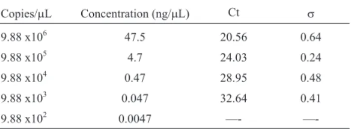
The average melting temperature for positive samples in the qPCR forM. tuberculosiswas 94.11 °C with a stan-dard deviation of 1.31 °C. The qPCR was able to detect up to 9.88 x 10³ copies/mL in a standard curve with an R2of
99% and with 78% efficiency (Table 1). The average melt-ing temperature for positive samples in the qPCR for ribo-somal DNA was 87.2 °C.
Repeatability and robustness
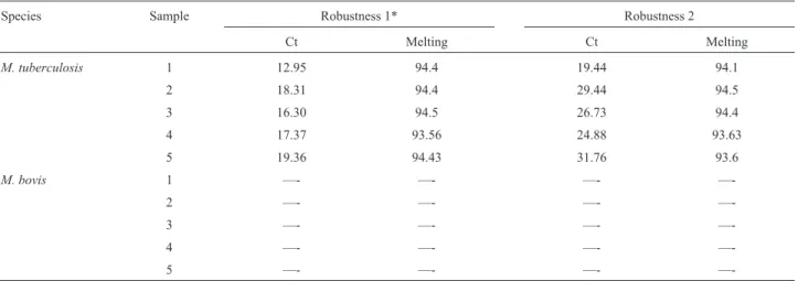
The qPCR had similar results when performed on dif-ferent days and by a difdif-ferent analyst. The average melting temperature was 94.72 °C with a standard deviation of 0.26 °C. No amplification was detected when M. bovis DNA was used. Variations in cycle thresholds (Ct) and melting temperatures detected in the robustness tests are described in table 2 and Figure 1.
Statistical analyses
The McNemar test showed no significant difference between the qPCR (p = 0.3173) and microbiological isola-tion methods, and the Kappa test showed perfect agreement (0.98) between the two tests.
Sensitivity and specificity
The qPCR detected only DNA extracted fromM. tu-berculosis. All other nucleic acid from other
Mycobacte-Table 1- Mean valuessof Ct and standard deviations of qPCR.
Copies/mL Concentration (ng/mL) Ct s
9.88 x10⁶ 47.5 20.56 0.64
9.88 x10⁵ 4.7 24.03 0.24
9.88 x10⁴ 0.47 28.95 0.48
9.88 x10³ 0.047 32.64 0.41
—-Table 2- Variation of melting temperature and cycle threshold in robustness tests for qPCR.
Species Sample Robustness 1* Robustness 2
Ct Melting Ct Melting
M. tuberculosis 1 12.95 94.4 19.44 94.1
2 18.31 94.4 29.44 94.5
3 16.30 94.5 26.73 94.4
4 17.37 93.56 24.88 93.63
5 19.36 94.43 31.76 93.6
M. bovis 1 —- —- —-
—-2 —- —- —-
—-3 —- —- —-
—-4 —- —- —-
—-5 —- —- —-
—-*Robustness 1: increase in 1 °C in annealing temperature and 10% less enzyme, MgCl2and primers; Robustness 2: decrease by 1 °C temperature
anneal-ing and 10% more enzyme, MgCl2and primers.
riumand another bacterial species tested in this work were negative in all of the performed tests. The diagnostic sensi-tivity and specificity were 100% (CI = 95.94% - 100%) and 100% (CI = 93.98% - 100%), respectively.
Discussion
Phenotypic tests used to identifyMycobacterium iso-lates are time consuming and unreliable. The use of molec-ular tests to correctly identify microorganisms is fast, reliable and sensitive. The qPCR developed in this study did not amplify DNA from any of theMycobacteriumspp. other than theM. tuberculosissamples used in the analyti-cal specificity tests; positive results were only realized for theM. tuberculosisstrains used as positive controls.
The qPCR tests described here do not detect three species of the MTC:M. canettii,M. africanum1 andM. africanum2. However, these results do not invalidate the use of this qPCR assay outside of Africa.M. africanumand M. canettiicause human tuberculosis but are restricted to Africa or cases closely related to that continent. Medical treatment for infections caused by these species is the same as for M. tuberculosis (Frothingham et al., 1999, van Soolingenet al., 1997). There are a few cases related toM. africanum in countries closely related to West Africa. Spain, for example, has more than 7500 cases of tuberculo-sis per year but registered only 57 cases caused by M. africanumbetween 2000 and 2010 (Isea-Peñaet al., 2012). We focused on the differentiation ofM. tuberculosisandM. bovis. TB due toMycobacterium bovis, the causative agent of bovine tuberculosis, is clinically indistinguishable from human TB due toM. tuberculosisand is an important cause of infection in developing countries (Silva et al., 2013). This discrimination is possible only by the use of specific culture media and subsequent characterization by pheno-typic and molecular speciation methods. The treatment for M. bovis infection differs fromM. tuberculosisinfection due to the natural resistance to pyrazinamide (Ruggieroet al., 2007).
The efficiency of the qPCR was 78%, indicating the presence of inhibitors in the reaction, likely primer dimers or the formation of secondary structures in the amplicon. In fact, the primers have several repetitions of nucleotides, which increase the possibility of nonspecific reactions. The amplified region is rich in guanine and cytosine (G+C = 64%), which may hinder both the dissociation of amplicons and the formation of new amplicons due to the secondary structures (Rasoet al., 2011). Thus, this qPCR is not rec-ommended for quantification of samples; however, the number of samples used in this work show that it is a reli-able method to correctly identifyM. tuberculosis.
Repeatability and reproducibility tests are required to establish a routine technique in the laboratory as stated in ISO 17025 and Principles and methods of validation of di-agnostic assays for infectious diseases (OIE, 2010). This step is critical in the validation process and demonstrates
that the method can be easily duplicated by another analyst and does not suffer interference from continuous repetition. The robustness tests showed that the diagnosis may be in-fluenced by several factors together, such as a reduction in reagent amount and an increase in annealing temperature. The increase in reagent amount and the decrease in anneal-ing temperature improved the performance of the qPCR. These changes did not amplifyM. bovisused in the robust-ness tests, but it could reduce specificity, so we chose not to use it in our diagnostics.
This qPCR was developed with the goal of diagnos-ing the bacillus M. tuberculosis in bacterial suspension samples. Although not recommended for quantification, validation with a high number of positive and negative samples shows that it provides repeatability, reproduci-bility and diagnostic accuracy. It is important to validate the PCR with many samples. There are reports in literature that show unspecific results for detection of M. tuberculosis even when using regions considered safe for genetic char-acterization in MTC like RD9 and RD12 (Ueyamaet al., 2014). The method validated in this work could be used as an alternative to phenotypic tests in TB diagnosis, as it is a fast, reliable technique that uses a less expensive chemistry in real time PCR. It should also be taken into account that M. tuberculosisis the most prevalent agent in human TB. In addition to the fact that a large number of samples and tests (repeatability, reproducibility and robustness) have been used to validate these techniques, the qPCR developed in this work offers advantages over other PCR tests, which rely only on single nucleotide polymorphisms (Gohet al., 2006), require electrophoresis in agarose gels (Shahet al., 2002) or were tested on only few samples (Bakshiet al., 2005). Teoet al.(2013) and Leeet al.(2010) demonstrated the importance of a PCR to correct identifyM. bovisBCG infection, but used a low number of samples and a multi-plex PCR based on electrophoresis in agarose gels. Reddingtonet al.(2011) used a multiplex real time PCR based on hydrolysis probe, buts as this kind of PCR is more expensive, it is of limited use in developing countries where tuberculosis is more prevalent. The qPCR validated in this work has advantages over these cited PCR tests, as it does not require agarose gel electrophoresis to visualize the re-sults, it uses one of the least expensive qPCR fluorophores, and it is able to correctly identifyM. tuberculosisin less than three hours after bacterial isolation. The results indi-cate that the qPCR reported here can be used in clinical lab-oratories that are implementing a quality system aimed at improving their ability to consistently produce valid re-sults.
spe-cies. Finally, TB reference laboratories (health and agricul-ture sectors), public health programs and epidemiological studies will likely benefit from this method.
Acknowledgments
We would like to thank CNPq, Lanagro/MG and INCT-Pecuária. APL and MBH thank the CNPq for the fel-lowships.
References
Abrahão RMCM (1999) Tuberculose humana causada pelo My-cobacterium bovis: considerações gerais e a importância dos reservatórios animais. Arch Vet Sci 4:5-15.
Altschul SF, Gish W, Miller W, Myers EW, Lipman DJ (1990) Basic local alignment search tool. J Mol Biol 21:403-410. Altschul SF, Madden TL, Schäffer AA, Zhang J, Zhang Z, Miller
W, Lipman DJ (1997) Gapped BLAST and PSI-BLAST: a new generation of protein database search programs. Nu-cleic Acids Res 25:3389-3402.
Barouni AS, Augusto CJ, Lopes MT, Zanini MS, Salas CE (2004) A pncA polymorphism to differentiate between Mycobacte-rium bovis and Mycobacterium tuberculosis. Mol Cell Probes 18:167-170.
Brasil (1999) Fundação Nacional de Saúde. Vigilância epide-miológica de doenças e agravos específicos: tuberculose. Ministério da Saúde. Rio de Janeiro.
Brasil - Ministério da Saúde (2002) Manual Técnico para o Con-trole da Tuberculose: Cadernos de Atenção Básica. Minis-tério da Saúde, Brasília, 16 pp.
Campos R, Pianta C (2001) Tuberculose: histórico, epidemiologia e imunologia, de 1990 a 1999, e co-infecção TB/HIV, de 1998 a 1999, Rio Grande do Sul - Brasil. Bol Saúde 15:61-71.
Carvalho LGM (2006) Co-infecção porMycobacterium tubercu-losis e vírus da imunodeficiência humana: uma análise epidemiológica em Taubaté - SP. J Bras Pneumol 32:424-429.
Cole ST, Brosch R, Parkhill J, Garnier T, Churcher C, Harris D, Gordon SV, Eiglmeier K, Gas S, Barry CE, Tekaia F, Badcock K, Basham D, Brown D, Chillingworth T, Connor R, Davies R, Devlin K, Feltwell T, Gentles S, Hamlin N, Holroyd S, Hornsby T, Jagels K, Krogh A, Mclean J, Moule S, Murphy L, Oliver K, Osborne J, Quail MA, Rajandream MA, Rogers J, Rutter S, Seeger K, Skelton J, Squares R, Squares S, Sulston JE, Taylor K, Whitehead S, Barrell BG (1998) Deciphering the biology ofMycobacterium tubercu-losisfrom the complete genome sequence. Nature 393:537-544.
Costa DC (1985) Considerações sobre a tendência da tuberculose no Brasil. Cad Saúde Pública 1:313-326.
Cortez A, Castro AMMG, Heinemann MB, Soares RM, Leite RC, Scarcelli E, Genovez ME, Alfieri AA, Richtzenhain LJ (2006) Detecção de ácidos nucléicos de Brucella spp.,
Leptospiraspp., herpesvirus bovino e vírus da diarréia viral bovina, em fetos bovinos abortados e em animais mortos no perinatal. Arq Bras Med Vet Zootec 58:1226-1228. Daniel TM, Bates JH, Downes KA (1994) History of tuberculosis.
In: Tuberculosis: Pathogenesis, Prevention and Control. B.R. Bloom (ed) Am Soc Microbiol, Washington, DC.
Drosten C, Panning M, Kramme S (2003) Detection of Mycobac-terium tuberculosis by real-time PCR using pan-mycobacterial primers and a pair of fluorescence resonance energy transfer probes specific for theM. tuberculosis com-plex. Clin Chem 49:1659-1661.
Fonseca Jr AA, Carmagos MF, D’Ambros RMF, Braga AC, Ciacci-Zanella J, Heinemann MB, Leite RC, Reis JKP (2010) Diagnóstico e genotipagem do vírus da pseudoraiva por nested-PCR e análise de restrição enzimática. Ciência Rural 40:921-927.
Frothingham R, Strickland PL, Bretzel G, Ramaswamy S, Musser JM, Williams DL (1999) Phenotypic and genotypic charac-terization ofMycobacterium africanumisolates from West Africa. J Clin Microbiol 37:1921-1926.
Hall TA (1999) BioEdit: a user-friendly biological sequence alignment editor and analysis program for Windows 95/98/NT. Nucl Acids Symp Ser 41:95-98.
Huard RC, Lazzarini LC, Butler WR, van Soolingen D Ho JL (2003) PCR-based method to differentiate the subspecies of the Mycobacterium tuberculosis complex on the basis of genomic deletions. J Clin Microbiol 41:1637-1650. Isea-Peña MC, Brezmes-Valdivieso MF, González-Velasco MC,
Lezcano-Carrera MA, López-Urrutia-Lorente L, Martín-Casabona N, Monforte-Cirac ML, Palacios JJ, Penedo-Pallares A, Ramirez-Rosales A, Sánchez-Silos R, Tórtola-Fernández T, Viñuelas-Bayón J, Vitoria-Agreda A (2012) Red de Laboratorios de Microbiología Servicio de Salud del Principado de Asturias (SESPA), Esteban J. Mycobacterium africanum, an emerging disease in high-income countries? Int J Tuberc Lung Dis 16:1400-1404.
Job JRPP (1998) Comparação de dados epidemiológicos da tuber-culose pulmonar em Sorocaba, SP, Brasil, em uma década (1986-1996). Rev Saúde Pública 32:596-597.
Kent PT, Kubica GP (1985) Public Health Mycobacteriology: A Guide for the Level III Laboratory. Centers for Disease Con-trol and Prevention, Atlanta.
Kraemer HC (1992) Evaluating Medical Tests. Newbury Park, Sage.
Kumar P, Nath K, Rath B, Sen MK, Vishalakshi P, Chauhan DS, Katoch VM, Singh S, Tyagi S, Sreenivas V, Prasad HK (2009) Visual format for detection ofMycobacterium tuber-culosisand M. bovisin clinical samples using molecular beacons. J Mol Diagn 11:430-438.
Lee HR, Kim SY, Chang HE, Song SH, Lee HS, Park KU, Song J, Kim EC (2010) Novel multiplex PCR using dual-priming oligonucleotides for detection and discrimination of the My-cobacterium tuberculosis complex and M. bovis BCG. J Clin Microbiol 48:4612-4614.
Narayanan S (2004) Molecular epidemiology of tuberculosis. In-dian J Med Res 120:233-247.
Raso A, Mascelli S, Nozza P, Ugolotti E, Vanni I, Capra V, Biassoni R (2011) Troubleshooting fine-tuning procedures for qPCR system design. J Clin Lab Anal 25:389-394. Reddington K, O’Grady J, Dorai-Raj S, Niemann S, van
Soolingen D, Barry T (2011) A novel multiplex real-time PCR for the identification of mycobacteria associated with zoonotic tuberculosis. PLoS One 6:e23481.
resistanceto rifampin. Antimicrob Agents Chemother 41:2093-2098.
Rothschild BM, Martin LD, Lev G, Bercovier H, Bar-Gal GK, Greenblatt C, Donoghue H, Spigelman M, Brittain D (2001)
Mycobacterium tuberculosiscomplex DNA from an extinct bison dated 17,000 years before the present. Clin Infect Dis 33:305-311.
Ruggiero AP, Ikuno AA, Ferreira VCA, Roxo E (2007) Tubercu-lose bovina: alternativas para o diagnóstico. Arq Inst Biol 74:55-65.
Sales ML, Fonseca AA Jr, Orzil L, Alencar AP, Hodon MA, Issa MA, Soares Filho PM, Silva MR, Lage AP, Heinemann MB (2014a) Validation of two real-time PCRs targeting the PE-PGRS 20 gene and the region of difference 4 for the characterization of Mycobacterium bovis isolates. Genet Mol Res. 13:4607-4616.
Sales ML, Fonseca AA Jr, Sales EB, Cottorello AC, Issa MA, Hodon MA, Soares Filho PM, Ramalho AK, Silva MR, Lage AP, Heinemann MB (2014b) Evaluation of molecu-lar markers for the diagnosis of Mycobacterium bovis. Folia Microbiol (Praha) 59:433-438.
Silva MR, Rocha AS, Costa RR, Alencar AP, Oliveira VM, Fonseca Jr. AA, Sales ML, Issa MA, Soares Filho PM, Pereira OTV, Santos EC, Mendes RS, Ferreira AMJ, Mota PMPC, Suffys PN, Guimarães MDC (2013) Tuberculosis patients co-infected with Mycobacterium bovis and Myco-bacterium tuberculosis in an urban area in Brazil. Mem Inst Osw Cruz 108:321-327.
Sotiriadou I Pantchev N Gassmann D Karanis P (2013) Molecular identification ofGiardiaand Cryptosporidium from dogs and cats. Parasite 20:8.
Scorpio A, Zhang Y (1996) Mutations in pncA, a gene encoding pyrazinamidase/nicotinamidase, cause resistance to the
antituberculous drug pyrazinamide in tubercle bacillus. Nat Med 2:635-636.
Telenti A, Marchesi F, Balz M, Bally F, Böttger EC, Bodmer T (1993) Rapid identification of mycobacteria to the species level by polymerase chain reaction and restriction enzyme analysis. J Clin Microbiol 31:175-178.
Teo JW, Cheng JW, Jureen R, Lin RT (2013) Clinical utility of RD1, RD9 and hsp65 based PCR assay for the identification of BCG in vaccinated children. BMC Res Notes 6:434. Ueyama M, Chikamatsu K, Aono A, Murase Y, Kuse N,
Morimoto K, Okumura M, Yoshiyama T, Ogata H, Yoshimori K, Kudoh S, Azuma A, Gemma A, Mitarai S (2014) Sub-speciation of Mycobacterium tuberculosis com-plex from tuberculosis patients in Japan. Tuberculosis (Edinb) 94:15-19.
Untergasser A, Nijveen H, Ra X, Bisseling T, Geurts R Leunissen JAM (2007) Primer3Plus, an enhanced web interface to Primer3. Nucleic Acids Res 35:W71-W74.
van Embden JDA, Crawford JT, Dale JW, Eisenach KD, Gicquel B, Hermans P, Martin C, McAdam R, Shinnick TM (1993) Strain identification of Mycobacterium tuberculosis by DNA fingerprinting: recommendation for a standardized methodology. J Clin Microbiol 31:406-409.
van Soolingen D, Hoogenboezem T, de Haas PE, Hermans PW, Koedam MA, Teppema KS, Brennan PJ, Besra GS, Portaels F, Top J, Schouls LM, van Embden JD (1997) A novel pathogenic taxon of theMycobacterium tuberculosis com-plex, Canetti: characterization of an exceptional isolate from Africa. Int J Syst Bacteriol 47:1236-1245.
WHO (2013) Global Tuberculosis Report 2013. World Health Or-ganization Library Cataloguing-in-Publication Data, Geneva, 306 pp.