ALLOWS TO ESTIMATE THE STAGE OF INFECTION
Fernando Rosado Spilki1*, Ana Cláudia Franco2, Paulo Michel Roehe2,3
1
Laboratório de Microbiologia Molecular, Instituto de Ciências da Saúde, Universidade Feevale, Novo Hamburgo, RS, Brasil;
2
Laboratório de Virologia, Departamento de Microbiologia, Instituto de Ciências Básicas da Saúde, Universidade Federal do Rio Grande do Sul, Porto Alegre, RS Brasil; 3Fundação Estadual de Pesquisa Agropecuária, Saúde Animal, Instituto de Pesquisas
Veterinárias Desidério Finamor, Eldorado do Sul, RS, Brasil.
Submitted: April 26, 2011; Returned to authors for corrections: August 10, 2011; Approved: January 16, 2012.
ABSTRACT
Specific IgM, IgA, IgG1, IgG2, as well as neutralizing antibody responses were evaluated in sera of calves experimentally infected with two isolates of bovine herpesvirus type 1 (BoHV1) of distinct subtypes (subtype 1, BoHV1.1; subtype 2a, BoHV-1.2a). No significant differences were observed in the antibody responses induced by each BoHV-1 subtype. The antibody responses following primary acute infection were characterized by an increase in specific IgM and IgA levels between days 2 and 14 post inoculation (pi). IgG1 was detected from days 11 to 30 pi. IgG2 was detected on the sample taken on day 30 pi. Reactivation of infection following dexamethasone administration induced a significant rise in IgA levels, whereas IgG1 and IgG2 levels, which were at high levels from the beginning of the reactivation process, showed a slight alteration after corticosteroid treatment. These results suggest that it is possible to estimate the dynamics of BoHV-1 infections with basis on the analysis of class- and subclass-specific antibody responses. Such information may be particularly useful for the study of the kinetics of the infection in a herd and to aid in the adoption of appropriate control measures..
Key words: bovine herpesvirus type 1, infectious bovine rhinotracheitis, immunoglobulin subclasses, ELISA
Bovine herpesvirus type 1 (BoHV1) is a member of the order Herpesvirales (6). The infections are widespread on cattle populations, and BoHV1 is recognized as the causative agent of a number of clinical conditions including infectious bovine rinotracheitis (IBR), infectious pustular vulvovaginitis/ balanopostitis (IPV/IPB) (2, 4, 6). In addition, BoHV1 is a
important cause of abortion in cattle (5, 10, 11). BoHV1 has been subdivided into subtypes 1.2a and 1.2b with basis on restriction fragment length positioning (RFLP) of viral DNA (2). Distinct subtypes have also been related to somewhat different clinical syndromes. Thus, typical or classical BoHV1 strains, presently classified as subtype 1 (BoHV1.1), have been
Spilki, F.R. et al. Antibody responses to bovine herpesviruses
associated to respiratory and genital disease and abortions (1, 9, 11), subtype 1.2a has been associated to respiratory disease and abortions (1, 4, 13), whereas BoHV1.2b has been associated to genital disease but to date never linked to abortions (2, 10, 11).
Primary BoHV1 infection induces strong humoral and cell-mediated immune responses in cattle. Class and subclass-specific immunoglobulin levels were studied following BoHV1 primary infection, reinfection and reactivation (3, 6-8). After a primary experimental infection -or vaccination - calves develop an immune response revealed by a transient rise in specific IgM and IgA antibodies, followed by IgG1 and IgG2 responses (3, 5, 7, 8). BoHV1.2b induces seems to induce a lower degree of stimulation of humoral immune response, as estimated by the comparative amount of specific antibody production when compared to BoHV1.1 (1). BoHV1.2a immune responses have not been studied in detail. Since BoHV1.1 and BoHV1.2a are highly prevalent in Brazil (2, 4), the present study was carried out to examine the antibody response profile by measuring specific IgM, IgA, IgG1, IgG2and neutralizing antibodies following experimental infections with BoHV1.1 and BoHV1.2a in cattle. Animals were experimentally infected and monitored from inoculation through acute disease, latency and following corticosteroid-induced reactivation. The antibody response profile was evaluated to determine its potential values as an indicative of any particular stage of infection.
Madin Darby bovine kidney cells (MDBK; ATCC CCL22) free of bovine herpesviruses and of bovine viral diarrhea virus (BVDV) were cultured following standard procedures (13) For virus multiplication, BoHV1.1 strain EVI 123/98 (2, 4) and BoHV1.2a strain SV265 (2, 4, 15) were multiplied as described (13) at a multiplicity of infection (m.o.i.) between 0.1 and 1. Viral stocks were used for serum neutralization assays as well as for the preparation of the ELISA antigen.
Nine calves, 3 to 4 months old, seronegative for BoHV1,
were kept in isolation units. After 12 days of acclimation, calves were infected intranasally by instillation of 8 mL of cell culture medium containing 108.3 TCID50/mL of BoHV1.1 strain
EVI 123/98 (n=4) or BoHV1.2a strain SV 265 (n=3). Other two calves were mock infected with virus-free culture medium. Six months after challenge, calves received dexamethasone (0.1 mg per kg of body weight) as described (13) for 5 consecutive days. A detailed description of the experimental design, clinical and virological findings is provided elsewhere (13). The serum neutralisation assay (SN) was performed as reported (13) with strain BoHV1 EVI 123/96 as challenge virus.
ELISA antigen was prepared as previously described (12, 14). For the detection of IgA, IgM, IgG1 and IgG2, class or subclass-specific indirect ELISAs were developed. For each ELISA, an appropriate anti-bovine class or subclass-specific peroxidase conjugate (Serotec, UK) was used. Test plates were coated with an appropriate dilution of the antigen (1:3200) in bicarbonate buffer overnight at 4 °C. After the adsorption of
the antigen, plates were washed once with 100 L of PBST-20
(0.5 % Tween 20 in PBS), filled with another 100 L of PBST-20 and allowed to stand 1 h at room temperature. The sera under test were diluted 1:5 in PBST-20 and tested in duplicate. After 1 h incubation at 37 °C, plates were washed three times with PBST-20 and incubated with the appropriate class or subclass-specific peroxidase conjugate (diluted in PBS) for 1 h
at 37 °C. After washing with PBST-20, 100 L of the substrate ortho-phenylenediamine (OPD; Sigma) with 0.03 % H2O2 were
added to plates. After 5 minutes of incubation at 37 °C, the reaction was stopped by the addition of 50 µL of 2M H2SO4.
Differences between infected and control groups were analysed with Minitab® Release 11.1 for Windows (Minitab Inc., USA). Percentual ODs equal to or greater than twice the reference %OD were considered significant.
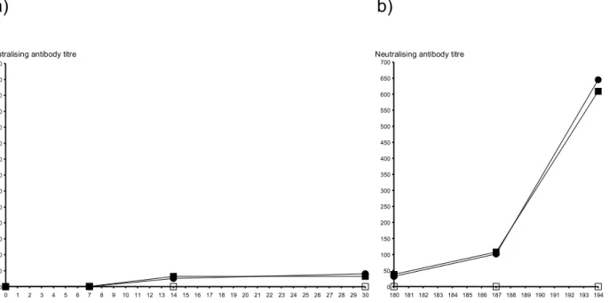
The analysis of the neutralizing antibody profile in sera of infected cattle revealed low to moderate neutralizing antibody titres from day 14 post -infection (pi) to until day 30 pi (1:16 to 1:64; Figure 1a). After reactivation, a significant rise in antibody titres (range 1:256 to 1:1024) was observed (Figure 1b). Uninfected control calves remained seronegative throughout.
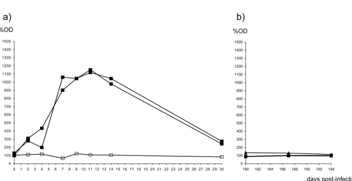
The results of the different ELISAs performed revealed that IgM, IgA, IgG1 and IgG2 responses did not significantly differ between calves infected with each of the two virus subtypes. IgM titres became detectable from day 2 pi and reached peaks between days 7 and 14 pi; subsequently, IgM levels started to decrease significantly towards day 30 pi (Figure 2a). IgA responses were detectable from day 2 pi and remained at high levels (as judged by %OD above 450) at least until day 30 pi, when sampling was discontinued (Figure 3a). IgG1 was initially detected on day 11 pi, rising to peak level on day 30 post inoculation (Figure 4a). IgG2 levels were detected only on samples collected on day 30 pi (Figure 5a).
At the beginning of the corticosteroid administration, 180 days pi, IgM was at trough levels and remained so for the next 14 days, when sampling was discontinued (Figure 2b). Antibodies of the IgA class (Figure 3b) remained at trough levels, whereas IgG1 (Figure 4b) and IgG2 (Figure 5b) remained at relatively high levels. Seven days later, IgA peaked with %OD values similar to those detected following primary acute infection. In addition, a slight, though not significant, increase in IgG1 and IgG2 levels was detected.
By comparing the patterns of antibody response, it was possible to estimate the stage of infection the calves were undergoing (Table 1). After primary infection, from day 2- 3 pi, until day 15 pi, only IgM and IgA antibodies could be detected. At days 11 to 14 pi, IgA levels remained elevated and
IgG1 antibodies became detectable. Neutralizing antibodies were initially detected on day 14 pi. Neutralizing antibodies were significantly elevated on day 194 pi. Antibodies of the IgG2 subtype were only detected on day 30 pi (Figure 5a). Thus, on day 30 pi, calves had IgA, IgG1, IgG2, and significantly decaying levels of IgM. On day 180 pi. when dexamethasone administration was started, IgG1 and IgG2 were still at levels which did not differ significantly from those detected on day 30 pi; IgM and IgA were at trough levels. On day 186, IgG1 and IgG2 remained at high levels and remained so, with no significant alteration.. On the other hand, IgA was significantly elevated on day 186 pi.
The analysis of the results revealed that that both BoHV1.1 and BoHV1.2a elicited similar patterns of humoral antibody responses in all classes and subclasses examined.
The neutralizing antibody profile detected was, as expected, a powerful indicative of infection, since these could be detected at any stage of the experiment (though at low levels) following the initial two weeks pi. Antibodies of all subclasses may be capable of inducing neutralization of BoHV1 in vitro (7, 8). However, early IgM and IgA antibodies, which here were detected earlier than neutralizaing antibodies (see below Figs 2 and 3) at least at this stage of infection seem not to contribute significantly to neutralization. Despite the usefulness of neutralizing antibodies as markers of infection, these could not be used to estimate whether infected animals were under acute primary acute infection or reactivation. Upon reactivation, neutralizing antibodies rose significantly; however, such increase was not followed by a corresponding rise in IgG1 or IgG2. On the other hand, IgA levels were raised at reactivation. Corticosteroid treatment apparently had no inhibitory effect on neutralizing antibodies, which at reactivation peaked to significantly higher titres than those obtained after primary infection.
Spilki, F.R. et al. Antibody responses to bovine herpesviruses
IgM was close to trough levels. During reactivation, no rise in IgM levels was detected, following an expected profile (5, 8). This may be useful for distinguishing the phases of infection, in that the analysis of the IgM profile can provide a way to differentiate acute infections from reactivation.
IgG1 levels were first detected on day 11 pi whereas IgG2 was evidenced on day 30 pi; from then on, during the subsequent phases of infection examined, both IgG1 and IgG2 were present concomitantly and to similar titres. Because the presence of IgG1 - but not IgG2 - is indicative of a recent primary infection, the determination of IgG1 and IgG2 levels may be used to estimate early acute infection.
Six months after infection, IgG1 and IgG2 levels were still similar to those detected on day 30 pi. Following reactivation, no significant increase in IgG1 and IgG2 levels were found. IgA levels were low at 180 days pi, but reactivation led to a new IgA peak. IgA was also found to rise upon reactivation by others (8). Therefore, the presence of elevated levels of IgA, concomitant with elevated IgG1 and IgG2 and absence of IgM
may provide additional evidence to indicate that calves have been through a recent reactivation process.
Based on the serological response obtained after the infection by BoHV1.1 and 1.2a, it could be possible to estimate the status of BoHV1 infection with basis on the serological analysis of the antibody responses induced in calves, provided that levels of IgM, IgA, IgG1 and IgG2 as well as neutralizing antibodies can be measured and compared (Table 1). It has been a hallmark in serology to request paired serum samples to identify the causes of acute infections. Here, in infections with both BoHV1 subtypes tested, it was shown that with simple class and subclass-specific ELISAs, it was possible to estimate with fair accuracy the stages of infection the animals were undergoing. The proposed scheme seems to fit adequately under the conditions of the present study, with a small number of animals and controlled conditions of infection. Although not evaluated here, at herd level, it may be possible to estimate the stages of infection, what could become particularly useful for monitoring the evolution of BoHV1 within the herd.
0 50 100 150 200 250 300 350 400 450 500 550 600 650 700
0 1 2 3 4 5 6 7 8 9 10 11 12 13 14 15 16 17 18 19 20 21 22 23 24 25 26 27 28 29 30
Neutralising antibody titre
0 50 100 150 200 250 300 350 400 450 500 550 600 650 700
180 181 182 183 184 185 186 187 188 189 190 191 192 193 194
Neutralising antibody titre
a)
b)
days post-infection
0 100 200 300 400 500 600 700 800 900 1000 1100 1200 1300 1400 1500
0 1 2 3 4 5 6 7 8 9 10 11 12 13 14 15 16 17 18 19 20 21 22 23 24 25 26 27 28 29 30
%OD
0 100 200 300 400 500 600 700 800 900 1000 1100 1200 1300 1400 1500
180 182 184 186 188 190 192 194
days post-infection %OD
a)
b)
Figure 2. Specific IgM antibody levels in sera of calves experimentally infected with BoHV-1.1 or BoHV1.2a. Data measured by
ELISA and expressed as mean percentual optical densities (%OD; see text for methods) for each group of calves. Plot a: at primary acute
infection; b: t reactivation. Full squares: BoHV-1.1 infected calves; full triangles: BHV-1.2a infected calves. Empty squares: control
uninfected calves.
%OD
0 100 200 300 400 500 600 700 800 900 1000 1100 1200 1300 1400 1500
0 1 2 3 4 5 6 7 8 9 10 11 12 13 14 15 16 17 18 19 20 21 22 23 24 25 26 27 28 29 30
0 100 200 300 400 500 600 700 800 900 1000 1100 1200 1300 1400 1500
180 182 184 186 188 190 192 194
days post-infection
a)
b)
%OD
Figure 3. ELISA analysis of IgA specific antibody in sera of calves experimentally infected with BoHV-1.1 or BoHV1.2a. Data measured by ELISA and expressed as mean percentual optical densities (%OD; see text for methods) for each group of calves. Plot a: at
primary acute infection; b: t reactivation. Full squares: BoHV-1.1 infected calves; full triangles: BHV-1.2a infected calves. Empty
Spilki, F.R. et al. Antibody responses to bovine herpesviruses
%OD
0 100 200 300 400 500 600 700 800 900 1000 1100 1200 1300 1400 1500
0 1 2 3 4 5 6 7 8 9 10 11 12 13 14 15 16 17 18 19 20 21 22 23 24 25 26 27 28 29 30 0 100 200 300 400 500 600 700 800 900 1000 1100 1200 1300 1400 1500
180 182 184 186 188 190 192 194
days post-infection
a)
b)
%OD
Figure 4. ELISA analysis of IgG1 specific antibody in sera of calves experimentally infected with BoHV-1.1 or BoHV1.2a. Data measured by ELISA and expressed as mean percentual optical densities (%OD; see text for methods) for each group of calves. Plot a: at primary acute infection; b: t reactivation. Full squares: BoHV-1.1 infected calves; full triangles: BHV-1.2a infected calves. Empty squares: control uninfected calves.
%OD
0 100 200 300 400 500 600 700 800 900 1000 1100 1200 1300 1400 1500
0 1 2 3 4 5 6 7 8 9 10 11 12 13 14 15 16 17 18 19 20 21 22 23 24 25 26 27 28 29 30 0 100 200 300 400 500 600 700 800 900 1000 1100 1200 1300 1400 1500
180 182 184 186 188 190 192 194
days post-infection %OD
a)
b)
Table 1. Estimate of the status of infection with basis on the patterns of isotype and subclass specific anti-bovine herpesvirus 1.1 (BHV-1.1) or 1.2a (BHV-1.2a) antibodies.
Status of infection Isotype-specific profile
Uninfected cattle IgM- IgA- IgG1- IgG2- SNAa -Up to day 15 post-infection IgM+ IgA+ IgG1- IgG2- SNA-or+ Between days 15 and 30 post- infection IgM+ IgA+ IgG1+ IgG2+ SNA+ During latency (after day 30 post infection) IgM- IgA- IgG1+ IgG2+ SNA+ After reactivation IgM- IgA+ IgG1+ IgG2+ SNA+
+ = present ; - = absent ; a = serum neutralizing antibodies
ACKNOWLEDGEMENTS
The authors thank Dr. Frans Rijsewijk (in memoriam) for review and comments. F.R.S., A.C.F, and P.M.R. are CNPq research fellows. Work supported by FEPAGRO, PRONEX, CNPq, CAPES and FAPERGS
REFERENCES
1. Bradshaw, B.J.F.; Edwards, S. (1996). Antibody isotype responses to experimental infection with bovine herpesvirus 1 in calves with colostrally derived antibody.Vet. Mic.53 (1-2), 143-151.
2. D'Arce, R.C.F.; Almeida, R.S.; Silva, T.C.; Franco, A.C.; Spilki, F.; Roehe, P.M.; Arns, C.W. (2002). Restriction endonuclease and monoclonal antibody analysis of Brazilian isolates of bovine herpesviruses types 1 and 5.Vet. Mic.88 (4), 315-324.
3. do Cilento, M.C.; Pituco, E.M.; Jordão, R.S.; Ribeiro, C.P.; Filho, M.M.; Montassier, H.J. (2011). Systemic and local antibodies induced by an experimental inactivated vaccine against bovine herpesvirus type 1.
Ciência Rural 41 (2), 307-313.
4. Esteves, P.A.; Dellagostin, O.A.; Pinto, L.S.; Silva, A.D.; Spilki, F.R.; Ciacci-Zanella, J.R.; Hübner, S.O.; Puentes, R.; Maisonnave, J.; Franco, A.C.; Rijsewijk, F.A.M.; Batista, H.B.C.R.; Teixeira, T.F.; Dezen, D.; Oliveira, A.P.; David, C.; Arns, C.W.Roehe, P.M. (2008). Phylogenetic comparison of the carboxy-terminal region of glycoprotein C (gC) of bovine herpesviruses (BoHV) 1.1, 1.2 and 5 from South America (SA). Virus Res. 131 (1), 16-22.
5. Guy, J.S.; Potgieter, L.N. (1985). Bovine herpesvirus-1 infection of cattle: kinetics of antibody formation after intranasal exposure and abortion induced by the virus.Am. J. Vet. Res. 46 (4), 893-898.
6. Davison, A.J.; Eberle, R.; Ehlers, B.; Hayward, G.S.; McGeoch, D.J.; Minson, A.C.; Pellett, P.E.; Roizman, B.; Studdert, M.J.; Thiry, E. (2009). The order Herpesvirales. Arch. Virol. 154 (1), 171–177.
7. Madic, J.; Magdalena, J.; Quak, J.Van Oirschot, J.T. (1995). Isotype-specific antibody responses in sera and mucosal secretions of calves experimentally infected with bovine herpesvirus 1. Vet. Immunol.
Immunopathol. 46 (3-4), 267-283.
8. Madic, J.; Magdalena, J.; Quak, J.Van Oirschot, J.T. (1995). Isotype-specific antibody responses to bovine herpesvirus 1 in sera and mucosal secretions of calves after experimental reinfection and after reactivation.
Vet. Immunol. Immunopathol. 47 (1-2), 81-92.
9. Miller, J.M.; van der Maaten, M.J. (1984). Reproductive tract lesions in heifers after intrauterine inoculation with infectious bovine rhinotracheitis virus.Am. J. Vet. Res. 45 (4), 790-794.
10. Miller, J.M.; Van der Maaten, M.J. (1986). Experimentally induced infectious bovine rhinotracheitis virus infection during early pregnancy: effect on the bovine corpus luteum and conceptus.Am. J. Vet. Res. 47 (2), 223-228.
11. Miller, J.M.; Van der Maaten, M.J.; Whetstone, C.A. (1988). Effects of a bovine herpesvirus-1 isolate on reproductive function in heifers: classification as a type-2 (infectious pustular vulvovaginitis) virus by restriction endonuclease analysis of viral DNA.Am. J. Vet. Res. 49 (10), 1653-1656.
12. Spilki, F.R.; Esteves, P.A.; Da Silva, A.D.; Franco, A.C.; Rijsewijk, F.A.M.; Roehe, P.M. (2005). A monoclonal antibody-based ELISA allows discrimination between responses induced by bovine herpesvirus subtypes 1 (BoHV-1.1) and 2 (BoHV-1.2).J. Virol. Methods 129 (2), 191-193.
13. Spilki, F.R.; Esteves, P.A.; De Lima, M.; Franco, A.C.; Chiminazzo, C.; Furtado Flores, E.; Weiblen, R.; Driemeier, D.; Roehe, P.M. (2004). Comparative pathogenicity of bovine herpesvirus 1 (BHV-1) subtypes 1 (BHV-1.1) and 2a (BHV-1.2a).Pesq. Vet. Bras. 24 (1), 43-49.
14. Teixeira, M.F.B.; Esteves, P.A.; Schmidt, C.S.; Spilki, F.R.; Silva, T.C.; Dotta, M.A.; Roehe, P.M. (2001). A monoclonal blocking ELISA for the serological diagnosis of bovine herpesvirus type 1 (BHV-1) infections. Pesq. Vet. Bras. 21 (1), 33-37.
Spilki, F.R. et al. Antibody responses to bovine herpesviruses
16. Welsh, M.D.; Cunningham, R.T.; Corbett, D.M.; Girvin, R.M.; McNair, J.; Skuce, R.A.; Bryson, D.G.; Pollock, J.M. (2005). Influence of
pathological progression on the balance between cellular and humoral immune responses in bovine tuberculosis.Immunology 114 (1), 101-111.


