Specific Antibody Levels and Antigenic Recognition of
Wistar Rats Inoculated with Distinct Isolates of
Trypanosoma evansi
Adriana O Queiroz, Ana P Legey, Samanta CC Xavier, Ana M Jansen
+Departamento de Protozoologia, Instituto Oswaldo Cruz-Fiocruz, Av. Brasil 4365, 21045-900 Rio de Janeiro, RJ, Brasil
“Mal de Cadeiras”, an enzootic disease caused by Trypanosoma evansi, is one of the most impor-tant trypanosomiases in the Brazilian Pantanal region. The disease affects mainly horses, which are widely used in extensive cattle production, an activity of greatest economical significance for the region. The parasite also infects sylvan (coatis and capybaras) and domestic (dogs) animals, respec-tively considered wild and domestic reservoirs of T. evansi. For a better understanding of the interac-tion of T. evansi with its rodent host, we evaluated the differences in the specific antibody level patterns and in the parasitic peptides recognition patterns of experimentally infected Wistar rats. The rats experimentally infected with T. evansi isolates obtained from coatis, dogs and horses were submitted to indirect immunofluorescence test (IgM e IgG) and Western blotting. The serological titers for IgM and IgG ranged between 1:40 and 1:160. The most recognized polypeptide profiles were in a range of 17 and 74 kDa. Our data suggest that the humoral immune response in Wistar rats is not sufficient for granting an effective control of T. evansi infections.
Key words: Trypanosoma evansi - Wistar rats - antigenic recognition - indirect fluorescent antibody test
Trypanosoma evansi is a widely distributed hemoflagellate, closely related to T. brucei. How-ever, unlike other salivarian trypanosomes, T. evansi is mechanically transmitted by blood suck-ing insects, especially by Tabanus spp. and Sto-moxys spp. In South and Central America, T. evansi is also transmitted by the vampire bat (Desmodus rotundus) which also acts as a carrier (Hoare 1972). Since only the slender forms have been de-scribed, T. evansi is considered a monomorphic try-panosome. Up to the present, many T. brucei re-lated species have been described based on differ-ences regarding virulence, morphology and hosts, among others T. equinum, T. venezuelense and T. hippicum in South America and T. marocanum in Morocco. The taxonomic significance of these spe-cies is at present considered dubious. Although T. evansi is presenting geographic and epidemiologi-cal peculiarities, only one taxon is accepted. This point of view was confirmed through recent mo-lecular and biochemical characterizations (Borst et al. 1987, Stevens et al. 1989, Lun et al. 1992a, b).
This investigation was supported by Capes, Faperj and Fiocruz.
+Corresponding author. Fax: +55-21-2598.4323. E-mail:
[email protected] Received 5 September 2000 Accepted 24 July 2001
In spite of the susceptibility of almost all spe-cies of domesticated livestock to T. evansi, the main host varies according to the geographical region. In Central and South America, the disease affects mainly horses, the principal hosts in Africa are the camels and the main host in China is the pig (Dia 1997).
we need biochemical, immunological, molecular and biological data of T. evansi derived from distinct hosts and geographical regions.
Only limited antigenic diversity has been de-scribed among T. evansi isolates. This characteris-tic reinforces the importance of the host’s humoral immune response and suggests the possibility of controlling this enzootic disease through immunoprophylactic methods (Jones & McKinnell 1995).
While there are many studies on T. brucei, only few data on T. evansi antigen recognition are avail-able and none, regarding Brazilian isolates (Uche et al. 1992, 1993, Uche & Jones 1994). The impor-tance of antibodies in the control of parasitaemia by T. evansi is poorly known and still controversial (Franke et al. 1994, Aquino et al. 1999). There is only one single study on antigenic variation in Wistar rats. This study, however, was carried out with T. evansi isolates from India and used para-sitic flagellar and cell membrane peptides (Singh et al. 1995).
In the present study we examined T. evansi antigen recognition and specific antibody levels of Wistar rats (Rattus norvegicus) experimentally in-fected with domestic and sylvatic T. evansi iso-lates from the Brazilian Pantanal region.
MATERIALS AND METHODS
Trypanosomes - T. evansi isolates from domes-tic and sylvadomes-tic animals (Table I) from the Pantanal of Mato Grosso were studied. The coati and dog isolates were obtained by one member of our group (AM Jansen) and horse isolates were kindly sup-plied by RAMS Silva (Embrapa, MT). The origin and codes of stocks used in the present work are given in the Table I. It is worth mentioning that Horse 4 and Coati 2 isolates are the most and the less virulent T. evansi isolates, respectively (Queiroz et al. 2000a).
All isolates were collected in the Southern re-gion of the Pantanal of Mato Grosso. After being collected, the isolates were maintained in liquid ni-trogen until use.
Preparation of trypanosome antigens - Total antigen was prepared from trypanosomes collected from Wistar rats experimentally infected and previ-ously immune-suppressed (cyclophosphamide, 200 mg/kg body weight). At the point of fulminating infection (109 parasites/ml), the rats for each iso-late were exsanguinated and the parasites were purified by separation in a DEAE cellulose column and re-suspended in ice-cold phosphate saline glu-cose (PSG) pH 8.0 (Lanham & Godfrey 1970). Try-panosomes were disrupted in five to six cycles of 2 min/4ºC in an ultrasonic disintegrator (Branson – 2510, USA). The parasites were then lysed through
freezing in Methanol and dry ice, alternately. After this, buffer containing protease inhibitors (Trizma base 20 mM, NaCl 40 mM, EDTA 10 mM, Iodoacetamyde 2 mM, 1,10 Phenantroline 1,6 mM and PMSF 1 mM) was added. The total antigen was aliquoted into 100 µl volumes and stored at -20ºC.
Inoculations - After purification of parasites from previously infected rats through an anion ex-changer column (DEAE cellulose), the inocula of each T. evansi isolate were adjusted in a Neubauer chamber so as to yield, 10 and 103 parasites/g of body weight respectively and injected into young, male Wistar rats weighing 100-150 g.
Two batches of six animals per inoculation schedule were inoculated intraperitoneally with 10 flagellates/g of body weight (low inoculum) and 103 flagellates/g of body weight (high inoculum). The follow-up included daily fresh blood smear examinations and weekly blood collection. The sera were aliquoted into 50 µl and stored at -20ºC. Sera from non-infected rats were used as negative con-trols.
Serological follow-up - An indirect fluorescent antibody test (IFAT), as described by Camargo (1966) was performed to follow-up T. evansi infected Wistar rats (Table). The antigen, consisting of tripomastigote forms of T. evansi, was adjusted to 40 parasites by microscopic field examination (40x) and stored at -20oC. Positive control serum was obtained from a rat experimentally infected with strain Horse 4. For the indirect immunofluorescene reaction a conjugate of anti-mouse IgG FITC con-jugate (Sigma F6257) and anti-mouse IgM FITC conjugate (Sigma F9259) was used. Due to the non-existence of anti-rat IgM we preferred to use anti mouse conjugate (IgM and IgG). The difference in the serological titers as tested by us was not sig-nificant.
SDS-PAGE - The trypanosome total antigens were solubilized by boiling them for 4 min in an equal volume of 1X Laemmli’s sample buffer, loaded into sample wells and separated by SDS-PAGE un-der reduced conditions in a 10% polyacrylamide gel containing SDS. The concentration of the stack-ing gel was of 4.5% polyacrylamide (Laemmli 1970). A mixture of calibration proteins with molecular weight ranging from 7.5 to 202 kDa (molecular weight markers, Broad range, BIORAD) was in-cluded in a separate well during each run. All gels were 1.5 mm thick and run at a constant current of 100 V for approximately 2 h.
the technique described by Towbin et al. (1979). The section of the nitrocellulose membrane con-taining the molecular weight markers and the sec-tion containing the strips was cut into strips of 5 mm width. The total strips were immediately stained for 10 min with 0.2% Ponceau S (Sigma, USA) in 3% acetic acid and 97% tri-distilled water to verify all transferred trypanosomal proteins and molecular weight markers. After that, the blots were washed for 30 min with tri-distilled water. The strips were then placed for 90 min in PBS solution (pH 7.2) containing 0.5% Tween 20 with 0.5% defatted milk to block non-specific binding sites. The strips were then washed again with PBS containing 0.05% Tween 20 (PBST). After the blocking, the strips were incubated overnight with sera obtained from ex-perimentally infected rats diluted in PBST (1:200). One strip was incubated with serum obtained from an uninfected rat as a negative control. After six 15 min washes with PBST the strips were incubated for 90 min with peroxidase conjugated anti-mouse IgG (Sigma, USA) diluted in a ratio of 1:4000. After six washes as described above, the strips were added to freshly prepared substrate solution (15 mg of 3,3'- diaminobenzidine tetrahydrochloride (Sigma, USA), 40 µl of H2O2 in 60 ml of citrate phos-phate buffer (C6H8O7, H2O – 0.01M and Na2HPO4 – 0.02M) pH 5.0 to visualize the antigenic compo-nents. The reaction was interrupted by washing the blots in distilled water. The molecular weight of the visualized components was measured by means of a calibration curve prepared for each isolate based on molecular weight standards (Singh et al. 1995).
RESULTS
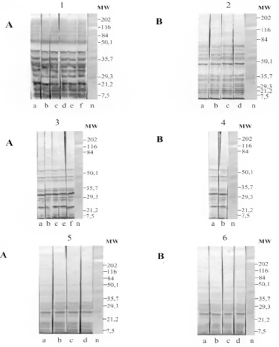
The recognition pattern of experimentally T. evansi infected rats is summarized in Fig. 1. Experi-mentally infected Wistar rats recognized polypep-tides of molecular weight ranging between 7 and 200 kDA, but the more intense recognition occurred in a range between 17 and 57 kDA. With exception of the rats inoculated with Coati 3 isolate, all tested animals recognized peptides ranging between 53 and 63 kDa – the MW of variant surface glycopro-teins of salivarian trypanosomes (Richards 1984).
No correlation between the number and/or in-tensity of peptide recognition and the serological titers could be observed in the infected rats, inde-pendently from the T. evansi isolate or inoculation schedule used.
No individual differences in the recognition pattern in a same batch of rats inoculated with the same T. evansi isolate were observed, but it dif-fered in the different rat groups according to the inoculated T. evansi isolate . No variation could be observed during the course of the experimental
in-fection. Actually, the profiles remained unchanged since the first testing (with seven days) (Fig. 1).
Rats infected with what we call high or low inocula of Horse 4 survived significantly longer and recognized a greater number of T. evansi anti-gens (Tables I, II). Moreover, no further differences were observed in the recognition pattern of rats infected with high or low inocula (Fig. 1). Rats in-fected with the Dog 2 isolate recognized antigens with the molecular weight of 18, 27, 48, 63 kDa and rats infected with Coati 3 isolate recognized sig-nificantly lesser antigens (22 kDa) (Fig. 1).
Beginning from the 7th day after infection, al-most all inoculated rats displayed specific IgM and IgG antibodies, independently from the inoculation schedule (high or low inoculum) or origin of the isolate (Fig. 2).
As refers to the specific antibody levels, impor-tant individual differences could be observed in the serological titers of the infected rats. However, no correlation between these differences and the inoculation schedule, the origin of the inoculated T. evansi isolate or the recognition pattern could be observed.
Most of the inoculated rats presented serologi-cal titers for IgM and IgG in the serum already from the 7th day after the inoculation, independently from the origin of the inoculum. No direct correla-tion could be established between antibody (IgM and IgG) levels and the survival time of the infected rats, independently from the origin of the isolate and/or the inoculation conditions.
Rats infected with high and low inocula of the ten T. evansi isolates presented IgM and IgG titers ranging between 1/40-1/160. The highest serologi-cal titers were observed on the 14th day after in-oculation in rats that received a high inoculum with Dog 3 isolate (IgM: 1/640 and IgG: 1/320) and on the 21st day after inoculation in rats that received a low inoculum with Horse 4 isolate (IgM:1/1280 and IgG: 1/640).
No clear evidence for the effectiveness of the immune response in controlling the parasite popu-lations could be observed. Although higher sero-logical titers for IgM and IgG were observed in rats that survived longer, some rats that survived for only a short time also displayed high serological titers as observed in the rats infected with Coati 2 isolate (Fig. 3).
DISCUSSION
Fig. 1: antigenic recognition of polypeptides of Trypanosoma evansi experimentally infected by Wistar rats with isolates: Horse 4 (1 and 2), Dog 2 (3 and 4) and Coati 3 (5 and 6) after respectively 1st, 2nd, 3rd, 4th, 5th and 6th weeks post experimental infection. A: low inoculum; B: high inoculum; a: 7 days; b: 14 days; c: 21 days; d: 28 days; e: 35 days; f: 42 days, n: negative control
ranging between 53 and 63 kDa (Richards 1984). In our experiments, all animals recognized a peptide in this MW except the rats inoculated with Coati 3 isolate that showed the shortest survival time. Fur-thermore, it is worth mentioning that rats inocu-lated with Coati 3 isolate, one of the most virulent isolates, recognized a polypeptide of 21 kDa, whose importance is not mentioned in the literature.
de-scribed for T. brucei and other T. evansi isolates. T. evansi is an interesting trypanosomatide. Despite its broad geographical distribution, wide range of hosts and distinct epidemiological features it is recognized as homogeneous species (Borst et al. 1987, Stevens et al. 1989). Indeed, genetic diver-sity as measured by the conventional biochemical and molecular markers has always led to irrelevant results (Masiga & Gibson 1990, Lun et al. 1992b).
In a previous work we described the homogeneity of ten T. evansi isolates derived from domestic and sylvatic mammals by means of biochemical (isoen-zymatic profiles) and molecular (schizodeme analy-sis) characterization. In spite of this homogeneity we observed significant differences in the virulence pattern of the T. evansi isolates, which could not be correlated with the species of the original host (Queiroz et al. 2000a, b).
Fig. 2: parasitaemia and immunoglobulin levels were detected by an indirect immunofluorescence antibody test in Wistar rats infected, and curves were plotted as follows: 0 (negative), 1 (dilution 1/ 10), 2 (1/ 20), 3 (1/ 40), 4 (1/ 80), 5 (1/ 160), 6 (1/ 320), 7 (1/ 640) and 8 (1/ 1280) with isolates of Trypanosoma evansi from Dog 2 (A), Horse 1 (B) and Coati 3 (C).
1,00E+00 1,00E+02 1,00E+04 1,00E+06 1,00E+08 1,00E+10
0 3 6 9 12 15 18 21 24 27 30 33 36 39 42
Time (days)
Parasites/ml
0 1 2 3 4 5 6 7 8
Reciprocal of
antibody titer (IFAT)
P AR AS IT AS /ML IgM IgG
0 102
104
106
108
1010
P aras ites /ml
A
1,00E+00 1,00E+02 1,00E+04 1,00E+06 1,00E+08 1,00E+10
0 3 6 9 12 15 18 21 24 27 30 33 36 39 42 45 48 51
Time (days)
Parasites/ml
0 1 2 3 4 5 6 7 8
Reciprocal of
antibody titer (IFAT)
0 102
104
106 108
1010 B
1,00E+00 1,00E+02 1,00E+04 1,00E+06 1,00E+08 1,00E+10
0 1 2 3 4 5 6 7 8 9 10 11 12 13 14 15 16 17
Time (days)
Parasites/ml
0 1 2 3 4 5 6 7 8
Reciprocal of
antibody titer (IFAT)
C
1010 108
106
104
102
In the present paper, we observed differences in the recognition pattern in the different batches of rats inoculated with the distinct T. evansi iso-lates. These differences are probably due to varia-tions of the polypeptide profile of the parasite iso-lates and not to individual variations in
recogni-TABLE I
Brazilian Trypanosoma evansi isolates and host species of ten stocks from Pantanal Matogrossense Region, Brazil
Code of stocks Isolation Host species Survival time - days ( x )
(year) (subgroups) High inocula Low inocula
MCAN/ CPAP - C 1995 Dog 1 5.83 9.67
MCAN/ ETRG E0507 1996 Dog 2 10.17 28.5
MCAN/ ETRG E0506 1996 Dog 3 24.33 23.17
MEQH/ CPAP - A 1994 Horse 1 9.33 31.67
MEQH/ CPAP - B 1994 Horse 2 7 9.83
MEQH/ CPAP - D 1995 Horse 3 8 19.5
MEQH/ CPAP - E 1996 Horse 4 30.67 23.25
NN3/ CPA - P 1996 Coati 1 8.17 7.83
NN4/ CPA - P 1996 Coati 2 23.33 25.33
NN5/ CPA - P 1996 Coati 3 9.17 10.5
tion since animals from the same batch displayed the same recognition pattern. These data show that a correlation of genotypic and phenotypic charac-teristics of parasites with distinct epidemiological features is still a challenge and indicate that the multiple influences of environmental factors on both, parasites and hosts, should also be taken into account.
Differences between resistant and susceptible rabbits with respect to their recognition pattern to non-surface components of T. evansi were ob-served: the recognition of polypeptides of 61 kDa, 67 kDa and 94 kDa, respectively, by the rabbits was associated to resistance (Uche et al. 1992). In our experiments, no recognition of peptides with this molecular weight was observed, suggesting pecu-liarities in the interaction of T. evansi with its differ-ent host species.
In T. congolense and T. brucei infections, a correlation in the recognition pattern between re-sistant N’Dama and susceptible Boran and Zebu cattle could also be noticed (Authié et al. 1993). This was not the case in this work, since all isolates of T.evansi were highly pathogenic for the Wistar rats and 100% mortality was observed.
It is worth mentioning that the rats inoculated with Horse 4 isolate survived longer and recog-nized more antigens, mainly in a high molecular weight range. Notwithstanding, all animals died with patent parasitaemia (Fig. 1, Table II).
The homogeneity in the recognition pattern in the early stages of infection in comparison to the final stages suggests that the fate of the infected animals was determined in the early stages of the infection.
It already was described for T. evansi and other salivarian trypanosomatids in other hosts, that the humoral immune response, mainly IgM, plays an important role in the control of the circulating para-sites (Roelants & Pinder 1984, Uche & Jones 1994). Fig. 3: parasitaemia and IFAT in Wistar rats infected with
isolate of Trypanosoma evansi from Coati 2. High sero-logical titers and short termed survival were observed.
1 100 10000 1000000 100000000 0000000000
0 1 2 3 4 5 6 7 8 9 10
Time (days)
Parasites/m
l
0 1 2 3 4 5 6 7 8
Reciprocal of
antibod
y
titer
(IFAT
)
0 102
104 106 108
1010
P AR AS IT AS /ML IgM
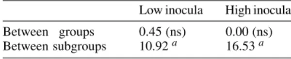
IgG P aras ites /ml TABLE II
Nested analysis of the course of experimental infection
(survival time) of Wistar rats with Trypanosoma
evansi isolates derived from horses, coatis and dogs
Low inocula High inocula
Between groups 0.45 (ns) 0.00 (ns)
Between subgroups 10.92 a 16.53 a
ns: not significant; a: highly significative. Level of
significance 0.05; F values in the comparative study
performed among the isolates of T. evansi considering
In our experiments, this correlation was not so clear since the infected rats displayed comparable anti-body titers. Our data confirm the findings of Aquino et al. (1999) who observed that high levels of parasitaemia persisted throughout the experiments in spite of the high antibody titers for IgM and IgG. Moreover, in spite of a longer survival, all animals died even when injected with low inocula. This suggests the humoral immune response, at least in this host model, to be far from being sufficient for controlling the parasite populations. It is worth mentioning that the Wistar rats responded earlier and with higher serological titers than previously reported for other animal models such as dogs (Aquino et al. 1999), rabbits (Luckins et al. 1978), camels (Luckins et al. 1979) and guinea pigs (Oliveira et al. 1989). Probably there exist characteristic mechanisms for controlling the parasitaemia in each animal species. Uche and Jones (1994) described that rabbits recognized a smaller spectrum of anti-gens than rats, reinforcing the idea of characteris-tic control mechanisms for T. evansi infection of each animal species.
These peculiarities could be at least one of the reasons for the distinct aspects of the disease. In-deed, the enzootic disease caused by T. evansi dis-plays distinct features according to the geographi-cal region. Pigs are the main reservoir in Asia, cam-els in Africa and horses in South America. The Chinese isolates, on the contrary to the others, are cultivable in axenic media (Zweygarth et al. 1991). Distinct virulence patterns were also described (Queiroz et al. 2000a). All these factors should be taken into account in case an immunoprophylaxis program is to be planned. It will also be necessary to evaluate the role played by cross recognition and cross reactivity among T. evansi isolates of a same geographical region.
ACKNOWLEDGEMENTS
To R Mexas for help with the photographs and A Ivo for technical help.
REFERENCES
Aquino TLPC, Machado RZ, Alessi AC, Marques LC, Castro MB, Malheiros EB 1999. Clinical, parasito-logical and immunoparasito-logical aspects of experimental
infection with Trypanosoma evansi in dogs. Mem
Inst Oswaldo Cruz94: 255-260.
Authié E, Muteti DK, Williams DJL 1993. Antibody
responses to invariant antigens of Trypanosoma
congolense in cattle of differing susceptibility to
try-panosomiasis. Parasite Immunol15: 101-111.
Borst P, Fase-Fowler F, Gibson W 1987. Kinetoplast
DNA of Trypanosoma evansi. Mol Biochem
Parasitol23: 31-33.
Camargo ME 1966. Fluorescent antibody test for the serodiagnosis of American trypanosomiasis.
Tech-nical modification employing preserved cultured
forms of Trypanosoma cruzi in a slide test. Rev Inst
Med Trop de São Paulo8: 227-234.
Dempsey WL, Mansfield JM 1983. Lymphocyte func-tion in experimental African trypanosomiasis. V. Role of antibody and the mononuclear phagocyte system
in variant – specific immunity. J Immunol130:
405-411.
Dia ML 1997. Epidémiologie de la Trypanosomose
Cameline à Trypanosoma evansi en Mauritanie. Universite Montpellier, PhD Thesis, 34 pp. Franke CR, Greiner M, Mehlitz D 1994. Investigations
on naturally occurring Trypanosoma evansi
infec-tions in horses, cattle, dogs and capybaras (Hydrochaeris hydrochaeris) in Pantanal de Poconé
(Mato Gross, Brazil). Acta Trop58: 159-169.
Hoare CA 1972. The Trypanosomes of Mammals. A
Zoological Monograph, Blackwell Scientific Publi-cations, Oxford-Edinburg, 749 pp.
Jones TW, McKinnell CD 1985. Antigenic variation in
Trypanosoma evansi: a comparison of the predomi-nant variable antigen type repertoires of stocks from
the Sudan. Trop Med Parasitol36: 205-209.
Laemmli UK 1970. Cleavage of structural proteins
dur-ing the assembly of the head of bacteriophag T4.
Nature227: 680-685.
Lanham SM, Godfrey DG 1970. Isolation of salivarian trypanosomes from man and other mammalians
us-ing DEAE-cellulose. Exp Parasitol28: 521-534.
Luckins AG, Gray AR, Rae P 1978. Comparison of the diagnostic value of serum immunoglobulin levels, an enzime-linked immunosorbent assay and a fluores-cent antibody test in experimental infections with
Trypanosoma evansi in rabbits. Ann Trop Med Parasitol72: 429-441.
Luckins AG, Mcintyre N, Rae PF 1992. Multiplication of Trypanosoma evansi at the site of infection in
skin of rabbits and cattle. Acta Trop50: 19-27.
Luckins AG, Boid R, Rae P, Mahmoud MM, El Mazik KH, Gray AR 1979. Serodiagnosis of infection with
Trypanosoma evansi in camels in the Sudan. Trop Anim Hlth Prod11: 1-12.
Lun ZR, Allingham R, Brun R, Lanham SM 1992a. The
isoenzyme characteristics of Trypanosoma evansi
and T. equiperdum isolated from domestic stocks in
China. Ann Trop Med Parasitol86: 333-340.
Lun ZR, Brun R, Gibson W 1992b. Kinetoplast DNA
and molecular karyotopes of Trypanosoma evansi
and T. equiperdum. Mol Biochem Parasitol50: 189-196.
Masiga DK, Gibson WC 1990. Specific probes for
Try-panosoma (Trypanozoon) evansi based on
kineto-plast DNA minicircles. Mol Biochem Parasitol40:
279-284.
Oliveira TCG, Sogayar R, Salata E 1989. Estudo
sorológico de infecções experimentais por
Trypano-soma evansi em cobaias. Rev Inst Med Trop São Paulo31: 95-99.
and biochemical characterization of isolates of Try-panosoma evansi from Pantanal of Matogrosso
-Brazil. Vet Parasitol92: 107-118.
Queiroz AO, Nehme-Russell NS, Brandão A, Jansen
AM 2000b. Homogeneity of Trypanosoma evansi
isolates from domestic and sylvatic mammals from
the Pantanal of Mato Grosso. Microbios103:
27-30.
Richards FF 1984. The surface of African
trypano-somes. J Protozool32: 60-64.
Roelants GE, Pinder M 1984. Immunobiology of
Afri-can trypanosomiasis. Contemp Top Immunobiol12:
255-274.
Singh V, Singh A, Chhabra MB 1995. Polypeptide pro-files and antigenic characterization of cell membrane
and flagellar preparations of different stocks of
Try-panosoma evansi. Vet Parasitol56: 269-279. Shapiro SZ, Murray M 1982. African trypanosome
an-tigens recognized during the course of infection in
N’Dama and Zebu cattle. Infect Immun35: 410-416.
Stevens JR, Nunes VLB, Lanham SM, Oshiro ET 1989.
Isoenzyme characterization of Trypanosoma evansi
isolated from capybaras and dogs in Brazil. Acta Trop
46: 213-222.
Towbin H, Staehelin T, Gordon J 1979. Electrophoretic transfer of proteins from polyacrylamide gels to ni-trocellulose sheets: procedure and some
applica-tions. Proc Natl Acad Sci USA76:4350-4354.
Uche UE, Jones TW 1994. Protection conferred by
Try-panosoma evansi infection against homologous and
heterologous trypanosome challenge in rabbits. Vet
Parasitol52: 21-35.
Uche UE, Jones TW, Boid R 1992. Antibody patters in rabbits showing different levels of susceptibility to
an experimental Trypanosoma evansi infection. Acta
Trop52: 139-147.
Uche UE, Jones TW, Boid R 1993. Class-specific anti-body response in rabbits experimentally infected with
Trypanosoma evansi. Trop Med Parasitol 44: 27-31.
Zweygarth E, Kaminsky R, Webter P 1991.
Trypano-soma brucei evansi: dyskinetoplastia and loss of
infectivity after long term in vitro cultivation. Acta