A
rt
ic
le
J. Braz. Chem. Soc., Vol. 20, No. 6, 1082-1088, 2009. Printed in Brazil - ©2009 Sociedade Brasileira de Química 0103 - 5053 $6.00+0.00
*e-mail: [email protected]
Dihydroflavonols from the leaves of
Derris urucu
(Leguminosae):
Structural Elucidation and DPPH Radical-Scavenging Activity
Lívia T. Lôbo,a Geilson A. da Silva,a Malisson Ferreira,a Milton N. da Silva,a Alberdan S. Santos,a
Alberto C. Arruda,a Giselle M. S. P. Guilhon,a Lourivaldo S. Santos,a Rosivaldo dos Santos Borgesb and Mara Silvia P. Arruda*,a
aPrograma de Pós-Graduação em Química, Instituto de Ciências Exatas e Naturais, Universidade Federal do Pará,
Campus Universitário do Guamá, 66075-970 Belém-PA, Brazil
bFaculdade de Farmácia, Instituto de Ciências da Saúde, Universidade Federal do Pará, Campus Universitário do
Guamá, 66075-970 Belém-PA, Brazil
Derris urucu é uma planta da Amazônia com propriedades inseticida e ictiotóxica. Estudos com esta espécie reportam a presença de flavonóides, principalmente rotenóides, bem como de estilbenos. A partir do extrato etanólico das folhas de Derris urucu (Leguminosae), três novos diidroflavonóides, denominados urucuol A (1), B (2) e C (3) e o diidroflavonol isotirumalina (4), foram isolados e identificados. As estruturas destes compostos foram elucidadas por uma extensiva análise espectroscópica de RMN uni e bidimensional, UV, IV e dados de EM, além de comparação com dados da literatura. Os compostos isolados (1-4) foram avaliados quanto ao seu potencial sequestrador do radical DPPH• e apresentaram baixo poder antioxidante quando comparados ao antioxidante comercial trans-resveratrol.
Derris urucu is an Amazonian plant with insecticide and ichthyotoxic properties. Studies with this species show the presence of flavonoids, mainly rotenoids, as well as stilbenes. The ethanol extract of the leaves of Derris urucu (Leguminosae) afforded three new dihydroflavonols named urucuol A (1), B (2) and C (3), and the dihydroflavonol isotirumalin (4). Their structures were elucidated by extensive analysis of 1D and 2D NMR, UV and IR spectra and MS data and comparison with literature data. The isolated compounds (1-4) were evaluated for DPPH•radical scavenging activity and showed a relatively lower antioxidant ability compared to the commercial antioxidant trans-resveratrol.
Keywords: Derris urucu, Leguminosae, dihydroflavonols, radical scavenging activity,
antioxidant
Introduction
Amazonian ecosystems are rich in plants with insecticide and piscicide properties, and those belonging to the Derris genus are the most used.1 These plants, in the
Amazon area, are called “timbó”. Some studies carried out on roots of Derris urucu extracts related the presence of rotenoids, especially rotenone, that show insecticide and ichthyotoxic activity.2-5 In addition to rotenoids, others
minor flavonoids, such as flavanones, isoflavanones and chalcones, together with stilbenes, have been also described from the roots of Derris urucu.6 Flavonoids belong to a
group of naturally occurring compounds with a number of
biological activities, such as antibacterial, antimutagenic, cytotoxic and anticarcinogenic,7-10 together with antioxidant
activity, which is one of the most studied.11,12 Antioxidant
activity arises from the ability of flavonoids to scavenge free radicals and thus eliminate reactive oxygen species.13,14
These phenolic compounds are known to possess an antioxidant character to various extents.15-17 Therefore,
the antioxidant activity of these natural compounds is related to a number of different mechanisms such as free radical scavenging, hydrogen donation, singlet oxygen quenching, metal ion chelation, and acting as a substrate for radicals such as superoxide and hydroxide.18 Because
Lôbo et al. 1083 Vol. 20, No. 6, 2009
Nevertheless, some phenolic compounds increase oxidative stress and toxicity because of their prooxidant properties.19
The balance between antioxidant or prooxidant properties can be determined by the scavenger capacity of radical oxygen or nitrogen species using spectrophotometric methods, such as DPPH, which has been applied to the phenolic compounds commonly present in natural products. The spectrophotometric technique employs the 1,1-diphenyl-2-picrylhydrazyl free radical (DPPH•), which shows a characteristic UV-Vis spectrum with a maximum of absorbance close to 517 nm in methanol. The addition of an antioxidant compound results in a decrease of absorbance proportional to the concentration and antioxidant activity of the compound.20 This method presents the advantage of
the use of a stable and commercially available free radical and has been extensively applied to the study of antioxidant activity of food items, such as olive oil, fruits, juices and wines.21-26 It is easy to perform, highly reproducible and
comparable with other methods such as ABTS, reduction of superoxide anion and inhibition of lipid peroxidation.27,28
In particular, DPPH• free radical has been used to assess the ability of phenolic compounds to transfer labile H atoms to radicals.29 Total H atom donating capacities are evaluated
in the EC50 index, defined as the concentration needed to reduce 50% of DPPH•free radical.
Thus, in this work we have investigated leaves of D. urucu for the first time, searching for compounds with potential antioxidant activity, resulting in the isolation of three new dihydroflavonols named urucuol A (1), B (2) and C (3), as well as dihydroflavonol isotirumalin (4) (Figure 1), which were evaluated for the ability of DPPH• radical-scavenging. Our purpose in this work was also contribute to a better understanding of the mechanistic features of antioxidant processes of the dihydroflavonols isolated from Derris urucu.
Results and Discussion
The dried leaves of Derris urucu were extracted with EtOH. The ethanolic extract was fractionated by silica
gel column chromatography affording six fractions. Chromatographic separation of the EtOAc-soluble fraction by semi-preparative HPLC led to the purification of pure substances 1-4 (Figure 1).
The UV spectra of these compounds showed similar behavior, with maxima of λ 229-235, 261-291, and 309-342 (sh) nm, corresponding to the π → π∗ andn → π∗
transitions, that matched the dihydroflavonol skeletons.30
The IR spectra of the isolated compounds showed a similar series of absorption bands at νmax 3458-3206 cm-1,
corresponding to OH vibrations; 2974-2933 cm-1,
corresponding to CH vibrations; 1633-1597 cm-1,
corresponding to C=O vibrations and 1574-1434 cm-1,
corresponding to C=C vibrations, of the aromatic ring.30
Compound 1 was obtained as a pale yellow powder. Its ESI mass spectrum in the positive mode exhibited a high intensity ion peak at m/z 407 [M+Na]+ and smaller
ion peaks at m/z 385 [M+H]+, 325 [M+Na-58(C
3H6O)-H]
+,
284 [M+H-58(C3H6O)-28(CO)-15(CH3)]+ and 236. The
molecular formula C21H20O7 was determined by HRESIMS at m/z 407.1126 [M+Na]+ (calc. for C
21H20O7Na, 407.1107).
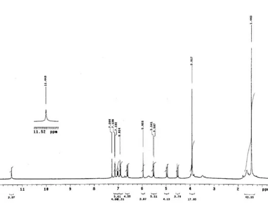
Its 1H NMR spectrum (Table 1) exhibited a typical AX
system due to H-2 and H-3 of a dihydroflavonol31 at d H
4.96 (d, J 12.0 Hz) and 4.51 (d, J 12.0 Hz), respectively. These assignments were confirmed by the 13C NMR
spectrum (Table 2), which showed three C-ring carbon signals at dC 83.1 (C-2), 72.3 (C-3) and 195.9 (C-4). The
configuration at C2-C3 was determined to be trans on the basis of the magnitude of 3J
H2-H3 12 Hz.
32 Besides, the 1H
NMR spectrum exhibited signals in the aromatic region at dH 7.14 (1H, d, J 1.8 Hz), 7.02 (1H, dd, J 8.1 e 1.8 Hz)
and 6.91 (1H, d, J 8.1 Hz), which indicated a AMX spin system of a 1,3,4-trisubstituted phenyl group, as well as one singlet at dH 5.96 attributed to a pentasubstituted
aromatic ring proton. The signals observed at dH 6.62 and
5.62 (1H each, d, J 10.2 Hz) and 1.44 (6H, s) revealed a 2,2-dimethylchromeno ring attached to an aromatic ring, and the singlets at dH 11.45 and 3.92 indicated the
presence of a quelated hydroxyl to carbonyl and one OMe group connected to the aromatic ring, respectively.
Dihydroflavonols from the leaves of Derris urucu (Leguminosae) J. Braz. Chem. Soc.
1084
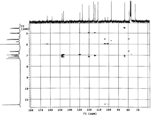
All the couplings were confirmed through the analysis of
1H-1H COSY spectrum. In addition to the signals related
to C-ring carbons, the 13C NMR spectrum of 1 exhibited
other 17 signals attributed to eighteen carbons with aid of the HETCOR and HMBC (Table 2) experiments. The 2,2-dimethylchromene ring attached to the A ring at C-6 and C-7 was deduced by 3J
C,H correlations from H-3′′, H-8
and OH-5 to C-6. The location of the OMe and OH groups at C-4′ and C-3′ of the aromatic B-ring, respectively, was supported by the combination of the substitution pattern on the aromatic ring (1,3,4-trisubstituted) observed in the
1H NMR spectrum with the 2,3J
C,H correlations from H-6′,
H-5′, H-2′ and OMe-4′ to oxidized aromatic carbon C-4′
(dC 147.3) and from both H-5′ and H-2′ to another oxidized
aromatic carbon C-3′ (dC 145.8). Therefore, the structure of
1 was determined as (2R,3R)-5,3′-dihydroxy-4′ -methoxy-6′′,6′′-dimethylpyrano[2′′,3′′:7,6]dihydroflavonol, which we named urucuol A. The spectral analysis clearly indicated that 1 is isomeric with (2R,3R)-5,4′-dihydroxy-3′ -methoxy-6′′,6′′-dimethyl pyrano[2′′,3′′:7,6]dihydroflavonol (eritrinol).33
Compound 2 was obtained as a yellow amorphous powder. Its positive ESI-MS fragmentation pattern is similar to that urucuol A (1), exhibiting ion peaks at m/z 421 [M+Na]+, 399 [M+H] +, 325 [M+Na-58 (C
3H6O)-15(CH3)]
+,
284 [M+H-58 (C3H6O)-28(CO)-28(CO)-H]+ and 236.
The ion peaks at m/z 421 and 399 are 14 mass units more than the corresponding peaks in 1, suggesting one additional Me group.These data, together with the 1H and 13C NMR data, have allowed us to consider a molecular
formula of C22H22O7 for 2, confirmed by HRESIMS at m/z 421.1285 [M+Na]+ (calc. for C
22H22O7Na, 421.1263).
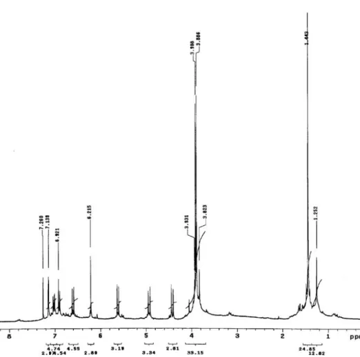
The 1H and 13C NMR data of compound 2 were very
similar to those reported for 1, indicating the presence of 1,3,4-trisubstituted and pentasubstituted aromatic rings, and still a gem-dimethylchromene ring. The main differences between the spectroscopic data of 1 and 2 are signals for two OMe groups at dH 3.91 and 3.88, instead
one OMe singlet as in 1, which showed correlations with the OMe signals at dC 55.9 and 62.6, respectively,
in the HETCOR spectrum. The last value is typical of a di-ortho-substituted OMe group,34 which was located at
C-5. This information and HMBC correlations between H-8 and two oxidized aromatic carbons (C-7 and C-9)and two substituted aromatic carbons (C-6 and C-10), as well the correlations between both H-3′′ and H-4′′ and C-6, confirmed the gem-dimethylchromene moiety placed at C-6/C-7. Analogous to compound 1, 2 possesses a 4′-OMe and 3′-OH substituted B-ring. This was confirmed by the
2,3J
C,H correlations from H-6′ and H-2′ to C-4′ (dC 147.2)
and from H-5′ and H-2′ to C-3′ (dC 145.8). The trans
configuration at C2-C3 was deduced from the coupling constant (J 12.0 Hz) between H-2 and H-3. On this basis
2 was unambiguously identified as (2R,3R)-3′ -hydroxy-5,4′-dimethoxy-6′′,6′′-dimethylpyrano[2′′,3′′:7,6] dihydroflavonol, named urucuol B.
Compound 3 was also isolated as a yellow amorphous powder and showed spectral characters similar to urucuol A (1) and urucuol B (2). The ESI-MS in the positive mode of 3 presented a fragmentation pattern analogous to both
1 and 2, exhibiting an ion peak at m/z 435 [M+Na]+and a
quasi molecular ion at m/z 413 [M+H]+, which are 28 and
14 mass units bigger than the respective peaks of 1 and 2, suggesting two and one additional OMe groups from 1 and
2, respectively, as well as the molecular formula (C23H24O7) for 3,confirmed by HRESIMS at m/z 435.1440 [M+Na]+
(calc. for C23H24O7Na, 435.1420). Comparison of the 1H
and 13C NMR spectral data of 3 with 2 (Table 1 and 2)fully
supported the same groups and substitution pattern for the A and C rings in both compounds.
The difference between compounds 2 and 3 is an extra OMe group located at C-3′, confirmed by the 3J
C,H
correlations observed between H-6′ (dH 7.09) and
OMe-4′ (dH 3.90) with C-4′ (dC 149.7), and from both H-5′
(dH 6.93) and OMe-3′ (dH 3.92) with C-3′ (dC 149.1).
Compound 3 showed a C2-C3 configuration similar to 1
and 2. This allowed the identification of 3 as (2R,3R)-5,3′,4′-trimethoxy-6′′,6′′-dimethylpyrano[2′′,3′′:7,6] dihydroflavonol, named urucuol C.
Compound 4 was also isolated as a white amorphous powder and its molecular formula was determined to be C22H24O7 based on its HRESI-MS at m/z 423.1439 [M+Na]+ (calc. for C
22H24O7Na, 423.1420). Its positive
ESI-MS showed a ion peak at m/z 423 [M+Na]+ and
small peaks at m/z 401[M+H]+, 345 [M+H-55(C
4H7)]
+,
325 [M+H-32(CH3OH)-42(C3H6)-H]+, 284
[M+H-32(CH3OH)-42(C3H6)-28(CO)-15(CH3)]+. The presence
of a γ,γ-dimethylallyl group was evidenced by 1H NMR
signals at dH 3.26 (d, J 7.2 Hz, 2H-1′′ ), 5.17 (t, J 7.2
Hz, H-2′′), 1.68 (s, 3H-4′′) and 1.77 (s, 3H-5′′).31,34 An
OH-5 quelated signal at dH 11.23 was also observed. The
aromatic and the C-ring proton signals of 4 (Table 1), as well as B and C-ring carbon signals of 4 (Table 2) were very similar to those of 1. Thus, the difference between
1 and 4 is in ring A. The γ,γ-dimethylallyl moiety and one OMe group were confirmed to be attached at C-6 and C-7 (A-ring), respectively, based on the long-range correlations from 2H-1′′, H-8 and OH-5 to C-6, and 2H-1′′
and OMe-7 to C-7. From these results, the structure of 4
Lôbo et al. 1085 Vol. 20, No. 6, 2009
Table 1.1H NMR Chemical Shifts (d
H in ppm) and Coupling Constants (J in Hz) of Compounds 1-4 in CDCl3 a H 1 dH 2 dH 3 dH 4 dH
2 4.96 (d, 12.0) 4.93 (d, 12.0)b 4.97 (d, 12.0) 4.97 (d, 12.0)
3 4.51 (d, 12.0) 4.42 (d, 12.0) 4.46 (d, 12.0) 4.53 (d, 12.0)
8 5.96 (s) 6.21 (s) 6.23 (s) 6.08 (s)
2′ 7.14 (d, 1.8) 7.14 (d, 2.1) 7.07 (d, 2.4) 7.15 (d, 1.8)
5′ 6.91 (d, 8.1) 6.90 (d, 8.1) 6.93 (d, 8.7) 6.91 (d, 8.1)
6′ 7.02 (dd, 8.1 and 1.8) 7.02 (dd, 8.1 and 2.1) 7.09 (dd, 8.7 and 2.4) 7.03 (dd, 8.1 and 1.8)
1′′ 3.26 (d, 7.2)
2′′ 5.17 (t, 7.2)
3′′ 5.52 (d, 10.2) 5.62 (d, 10.2) 5.62 (d, 10.0)
4′′ 6.62 (d, 10.2) 6.61 (d, 10.2) 6.61 1H, (d, 10.0) 1.68 (s)
5′′ 1.77 (s)
2Me-2′′ 1.44 (s) 1.44 (s) 1.44 (s)
OMe-5 3.88 (s) 3.89 (s)
OMe-6
OMe-7 3.83 (s)
OMe-3′ 3.92 (s)
OMe-4′ 3.92 (s) 3.91 (s) 3.90 (s) 3.91(s)
OH-5 11.45 (s) 11.23 (s)
OH-3′ 5.73 (brs)
a 1H NMR data were recorded at 300 MHz. bMultiplicity and coupling constant (J, Hz) are in parenthesis.
Table 2. 13C NMR chemical shifts (d
C in ppm) of compounds 1-4 in CDCl3 a
C
1 2 3 4
dC HMBC
a d C HMBC a d C HMBC a d C HMBC a
2 83.1 3, 2′, 6′ 82.9 3, 2′, 6′ 83.0 3, 2′ 83.5 3, 2′, 6′
3 72.3 72.9 72.9 2 72.5
4 195.9 190.8 2 190.7 2 196.1 2
5 157.7 156.8 OMe-5 156.8 4′′, OMe-5 159.8 OH-5
6 103.2 8, 3′′, OH-5 110.8 8, 3′′, 4′′ 110.1 8, 3′′ 110.7 8, 1′′
7 162.3b 8 161.3b 8 161.3 8, 4′′ 166.5 OMe-7
8 96.7 101.1 101.1 91.7
9 162.9b 8 163.7b 8 163.6 8 161.7 8
10 100.4 8, OH-5 106.3 8 106.3 8 100.7 8, OH-5
1′ 129.1 2, 5′ 129.5 2, 3, 5′ 128.7 2, 3, 5′ 129.5 2, 3, 5′
2′ 113.5 2, 6′ 113.6 2, 6′ 110.2 6′ 113.7 2, 6′
3′ 145.8 2′, 5′ 145.8 2′, 5′ 149.1 5′, OMe-3′ 146.1 2′, 5′
4′ 147.3 2′, 5′,6′, OMe-4′ 147.2 2′, 6′ 149.7 2′, 6′, OMe-4′ 147.6 2′, 6′, OMe-4′
5′ 110.5 110.5 111.0 110.8
6′ 119.7 2, 2′ 119.7 2 120.3 2, 2′ 119.9 2, 2′
1′′ 21.2
2′′ 78.6 3′′, 4′′, 2Me-2′′ 78.1 3′′ 78.1 3′′, 4′′, 2Me-2′′ 122.2 1′′, 4′′
3′′ 126.6 2Me-2′′ 129.0 129.0 2Me-2′′ 132.1 1′′, 4′′, 5′′
4′′ 114.9 115.6 115.6 26.1 5′′
5′′ 17.9 4′′
2Me-2′′ 28.4 Me-2′′ 28.4 28.3 Me-2′′
OMe-5 62.6 62.6
OMe-7 56.3
OMe-3′ 55.8
OMe-4′ 55.9 55.9 55.9 56.2
Dihydroflavonols from the leaves of Derris urucu (Leguminosae) J. Braz. Chem. Soc.
1086
Free-radical (DPPH) scavenging activity
The radical scavenging activity of compounds 1-4 was evaluated towards the stable free radical DPPH, which exhibits an absorption maximum at 517 nm, evidencing poor activities for compounds 1-4 compared with the positive control trans-resveratrol (Figure 2). Urucuol A (1) showed an EC50 of 124.09 µg mL-1, followed by
isotirumalin (4) (EC50 142.31 µg mL-1), urucuol B (2) (EC 50
154.62 µg mL-1) and urucuol C (3) (EC
50 405.86 µg mL
-1).
These results showed that compounds 1-4 were 7-23 times less active than positive control trans-resveratrol (EC50 value of 17.69 µg mL-1).
The free radical scavenging activity of flavonoids and other phenols is mostly due to their aromatic hydroxyl groups, which afford greater stability to the phenolic radical as soon as it is formed, after one hydrogen radical donation to DPPH,36 so the dihydroxylated dihydroflavanols
1 and 4 were more effective in promoting DPPH reduction, compared with the monohydroxylated 2 and the unhydroxylated 3, the least active.
Dihydroflavonols show a different behavior compared with flavones or flavonols. The methylation in the hydroxyl group at the para-position decreased DPPH scavenging activity. Other studies show that a C2-C3 double bond and the catechol absence decrease scavenging activity.37-39 An additional methylation in the meta-position
significantly decreased DPPH scavenging capacity. Based on all the information cited, it is possible to justify the lesser antioxidant activity of the compounds tested compared to the resveratrol, whose structure presents trihydroxylation at the para- and meta-positions and an all conjugated system.
Experimental
General
IR spectra were obtained in a Bomen MB-102 spectrophotometer, using the thin solid film method. UV spectra were obtained from a Shimadzu Prominence 20A LC equipped with DAD. NMR spectra, including 1H-1H
COSY, HETCOR, HMBC experiments, were recorded on a Varian Mercury-300 spectrometer, operating at 300 MHz for 1H and 75 MHz for 13C, using d-chloroform as solvent
and internal standard. Mass spectral analyses were performed at low resolution on a Quattro-LC instrument (Micromass, Manchester, UK) provided with an ESI ion source and a triple quadrupole mass analyzer. High resolution analyses were performed on UltrOTOF-Q (Brucker, Daltonics, Billerica MA, USA) only in the cationized ion region. After a systematic investigation, the heated capillary and the voltage were maintained at 250 °C and 3 kV, respectively. A 20 V (cone energy) was applied for the ion extraction and the mass spectrometry data were acquired in the positive mode for all compounds. HPLC was carried out in a preparative LC-8A Shimadzu system with SPD-10AV Shimadzu UV detector (Tokyo, Japan); using a Phenomenex Gemini C18 column (250 mm × 10 mm, 5µm), an isocratic system of water/acetonitrile (46:54) and a flow rate of 4.7 mL per min. Detection was performed at 270 and 320 nm. All solvents were filtered through a 0.45 µm membrane filter prior to use. Absorbance measurements were recorded on a Spectrum UV SP-220 spectrophotometer.
Plant material
The leaves of Derris urucu were collected in January 2006, in the forest reserve of EMBRAPA-Amazônia Oriental in Belém, Pará State, Brazil. A voucher specimen (IAN 179599) was deposited at the herbarium of this institution.
Extraction and isolation
The dried and powdered leaves of Derris urucu (300 g) were extracted with ethanol at room temperature. The solvent was removed under vacuum furnishing a residue (50 g). The crude ethanol residue (30 g) was passed through a silica gel column with gradient elution: hexane-ethyl acetate (9:1, 7:3, 5:5 and 0:10) and ethyl acetate-methanol (5:5 and 0:10), yielding, after removal of the organic solvent, six fractions named DU-1(1.28 g), DU-2 (2.37 g), DU-3 (5.17 g), DU-4 (5.36 g), DU-5 (3.53 g) and DU-6(3.75 g), respectively. The
1HNMR and HPLC analysis of these fractions showed that
Lôbo et al. 1087 Vol. 20, No. 6, 2009
the DU-4 was the most interesting as it had aromatic proton signals and a chromatogram with intense peaks, suggesting the presence of the aromatic compounds. Fraction DU-4 (1 g) was purified by semi-preparative HPLC yielding four dihydroflavonols: 1 (20 mg), 2 (28 mg), 3 (80 mg) and 4 (20 mg), which showed peaks in the chromatogram with retention times of 9.10, 7.84, 13.52 and 16.28 min, respectively.
DPPH assays
A methanolic solution (25 mg L−1) of the radical DPPH• was prepared daily and protected from light. Absorbance was recorded to check the stability of the radical throughout the time of analysis. The effect of phenolic compounds on the DPPH• absorbance was estimated by using the procedure described in the literature.20Different sample
concentrations dissolved in methanol were added to DPPH• methanolic solutions. Absorbance at 517 nm was recorded at different time intervals until the reaction reached an equilibrium. The initial absorbance was close to 1.100-1.150 in all cases. The blank reference cuvette contained methanol. All measurements were performed in duplicate. Six different concentrations of each phenolic compound studied were assayed in order to check the linearity of response and to establish the antioxidant activity values in an adequate linear range. All phenolic compounds were properly dissolved in methanol.
Data analysis
Reaction kinetics of phenols with DPPH• were registered for each antioxidant concentration tested. From these plots, the percentage of DPPH• remaining at the steady state (DPPH• rem) was determined as %DPPH• rem = (Af / A0) × 100. A0 and Af correspond to the absorbances at 517 nm of the radical at the beginning and at the steady state, respectively. Time at steady state was used in order to ensure that reaction did not progress further. Concentrations of the phenolic compounds in the reaction medium were plotted against the percentages of the remaining DPPH•at the end of the reaction in order to obtain the EC50 index, defined as the amount of antioxidant needed to decrease the initial DPPH•concentration by 50%. Analysis of variance and linear correlations tests were performed using the BIOSTAT® version software package.
Urucuol A (1)
Pale yellow powder; IR (thin solid film) νmax/cm-1:
3345, 2974, 1633, 1514, 1434, 1282, 1129, 1016. UV (water:acetonitrile) λmax/nm: 229, 272, 309 (sh);
ESI-MS m/z: 407, 385 [M+H]+, 325 [M+Na-58 (C
3H6O)-H]
+,
284 [M+H-58(C3H6O)-28-15]+, 236; 1H and 13C NMR
spectral data: see Tables 1 and 2.
Urucuol B (2)
Yellow amorphous powder; IR (thin solid film) νmax/cm-1:
3397, 2933, 1597, 1511, 1445, 1266, 1127, 1028. UV (water:acetonitrile) λmax /nm: 235, 261, 342 (sh); ESI-MS m/z: 421 [M+Na]+, 399 [M+H] +, 325 [M+Na-58 (C
3H6
O)-15(CH3)]+, 284 [M+H-58 (C
3H6O)-28(CO)-28(CO)-H]
+,
236; 1H and 13C NMR spectral data: see Tables 1 and 2.
Urucuol C (3)
Yellow amorphous powder; IR (thin solid film) νmax/cm-1:
3418, 2934, 1606, 1529, 1467, 1261, 1129, 1082, 1022; UV (water/acetonitrile) λmax/nm: 235, 261, 342 (sh); ESI-MS m/z: 435 [M+Na]+, 413 [M+H]+, 325 [M+Na-58(C
3H6
O)-28(CO)-H]+, 284 [M+H-58 (C
3H6O)-28(CO)-15(CH3
)-28(CO)]+, 236; 1H and 13C NMR spectral data: see Tables
1 and 2.
Isotirumalin (4)
White amorphous powder; IR (thin solid film) νmax/cm-1:
3458, 3206, 2934, 1632, 1574, 1434, 1248, 1129, 1095, 1016; UV (water/acetonitrile) λmax/nm: 233, 291, 335 (sh); ESI-MS m/z: 423 [M+Na]+, 401[M+H]+, 345
[M+H-55(C4H7)]+, 325 [M+H-32(CH
3OH)-42(C3H6)-H] +, 284
[M+H-32(CH3OH)-42(C3H6)-28(CO)-15(CH3)]+, 236; 1H
and 13C NMR spectral data: see Tables 1 and 2.
Acknowledgments
The authors are grateful to the Conselho Nacional de Desenvolvimento Científico e Tecnológico (CNPq) for financial support and to the Coordenação de Aperfeiçoamento de Pessoal de Nível Superior (CAPES) for scholarships. We are also thankful to Dr. Norberto P. Lopes of Faculdade de Farmácia-USP-Ribeirão Preto for the ESI-MS and HRESI-MS.
Supplementary Information
Supplementary data is available free of charge at http://jbcs.sbq.org.br, as a PDF file.
References
1. Conceição, H. E. O.; Pinto, J. E. B. P.; Santiago, E. J. A.; Gonçalves, A. A. S.; Cienc. Agrotec. 2002, 26, 472.
Dihydroflavonols from the leaves of Derris urucu (Leguminosae) J. Braz. Chem. Soc.
1088
3. Gusmão, S. D.; Páscoa, V.; Mathias, L.; Vieira, I. J. C.; Braz-Filho, R.; Lemos, F. J. A.; Mem. Inst. Oswaldo Cruz2002, 97, 371.
4. Mors, W. B.; Do Nascimento, M. C.; Ribeiro do Valle, J.; Aragão, J. A.; Cienc. Cult.1973, 25, 647.
5. Fang, N.; Casida, J. E.; J. Agric. Food Chem.1999, 47, 2130. 6. Fang, N.; Casida, J. E.; J. Nat. Prod.1999, 62, 205.
7. Russo, A.; Acquaviva, R.; Campisi, A.; Sorrentini, V.; Di Giacomo, C.; Cell Biol. Toxicol. 2000, 63, 1035.
8. Hirono, I.; Biol. Pharm. Bull.1987, 2, 120.
9. Alldrick, A.;Flynn, J.; Rowland, I.; Mutat. Res. 1986, 163, 225.
10. Kawaii, S.; Tomono, Y.; Katase, E.; Ogawa, K.; Yano, M.; Biosci. Biotechnol. Biochem. 1999, 63, 896.
11. Sakihama, Y.; Cohen, M.; Grace, S.; Yamasaki, H.; Toxicology
2002, 177, 67.
12. Vaya, J.; Mahmood, S.; Goldblum, A.; Aviram, M.; Volkova, N.; Salan, A.; Musa, M.; Tamir, S.; Phytochemistry2003, 62, 89.
13. Pietta, P.; J. Nat. Prod.2000, 63, 1035.
14. Morel, I.; Lescoat, G.; Cogrel, P.; Sergent, O.; Pasdeloup, N.; Brissot, P.; Cillard, P.; Biochem. Pharmacol.1993, 45, 13. 15. Teguo, P. W.; Fauconneau, B.; Deffieux, G.; Huguet, F.;
Vercauteren, J.; Mérillon, J. M.; J. Nat. Prod.1998, 61, 655. 16. Escobar, C. L. M.; Braga, A.; Tommasi, N. D.; J. Nat. Prod.
2003, 66, 1061.
17. Nakamura, Y.; Watanabe, S.; Miyake, N.; Kohno, H.; Osawa, T.; J. Agric. Food Chem.2003, 51, 3309.
18. Robards, K.; Prenzlere, P. D.; Tucker, G.; Swatsitang, P.; Glover, W.; Food Chem.1999, 66, 401.
19. Galati, G.; Sabzevari, O.; Wilson, J. X.; O’Brien, P. J.;
Toxicology2002,177, 91.
20. Brand-Williams, W.; Cuvelier, M. E.; Berset, C.; Food Sci. Technol. 1995, 28, 25.
21. Arnous, A.; Makris, D. P.; Kefalas, P.; J. Agric. Food Chem.
2001, 49, 5736.
22. Da Porto, C.; Calligaris, S.; Celotti, E.; Nicoli, M. C.; J. Agric. Food Chem,2000, 48, 4241.
23. Gorinstein, S.; Martin-Belloso, O.; Katrich, E.; Lojak, A.; Ciz, M.; Gligelmo-Miguel, N.; Haruenkit, R.; Park, Y.-S.; Jung, S.-T.; Trakhtenberg, S.; J. Nutr. Biochem.2003, 14, 154.
24. Heinonen, I. M.; Lehtonen, P. J.; Hopia, A. I.; J. Agric. Food Chem.1998, 46, 25.
25. Llorach, R.; Espín, J. C.; Tom´as-Barberán, F.A.; Ferreres, F.;
J. Agric. Food Chem.2003, 51, 2181.
26. Sánchez-Moreno, C.; Plaza, L.; De Ancos, B.; Cano, M. P.;
J. Sci. Food Agric.2003, 83, 430.
27. Gil, M. I.; Tom´as-Barber´an, F. A.; Hess-Pierce, B.; Holcroft, D. M.; Kader, A. A.; J. Agric. Food Chem.2000, 48, 4581. 28. Lu, Y.; Foo, L.Y.; Food Chem. 2000, 68, 81.
29. Goupy, P.; Dufour, C.; Loonis, M., Dangles, O.; J. Agric. Food Chem.2003, 51, 615.
30. Mabry, T.; Markham, K. R.; Thomas, M. B.; The Systematic Identification of flavonoids, Springer-Verlag: New York, 1970, p. 166.
31. Mbwambo, Z. H.; Kapingu, M. C.; Moshi, M. J.; Machumi, F.; Apers, S; Cors, P.; Ferreira, D.; Marais, J. P. J.; Vanden Berghe, D.; Maes, L.; Vlietinck, A.; Pieters, L.; J. Nat. Prod.2006, 69, 369.
32. Rao, K. V.; Gunasekar, D.; Phytochemistry1998, 48, 1453. 33. Nkengfack, A. E.; Kouam, J.; Voufoo, W. T.; Fomum,
Z. T.; Dagne, E.; Sterner, O.; Browne, L. M.; Guijun, J.;
Phytochemistry1993, 32, 1305.
34. Agrawal, P. K.; Rastogi, R. P.; Heterocycles1981, 16, 2181. 35. Rao, K. V; Gunasekar, D.; Indian J. Chem., Sect. B: Org. Chem.
Incl. Med. Chem. 1988, 27, 383.
36. Ribeiro, A. B.; Bolzani, V. S., Yoshida, M.; Santos, L. S.; Eberlin, M. N.; Silva, D. H. S.; J. Braz. Chem. Soc.2005, 16, 526.
37. Trouillas, P.; Marsal, P; Siri, D.; Lazzaroni, R.; Duroux, J.-L.;
Food Chem.2006, 97, 679.
38. Van Acker, S. A. B. E.; Van Den Berg, D.-J.; Tromp, M. N. J. L.; Griffioen, D. H.; Van Bennekom, W. P.; Van Der Vijgh, W. J. F.; Bast. A.; Free Radical Biol. Med.1996, 20, 331. 39. Leopoldini, M.; Russo, N.; Toscano, M.; J. Agric. Food Chem.
2006, 54, 3078.
S
u
p
p
le
m
e
n
ta
ry
In
fo
rm
a
ti
o
n
J. Braz. Chem. Soc., Vol. 20, No. 6, S1-S14, 2009. Printed in Brazil - ©2009 Sociedade Brasileira de Química 0103 - 5053 $6.00+0.00
*e-mail: [email protected]
Dihydroflavonols from the leaves of
Derris urucu
(Leguminosae):
Structural Elucidation and DPPH Radical-Scavenging Activity
Lívia T. Lôbo,a Geilson A. da Silva,a Malisson Ferreira,a Milton N. da Silva,a Alberdan S. Santos,a
Alberto C. Arruda,a Giselle M. S. P. Guilhon,a Lourivaldo S. Santos,a Rosivaldo dos Santos Borgesb and Mara Silvia P. Arruda*,a
aPrograma de Pós-Graduação em Química, Instituto de Ciências Exatas e Naturais, Universidade Federal do Pará,
Campus Universitário do Guamá, 66075-970 Belém-PA, Brazil
bFaculdade de Farmácia, Instituto de Ciências da Saúde, Universidade Federal do Pará, Campus Universitário do
Guamá, 66075-970 Belém-PA, Brazil
Figure S1.1H NMR spectrum (in CDCl
Dihydroflavonols from the leaves of Derris urucu (Leguminosae) J. Braz. Chem. Soc.
S2
Figure S2.13C NMR spectrum (in CDCl
3, 75 MHz) of the compound 1 isolated from leaves of Derris urucu.
Lôbo et al. S3 Vol. 20, No. 6, 2009
Figure S4. COSY NMR experiment(in CDCl3, 300 MHz) of the compound 1 isolated from leaves of Derris urucu.
Dihydroflavonols from the leaves of Derris urucu (Leguminosae) J. Braz. Chem. Soc.
S4
Figure S6. HMBC NMR experiment(in CDCl3, 300 × 75 MHz) of the compound 1 isolated from leaves of Derris urucu.
Lôbo et al. S5 Vol. 20, No. 6, 2009
Figure S8.1H NMR spectrum (in CDCl
3, 300 MHz) of the compound 2 isolated from leaves of Derris urucu.
Figure S9.13C NMR spectrum (in CDCl
Dihydroflavonols from the leaves of Derris urucu (Leguminosae) J. Braz. Chem. Soc.
S6
Figure S10. ESI mass spectrum of the compound 2 isolated from leaves of Derris urucu.
Lôbo et al. S7 Vol. 20, No. 6, 2009
Figure S12. HMBC NMR experiment(in CDCl3, 300 x 75 MHz) of the compound 2 isolated from leaves of Derris urucu.
Figure S13.1H NMR experiment(in CDCl
Dihydroflavonols from the leaves of Derris urucu (Leguminosae) J. Braz. Chem. Soc.
S8
Figure S14.13C NMR spectrum (in CDCl
3, 75 MHz) of the compound 3 isolated from leaves of Derris urucu.
Lôbo et al. S9 Vol. 20, No. 6, 2009
Figure S16. COSY NMR experiment(in CDCl3, 300 MHz) of the compound 3 isolated from leaves of Derris urucu.
Dihydroflavonols from the leaves of Derris urucu (Leguminosae) J. Braz. Chem. Soc.
S10
Figure S18. HMBC NMR experiment(in CDCl3, 300 × 75 MHz) of the compound 3 isolated from leaves of Derris urucu.
Lôbo et al. S11 Vol. 20, No. 6, 2009
Figure S20.1H NMR experiment(in CDCl
3, 300 MHz) of the compound 4 isolated from leaves of Derris urucu.
Figure S21.13C NMR spectrum (in CDCl
Dihydroflavonols from the leaves of Derris urucu (Leguminosae) J. Braz. Chem. Soc.
S12
Figure S22. ESI mass spectrum of the compound 4 isolated from leaves of Derris urucu.
Lôbo et al. S13 Vol. 20, No. 6, 2009
Figure S24. HETCOR NMR experiment(in CDCl3, 300 x 75 MHz) of the compound 4 isolated from leaves of Derris urucu.
Dihydroflavonols from the leaves of Derris urucu (Leguminosae) J. Braz. Chem. Soc.
S14
Figure S26. HMBC NMR experiment of the compound 4 (expansion).