(Rio Maior, Portugal)
Kimmarie Murphy Kenyon College Bryan Tucker University of Florida Carlos M. C. Pereira Câmara Municipal de Rio Maior Humberto Nuno de Oliveira Universidade Lusíada de Lisboa Paul T. Thacker Wake Forest UniversityIntroduction
A team of archaeologists discovered numerous human remains during three sea-sons of excavations and architectural analysis at Capela de Nossa Senhora da Victória (Our Lady of Victory Chapel) in Rio Maior, Portugal between 1987 and 1989. The earliest exca-vation in 1987-88 occurred both inside of the standing chapel and outside along the south wall. These initial excavations, directed by Oliveira, Martins, and Pereira, uncovered the foundations of a medieval manor house and a few bones identified as human remains (Oliveira et al., 1989). As Figure 1 illustrates, in 1989 more extensive excavations in front of the Chapel uncovered a small cemetery containing at least seven burials and an ossuary (Oliveira et al., 1990).
This report focuses on the osteological analysis of skeletal remains recovered from the excavations at Capela de Nossa Senhora da Victória. Both during the fieldwork and in the years following the Chapel excavations, the skeletal collection was vandalized, unfor-tunately commingling many of the burials and losing their precise associations beyond general excavation units. Due to this imprecise provenience and the commingling of the remains, we analyze the skeletal material from outside of the Chapel structure (Burial Con-text IV) as an ossuary rather than discrete burials.
Background
The origins of the Capela De Nossa Senhora da Victória are uncertain but the earliest religious structure on the site may have been a small chapel associated with the
medieval manor house. The standing chapel was built or extensively reconstructed after AD 1640 and dedicated in commemoration of the end of the Spanish occupation of Portu-gal. The inclusion of “Victória” is both a commemoration and an offering of thanks for a favorable outcome of an a military event. The total assemblage of human remains from the Chapel and cemetery span a period of at least two hundred years, based on evidence from architectural construction, stratigraphy, and associated artifacts.
During the initial construction (or extensive renovation) after AD 1640, sections of the old manor house wall to the south of the Chapel were demolished in order to establish a large entrance area and cemetery for the new Chapel (see Figure 1). The new outer re-taining wall was built which facilitated the filling and leveling of the portico and cemetery (Figure 2). The small cemetery could have received new interments as late as AD 1835, the date of Portuguese government legislation regulating church cemeteries including those in Rio Maior:
…From the middle half of the Eighteenth century, some doctors, enlightened intel-lectuals, and some liberal clergy began a protest against burial interment in churches. …the beginning of our cemetery legislation came from more moderate liberal go-vernments: decreed from September 21, 1835 to October 8 of the same year… ”a partir de meados do séc. XVIII, alguns médicos, intelectuais iluministas e certos ecleseásticos mais esclarecidos iniciaram a contestação dos enterramentos nas Igrejas.”
“(…) o arranque da nossa legislação cemiterial partiu dos governos liderados pela facção moderada do liberalismo: decretos de 21 de Setembro de 1835 e de 8 de Outubro do mesmo ano (…).”
Mattoso, 1993 (Vol. V): 595 Given these historical events, it is likely that the skeletal material spans a maximum range of 195 years from AD 1640 to AD 1835.
The meaning of burial location and its relationship with church architecture is com-plex at Nossa Senhora da Victória. Throughout most of Portugal, parishes allowed rich or powerful individuals to be buried within the formal architectural confines of churches. This practice was maintained:
“In virtue of (the) … belief that they can reach the glory of the souls, those whose bodies laid in the presence of the images of the Saints inside of the sacred temples.” “em virtude da … crença de que alcançariam a glória das almas aqueles cujos cor-pos jazessem em companhia das imagens dos santos, dentro dos templos sagrados.” Mattoso, 1993 (Vol. V): 596
The Congregation of the Souls that owned Capela de Nossa Senhora da Victória, however, did not allow interments within the Chapel (Oliveira et al., 1989). In Rio Maior, historical records reveal that the rich and powerful citizens who wanted to be buried within a Catholic sanctuary were buried within the nearby Igreja da Misericórdia. This local prac-tice suggests the burials recovered from the cemetery are middle class members of the congregation, possessing enough status to be interred in the limited space near the church but not influential enough to be interred inside the central Igreja da Misericórdia.
Human remains were recovered from four different burial contexts at Capela de Nossa Senhora da Victória (Figure 1). An isolated skeleton was recovered subfloor from immediately inside of the east doorway of the Chapel (Figure 1: Burial Context I). Asso-ciated artifacts including a button and bullets date this individual to the 19th century du-ring the period of the Peninsular War against Napoleon. It is probable that this youth was secretly buried during a period that the chapel was abandoned, perhaps as a consequen-ce of the French invasions. Unfortunately, many of these remains were lost and remain unstudied.
Two commingled assemblages of human remains were recovered in the excavations, one buried inside of the Chapel (Figure 1: Burial Context II) and the other comprised of the ossuary found just exterior of the southeast foundation corner (Figure 1: Burial Context III). The commingled remains from the interior of the Chapel, largely represented by cranial remains, were found in the builder’s trench along the Chapel wall. These individuals could have been associated with the manor house, or victims of a plague and/or the 1531 earth-quake, and were buried in the chapel of the manor house during a period of abandonment. These burials may have been disinterred during Seventeenth century construction activi-ties at the Chapel and subsequently reburied in the builder’s trench. The remains from the ossuary outside the Chapel were more numerous and may represent individuals disinter-red from the cemetery to make space for later internments.
The bulk of analyzed skeletal material was excavated from the small cemetery in front of the Chapel (Figure 1: Burial Context IV). The skeletons indicated in Figure 1 are only schematics for burial position and orientation and do not represent actual recovered skeletal elements. While this article is primarily focused on osteological analysis of the remains, archaeological excavation provides insight into post-medieval mortuary practices in Rio Maior. First, the burials in the cemetery and exterior ossuary lacked evidence for coffins or vault hardware. The dead individuals were wrapped in a shroud closed with sil-ver pins. All individuals excavated from distinct graves in the cemetery were buried without personal effects or jewelry and in a common position on their back. The bodies were posi-tioned with arms flexed, usually crossed on the chest or abdomen. All individuals were buried with rosaries with the exception of the isolated child, which probably indicates that the child had not been baptized.
For analysis, two populations were delineated. First, because of chronological and contextual differences, we treat the remains from inside the Chapel as a distinct population from the outside burials. Second, because some of the skeletal elements from the outside ossuary (Burial Context III) have been commingled with the formerly discreet burials (Burial Context IV), this material is treated as a single population under the assumption that the ossuary represents secondary burials of former occupants of the cemetery.
Methods
We determined MNI on the basis of age, sex, color, size, and texture following pro-cedures outlined by Ubelaker (1974). We also used basic osteological protocol as outlined by Buikstra & Ubelaker (1994) in order to estimate sex and age at death for the population. Sex estimates were based on element robusticity and/or pelvic or cranial morphology. Age estimates were based on one or more of the following features: degree of epiphyseal clo-sure, dental development and eruption, suture cloclo-sure, and changes associated with the pubic symphasis and auricular surface of the pelvis. Distinctions between adults and sub-adults were assigned and subsub-adults were grouped in 5-year increments. We used the cri-teria outlined in Ortner & Putschar (1985) to note the presence of dental and skeletal pa-thology. Observations of skeletal pathology included presence or absence and patterning of proliferative (bone formation) and/or resorptive (bone destruction) responses. Dental re-mains were assessed for the presence or absence of the following: caries, defects of den-tal enamel, antemortem tooth loss (AMTL) and abscesses. Such skeleden-tal and denden-tal chan-ges may be associated with 1) environmental stressors during growth and development, 2) exposure to infectious pathogens, and/or 3) traumatic injuries and activity – related changes (Larsen, 1997).
Data for this analysis were collected in Rio Maior, Portugal as part of a research pro-ject supported by PARM (Equipa Inter-Disciplinar para o Património e Arqueologia de Rio Maior), the Sector de Arqueologia de Câmara Municipal de Rio Maior, and the respective institutions of the authors.
Results
Burial contexts from the interior of Nossa Senhora da Victória
Within the Chapel, human remains were recovered from Trenches 2, 3, 4, and 6 in the builder’s trench around the base of the Chapel’s wall. Photographs of the excavation
allow the positive identification of the two crania in trench 3 (Figure 3). These crania are partially burned post-mortem and represent the only burned bones in the entire assemblage, other than a single parietal fragment from excavation unit A1. No charcoal or other evi-dence of burning was recovered from the builder’s trench or any part of the excavation and, since Catholics do not commonly practice cremation, no explanation for the burning is apparent.
Seventy-five tooth and bone fragments were recovered from Trenches 2, 3, 4, and 6 (Table 1) within the Chapel. The remains from Trench 2 are those of a juvenile between 5-10 years based on dental development and epiphyseal closure. Two individuals are present in the trench 3, an adult female aged 20-34 years and a juvenile estimated be-tween 10-15 years. Isolated permanent dentition are associated with Trenches 4 and 6, and two of the three permanent teeth from Trench 6 exhibit carious lesions.
Exterior burial contexts associated with the Chapel
Using MNI estimates from femora recovered from the cemetery and exterior ossuary (Table 2), a minimum number of 19 individuals account for remains buried in archaeologi-cal contexts III and IV. The second highest postcranial elements represented are tibiae and scapulae, each at ca. 50% of the MNI, or 10 individuals. Table 3 illustrates the skeletal representation of this population based on skull and/or dental remains, with a resulting MNI of 17 individuals.
The population from outside of the Chapel is composed of almost equal numbers of adults (N = 8) and juveniles (N = 9) based on age estimates from the cranial material (Ta-ble 3). As a check, standardized age estimates (Buikstra and Ubelaker 1994) from epiphy-seal union and femora size (N = 20) were plotted against the cranial and dental estimates (Figure 4). While not as precise, age estimates from femora roughly correspond to those derived from the cranial/dental elements at Nossa Senhora da Victória.
Identification and frequency of skeletal pathologies
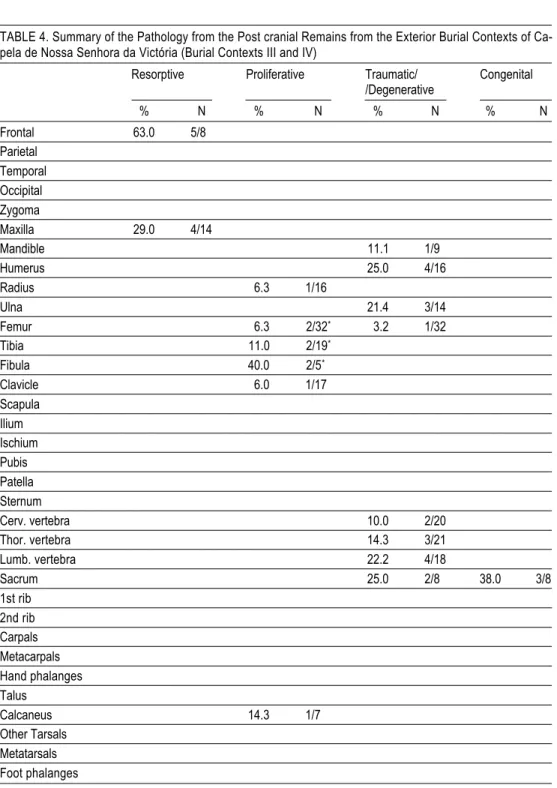
The human skeleton responds to various insults in a limited number of ways, by forming new bone (proliferative response), destroying bone (resorptive response) or a com-bination of proliferation and resorption. A summary of skeletal lesions by bone response for the remains outside the Chapel is found in Table 4. 63% of frontals, or 5 of 8 individuals with cranial remains displayed resorptive lesions along the superior orbital margins des-cribed in the literature as cribra orbitalia (Stuart-Macadam, 1991, 1992). In addition, resor-ptive lesions associated with dental abscessing are seen in 4 out of 14 maxillary elements. Proliferative lesions are found in a small number of postcranial elements. While the remains
outside the Chapel are commingled, the size, shape, color and robusticity of the affected femora (2/32), tibia (2/19), and fibulae (2/5) could feasibly be associated with the same individual. In addition, new bone formation is also seen on a clavicle, radius, and calcaneus. Degenerative changes associated with either traumatic/degenerative conditions such as arthritis are found on 25% of humeri, 21.4% of ulnae, 11% of mandibular condyles, and 3.2% of femora. In addition, a number of vertebral and sacral elements exhibit increased porosity and marginal lipping. Congenital malformations such as spina bifida occulta were identified in 38% or 3 of 8 sacra.
Each tooth type was analyzed separately, but maxillary and mandibular tooth classes are combined for purposes of this analysis (Table 5). Out of a total of 103 permanent tition only 5 teeth were carious (5%). No caries were present in any of the deciduous den-tition. 117 tooth positions were available for analysis; 5.1% had evidence of abscessing and 33% were affected by antemortem tooth loss (AMTL). Analysis of dental pathology at the individual level reveals that five of eight adults (62%) with teeth had caries and AMTL, and half of the individuals suffered from dental abscesses.
Discussion
A mortality curve for an urban population is expected to show a high number of deaths in all age groups, however, infant and children are expected to be the most suscep-tible to disease. Therefore, we expect to see a high rate of child mortality followed by a decrease in sub-adults and then an increase in the deaths of older adults. In these socie-ties, if an individual lived through the dangerous years of weaning and childhood, they could expect to live into adulthood, before the rigors of advancing age began to take their toll. The sample from the exterior burial contexts of the Chapel consists of 17 individuals: 4 infants, 4 juveniles, 1 sub-adults, and 8 adults. The interpretation of these results is ambi-guous due to the broad age ranges assigned to the remains and the small sample size. Had the remains been less fragmentary or retained more of their original provenience, nar-rower age ranges may have been possible. Even so, if the infants and juveniles are col-lapsed into a single category to balance the combination of the younger and older adults, the population structure takes on the expected parabolic curve: 8 infants/juveniles, 1 sub-adults, and 8 adults. These results generally fit the trend of the hazard of death model for a non-industrial society presented by Wood (1994). This model shows an initial hazard of death rate of nearly 20% at birth which then decreases to almost 0.0% around 13-14 years of age, only climb again until a near 100% is reached around 78 years of age.
It is important to note that this cemetery is not a random sample of the historic popu-lation in Rio Maior. Only middle class individuals were buried at Nossa Senhora da Victória
and the small size of the cemetery necessitates that many of the members of the congre-gation were buried elsewhere. Also, though preservation was relatively good, the smaller more fragile bones of infants and juveniles are less likely to be preserved in the archaeo-logical record. Additionally, many newborn infants may not be included in cemeteries due to cultural practices.
The most common bony responses among the cemetery populations are cribra or-bitalia (n = 6/20) and dental abscesses (discussed with dental pathology). In the past, cribra orbitalia has been variously attributed to anemia due to dietary deficiencies and/or patho-gen loads. More recently, research indicates that these lesions are merely a symptom or a bony response that may be attributable to a variety of etiologies including scurvy, infec-tions, hypervascularization, anemia, and pseudopathology (Ortner et al., 2001; Wapler et al., 2004). Histological examination might provide us with a means of refining the potential etiology of cribra orbitalia in the present study, but invasive analyses were not undertaken. Regardless, the high rate of cribra orbitalia in the remains outside the Chapel likely indi-cates some health stressor on this population.
Proliferative bone lesions are often attributable to infectious disease processes, trau-ma, neoplasms, vascular problems, and excess vitamin A (Ortner, 2003: 53). The periosto-sis associated with the several long bones potentially belonging to a single individual sug-gest response to a chronic disease condition. However, given the commingled nature of the sample and the lack of any distinctive patterning, a more specific differential diagnosis is not possible. The degenerative changes seen in the vertebral column and in the elements from the upper body are consistent with patterns associated with habitual movement and strenuous labor (Larsen, 1997).
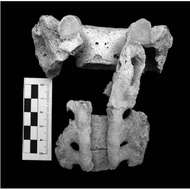
Three cases of spina bifida occulta were found in the assemblage (Figure 5). Spina bifida occulta is a condition in which there is incomplete fusion of the posterior neural arch segments. Commonly, only 1-2 of the sacral elements are affected (Aufderheide and Ro-driguez-Martin, 1998: 61). Spina bifida occulta is generally considered to be asymptomatic and results in few if any negative side effects (Ortner, 2003: 463). The three cases of spina bifida occulta from this analysis follow a similar pattern, with incomplete fusion ranging from S5-S3. The etiologies of spina bifida are varied and comprise both genetic and environ-mental causes (Frey and Hauser, 2003). One of the most commonly reported environmen-tal factors associated with spina bifida is inadequate folic acid intake (Locksmith and Duff, 1998).
The rate of dental caries supports the assumption that these individuals were sub-sisting on a largely agricultural diet which is expected for the time period and region. The overall rate of teeth with at least one carious lesion is around 5%, close to rates reported in early-medieval Britain (Roberts and Manchester, 1997). Early medieval populations, sub-sisting on local agriculture without the addition of imported sugar, exhibit similar caries rates,
around 4.4 to 7.3% (Roberts and Manchester, 1997). During the 17th century, sugar im-ports increased to Europe, mainly northern Europe and Britain. During this time caries rates began a marked increase (Roberts and Manchester, 1997). Data from 16th and 17th cen-tury British populations have caries rates of 8.4% and 10.2% of the total teeth (Miles, 1989). These data suggest the middle class residents of Rio Maior did not have access to im-ported sugar or other caries causing foodstuffs at a rate comparable to the residents in Britain.
Antemortem tooth loss (AMTL) and abscesses are found in the majority of adults with dentition. Both of these dental pathologies are likely due to the combination of heavy dental attrition and caries. There are not many first molars present due to the combination of AMTL and postmortem loss, but individuals ranging from ages 10-20 already have den-tal wear scores (based on methods in Scott, 1979) that range from 6-14. Adults older than 20 years have dental wear scores ranging from 14-35.
Interestingly, no defects of dental enamel (DDE), including liner enamel hypoplasias (LEH), were detected on the teeth from this population. Various environmental stressors such as; infections, nutrition, genetic disorders, neonatal disturbances, toxins and even allergies can slow or cause complete cessation of enamel formation on the tooth surface (Pindborg, 1982; Cutress & Suckling, 1982). DDE can manifest as horizontal grooves or pits that form in the tooth enamel as a result of environmental stressors. The lack of iden-tifiable DDE in this population may be due to the heavy dental wear, high frequency of AMTL, and/or decreased stressors occurring during childhood development.
Conclusion
The excavations at Nossa Senhora da Victória yielded a diachronic sample of hu-man remains buried under very different conditions. The remains recovered inside of the Chapel consisted of a small assemblage of relatively undiagnostic human remains, likely dating to the medieval period. These burials do not represent a systematic burial practice by the church congregation. The excavations outside of the Chapel revealed graves con-taining a minimum of 19 individuals. Due to the vandalism and resultant commingling of the remains, analysis of individual burials is impossible. The entire assemblage indicates the interment of 19 or more individuals across a chronological period spanning nearly 200 years. These 19 individuals included at least 4 infants, 4 juveniles, 1 sub-adult, and 8 adults and the high rate of child mortality is similar to those encountered in other urban populations in the 17th to 19th centuries. The presence of cribra orbitalia may indicate an underlying health stressor. Caries rates are consistent with other European populations prior to the introduction of cane sugar in the 17th century. High frequencies of AMTL and abscesses
may be the result of heavy dental attrition. The presence of three cases of spina bifida occulta warrants further investigation into possible genetic and/or environmental factors responsible. Finally, the work and stress related pathologies are consistent with life in a non-industrialized society.
Despite the limitations imposed by curation issues, the bioarchaeological investiga-tion of human remains from Nossa Senhora da Victória has provided valuable informainvestiga-tion about rural, central Portuguese populations. The analyses indicate the middle class inhab-itants of Rio Maior during the late 17th, 18th, and early 19th centuries enjoyed relatively good health and nutrition. More archaeological research on historic sites from this period coupled with archival research could further contextualize social practice and change in Rio Maior during the post-medieval period.
References cited
AUFDERHEIDE, A. C. & RODRIGUEZ-MARTIN, C. (1998) – Cambridge Encyclopedia of Human Paleopatho-logy. Cambridge University Press, Cambridge, UK.
BUIKSTRA, J. F. & UBELAKER, D. H. (Eds.) (1994) – Standards for Data Collection from Human Skeletal Remains. Arkansas Archaeological Survey, 44. Fayetteville.
CUTRESS, T. W. & SUCKLING, G. W. (1982) – The Assessment of Non-Carious Defects of Enamel. Interna-tional Dental Journal, 32 (2): 117-22.
FREY, L. & HAUSER, W. A. (2003) – Epidemiology of Neural Tube Defects. Epilepsia, 44: 4-13.
LARSEN, C. S. (1997) – Bioarchaeology: Interpreting Behavior from the Human Skeleton. Cambridge Studies
in Bioarchaeology. Cambridge University Press, Cambrige.
LOCKSMITH, G. J. & DUFF, P. (1998) – Preventing Neural Tube Defects: The Importance of Periconceptional Folic Acid Supplements. Obstet Gynecol, 91 (6): 1027-1034.
MATTOSO, J. (1993) – História de Portugal. Volume V – O Liberalismo (1807-1890). Lisboa, Editorial Estampa.
MILES, A. E. W. (1989) – An Early Christian Chapel and Burial Ground on the Isle of Ensay, Outer Hebrides, Scotland with a Study of the Skeletal Remains. BAR, Oxford, England.
OLIVEIRA, H. N. de; MARTINS, M.; PEREIRA, C. & TELLES, F. J. (1989) – Relatório dos Trabalhos Arqueo-lógicos de Emergência realizados na Capela de N.ª Sr.ª da Victória, Rio Maior. On file: IPPC (Instituto
Portu-guês do Património Cultural) and CMRM (Câmara Municipal de Rio Maior).
OLIVEIRA, H. N. de; MARTINS, M.; PEREIRA, C. & TELLES, F. J. (1990) – Relatório dos Trabalhos Arqueoló-gicos realizados na Capela de N.ª Sr.ª da Victória, Rio Maior, 1989. On file: IPPC (Instituto Português do
Patri-mónio Cultural) and CMRM (Câmara Municipal de Rio Maior).
ORTNER, D. J. (2003) – Identification of Pathological Conditions in Human Skeletal Remains. Second Ed.
Academic Press, Amsterdam.
ORTNER, D. J.; BUTLER, W.; CAFARELLA, J. & MILLIGAN, L. (2001) – Evidence of Probable Scurvy in Sub-adults from Archeological Sites in North America. American Journal of Physical Anthropology, 114 (4): 343-351.
ORTNER, D. J. & PUTSCHAR, W. G. (1985) – Identification of Pathological Conditions in Human Skeletal Remains. Smithsonian Institution Press, Washington, D.C.
PINDBORG, J. J. (1982) – Aetiology of Developmental Enamel Defects Not Related to Fluorosis. International Dental Journal, 32 (2): 123-34.
ROBERTS, C. A. & MANCHESTER, K. (1997) – The Archaeology of Disease. Second Ed. Cornell University
Press, Ithaca, New York.
SCOTT, E. C. (1979) – Dental Wear Scoring Technique. American Journal of Physical Anthropology, 80: 11-24.
STUART-MACADAM, P. (1991) – Porotic Hyperostosis: Changing Interpretations. In Donald J. Ortner and Arthur C. Aufderheide (Eds.), Human Paleopathology. Current Syntheses and Future Options. Symposium. Zagreb 24-31.7.1988, pp. 36-39. Smithsonian Institution Press, Washington, D.C.
STUART-MACADAM, P. (1992) – Porotic Hyperostosis: A New Perspective. American Journal of Physical An-thropology, 87: 39-47.
UBELAKER, D. H. (1974) – Reconstruction of Demographic Profiles Ossuary Skeletal Samples: A Case Study from the Tidewater Potomac. Smithsonian Institution Press, Washington, D.C. (Smithsonian Contributions to
Anthropology, 18).
WAPLER, U.; CRUBÉZY, E. & SCHULTZ, M. (2004) – Is Cribra Orbitalia Synonymous with Anemia? Analysis and Interpretation of Cranial Pathology in Sudan. American Journal of Physical Anthropology, 123 (4): 333-339.
WOOD, J. W. (1994) – Dynamics of Human Reproduction: Biology, Biometry, Demography. Aldine-De Gruyter,
TABLE 1. Skeletal inventory from interior of Capela de Nossa Senhora da Victória (Burial Contexts I and II) Provenience Elements Present Age Sex Pathology/Comments
Estimate Estimate Trench 2 < 25% dental remains 5-10 Unknown
< 25% postcrania
Trench 3 Cranium 10-15 Unknown Burning present on cranium
Trench 3 Skull 21-34 Female? Burning present on skull
Trench 4 Isolated dentition Adult Unknown
Trench 6 Isolated dentition Adults Unknown Two carious lesions, lower Rt. canine, lower Rt. lateral incisor
TABLE 2. Skeletal representation by element of human remains recovered from the exte-rior of Capela de Nossa Senhora da Victória (Burial Contexts III and IV; Total MNI = 19)
Element Left Right Total na % of MNI
Frontal 8 8 40.0 Parietal 8 9 17 9 45.0 Temporal 7 9 16 9 45.0 Occipital 8 8 40.0 Zygoma 5 5 10 5 25.0 Sphenoid 6 6 30.0 Ethmoid 4 4 20.0 Maxilla 6 8 14 8 40.0 Mandible 4 8 12 8 40.0 Humerus 9 7 16 9 45.0 Radius 9 7 16 9 45.0 Ulna 5 9 14 9 45.0 Femur 13 19 32 19 100.0 Tibia 9 10 19 10 50.0 Fibula 4 1 5 4 20.0 Clavicle 9 8 17 9 45.0 Scapula 9 10 19 10 50.0 Ilium 8 9 17 9 45.0 Ischium 7 8 15 8 40.0 Pubis 8 9 17 9 45.0 Patella 1 2 3 2 10.0 Sternum 2 2 10.0 Cerv. vertebra 20 Thor. vertebra 21 Lumb. vertebra 18 1st rib 2 2 4 2 10.0 2nd rib 2 2 2 10.0 3rd rib 2 2 2 10.0 Sacrum 8 8 40.0 Carpals 4 Metacarpals 1 15 14 29 Hand phalanges 15 Talus 4 3 7 4 20.0 Calcaneus 6 5 11 6 30.0 Other Tarsals 5 8 13 Metatarsals 39 Foot phalanges 21
TABLE 3. Summary of the demographic characteristics and associated pathology from the exterior burial con-texts of Capela de Nossa Senhora da Victória
Provenience Elements of the Skull Present Sex Age Pathology Burial Context III: A1 Skull, dentition Unknown 5-10 Cribra orbitalia Burial Context III: A1 Cranium, dentition Unknown 10-15 Cribra orbitalia Burial Context III: A1 Calvaria Unknown 10-15 Cribra orbitalia
Burial Context III: A1 Calvaria Unknown 20+
Burial Context IV: B0 Cranium, dentition Unknown 0-5
Burial Context IV: B0 Mandible, dentition Unknown 15-20 Caries, AMTL Burial Context IV: B1 Skull, dentition Unknown 10-15
Burial Context IV: B1 Skull, dentition Unknown 20+ Cribra orbitalia, AMTL, abscess Burial Context IV: B2 Calvaria, mandible, dentition Unknown 0-5
Burial Context IV: C0 Cranium, dentition Unknown 20+ Caries with abscess Burial Context IV: C0 Cranium, dentition Female? 20+ Caries, abscess Burial Context IV: C1 Skull, dentition, Unknown 0-5
Burial Context IV: ? Calvaria, dentition Unknown 0-5 Cribra orbitalia Burial Context IV: ? Skull, dentition Unknown 20+ Caries, AMTL,
abscess Burial Context IV: ? Skull, dentition Male? 20+ Caries, AMTL
Burial Context IV: ? Dentition Unknown 20+
TABLE 4. Summary of the Pathology from the Post cranial Remains from the Exterior Burial Contexts of Ca-pela de Nossa Senhora da Victória (Burial Contexts III and IV)
Resorptive Proliferative Traumatic/ Congenital /Degenerative % N % N % N % N Frontal 63.0 5/8 Parietal Temporal Occipital Zygoma Maxilla 29.0 4/14 Mandible 11.1 1/9 Humerus 25.0 4/16 Radius 6.3 1/16* Ulna 21.4 3/14 Femur 6.3 2/32* 3.2 1/32 Tibia 11.0 2/19* Fibula 40.0 2/5* Clavicle 6.0 1/17* Scapula Ilium Ischium Pubis Patella Sternum Cerv. vertebra 10.0 2/20 Thor. vertebra 14.3 3/21 Lumb. vertebra 22.2 4/18 Sacrum 25.0 2/8 38.0 3/8 1st rib 2nd rib Carpals Metacarpals Hand phalanges Talus Calcaneus 14.3 1/7* Other Tarsals Metatarsals Foot phalanges
TABLE 5. Dental pathology frequency (%) per tooth type, Exterior Burial Contexts of Capela de Nossa Senhora da Victória (Burial Contexts III and IV)
Tooth Class N1 Caries N2 AMTL Abscesses
Incisors 29 0.0 28 29.0 0.0
Canines 16 6.3 14 14.3 7.1
Premolars 25 4.0 29 35.0 7.0
Molars 33 9.1 46 39.1 7.0
TOTAL 103 5.0 117 33.0 5.1
N1 = total number of teeth. N2 = total number of tooth positions.
FIGURE 1. Plan map of Capela de Nossa Senhora da Victória (Rio Maior, Portugal), indicating major architecture, excava-tion units and burial contexts. Skeleton diagrams are only schematics for burial posiexcava-tion and orientaexcava-tion and do not repre-sent actual recovered skeletal elements.
FIGURE 2. Composite profile of east excavation wall, exterior cemetery of Capela de Nossa Senhora da Victória (Rio Maior, Portugal).
FIGURE 3. Crania encountered in excavation Trench 3 (Burial Context II), Capela de Nossa Senhora da Victória (Rio Maior, Portugal).
FIGURE 4. Demographic age profile of human remains recovered from Burial Contexts III and IV, exterior of Capela de Nossa Senhora da Victória (Rio Maior, Portugal).
FIGURE 5. Example of spina bifida occulta from Burial Context III, Capela de Nossa Senhora da Victória (Rio Maior, Portugal). Note lack of fusion along median crest.