974 Mem Inst Oswaldo Cruz, Rio de Janeiro, Vol. 110(8): 974-980, December 2015
online | memorias.ioc.fiocruz.br
Opisthorchiasis in infant remains from
the medieval Zeleniy Yar burial ground of XII-XIII centuries AD
Sergey Mikhailovich Slepchenko1/+, Alexander Vasilevich Gusev2,
Sergey Nikolaevich Ivanov1, Evgenia Olegovna Svyatova3
1Siberian Branch of the Russian Academy of Sciences, Institute for Problems of the Development of the North, Tyumen, Siberia, Russia 2YaNAO Arctic Research Center, Archeology Department, Archeology and Ethnology Sector, Salekhard, YaNAO, Russia 3Institution of Culture of Sverdlovsk Region, Center for Protection and Use of Monuments of History and Culture of Sverdlovsk Region,
Scientific and Production Center, Ekaterinburg, Urais, Russia
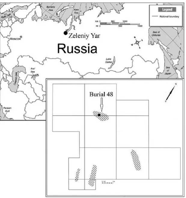
We present a paleoparasitological analysis of the medieval Zeleniy Yar burial ground of the XII-XII centuries AD located in the northern part of Western Siberia. Parasite eggs, identified as eggs of Opisthorchis felineus, were found in the samples from the pelvic area of a one year old infant buried at the site. Presence of these eggs in the soil samples from the infant’s abdomen suggests that he/she was infected with opisthorchiasis and imply consumption of undercooked fish. Ethnographic records collected among the population of the northern part of Western Siberia reveal numerous cases of feeding raw fish to their children. Zeleniy Yar case of opisthorchiasis suggests that this dietary custom has persisted from at least medieval times.
Key words: Opisthorchis felineus - paleoparasitology - paleopathology - Western Siberia - health status - food processing
doi: 10.1590/0074-02760150156
Financial support: RHCF (13-01-00097 - Ethnodemographic, bioanthro-pological and archaeological complex study Taz Nenets and Selkup), RFBR (13-06-00158 А - Features of physical development of Aboriginal and old residents of Siberia, in the light of adaptation options)
+ Corresponding author: s_slepchenko@list.ru Received 21 April 2015
Accepted 23 October 2015
Paleoparasitological analysis can be carried out using a wide range of archaeological and biological
objects, including samples from the pelvic/abdominal areas, coprolites, hair, mummified tissues, and even skeletal remains (Araújo & Ferreira 1996, Bouchet 1997, Le Bailly et al. 2005, Araújo et al. 2008). Identifi -cation of endoparasites can complement archaeological data on the paleodiet, subsistence practices, and food processing techniques, as well as trace migration pat-terns and help to reconstruct community health of pre-historic populations (Reinhard 1988, 1992a, b, Araújo
& Ferreira 1996, 2000, Horne & Tuck 1996, Bouchet et al. 2001, Reinhard & Araújo 2008, Jaeger et al. 2013, Slepchenko & Ivanov 2015, Yeh et al. 2015).
Paleoparasitological research is still nascent in
Rus-sia. Arkadiy Savinetsky and Aleksandr Khrustalev in
2013 presented a meta-analysis of endoparasites found
in the animal faecal depositions from caves with ar -chaeological deposits dating from as early as 40,000 years ago to XX century. Their study identified hel-minth eggs belonging to Nematodirus sp., Oxyuris
(Oxyurida), Fasciola sp., Dicrocoelium sp., Trichuris,
Capillaria sp., Diphyllobothrium sp., Opisthorchis felineus, Alaria alata, and Dioctophyma renale. The
diversity of helminths in the animal faeces provided
insights into the subsistence practices of the early hu-mans and hazards of animal farming in those regions.
Paleoparasitological analysis of dog coprolites from
Maray I settlement (dating to 2645 ± 30 BP), located in the forest-steppe zone of West Siberia, revealed larvae
of Strongyloides papillosus, Strongyloides westeri, and
Strongyloides stercoralis (Zach et al. 2011, Tsembalyuk
2013), suggesting heavy parasitic loads in the human set
-tlements of the region. Recent analysis of the XVII cen -tury soil and dog coprolites samples from Mangazeya, the
earliest Russian town located beyond the Arctic Circle in
West Siberia. The eggs of O. felineus, Diphillobathrium latum, Trichocephalus sp., Toxocara canis, and Fasciola hepatica were identified there (Vizgalov et al. 2013).
The only paleoparasitological investigation of the re -cent indigenous populations from Western Siberia was
carried out on the samples from the pelvic area from the XVII-XIX centuries Selkup Kikki-Akki burial site.
Tapeworm eggs of Diphyllobothrium sp., a typical fish endoparasite, showed that uncooked or undercooked fish
served as the primary source of intestinal parasites in Selkup communities (Slepchenko & Ivanov 2015). This
finding corroborated the ethnographic records that the Selkups consumed large quantities of raw or minimally
processed fish (Khomich et al. 2002, Tuchkova 2013).
The absence of O. felineus, a fish endoparasite
com-mon in Ob and Irtysh river basins, in samples from the Kikki-Akki burial ground suggested that Selkups from Kikki-Akki rarely migrated to the Ob and Irtysh region for fishing (Yossepowitch et al. 2004, Bonina & Fedorov 2010, Mordvinov et al. 2012). Thus, the Selkups migra
-tion routes were probably limited to the nearby Taz River Basin and, perhaps, the Yenisei and the Pur river basins
Here, we present an analysis of helminth eggs from
the soil samples collected from the pelvic and abdomi
-nal area of the child buried in grave 48 (hereafter M48) of the Zeleniy Yar cemetery dated to between XII-XIII centuries. The Zeleniy Yar archaeological site is located in the Cisuralian area of Yamalo-Nenets Autonomous Okrug in the north of Western Siberia (Fig. 1). It is situ
-ated on a flood plain island between the Poluy River and
the Gorny Poluy anabranch. Remains of an early metal-lurgy workshop, including two melting furnaces dating
to VI-VII centuries AD, have been recovered from the site. Two associated burial grounds have been dated to the VIII-IX centuries and the XII-XIII centuries.
MATERIALS AND METHODS
The site was excavated by the authors during the 2014 field season. Four burials dating from the XII-XIII centu
-ries were excavated and analysed at the same time. Sam -ples were collected from M48, since it was the only burial
that remained intact and undisturbed by carnivores.
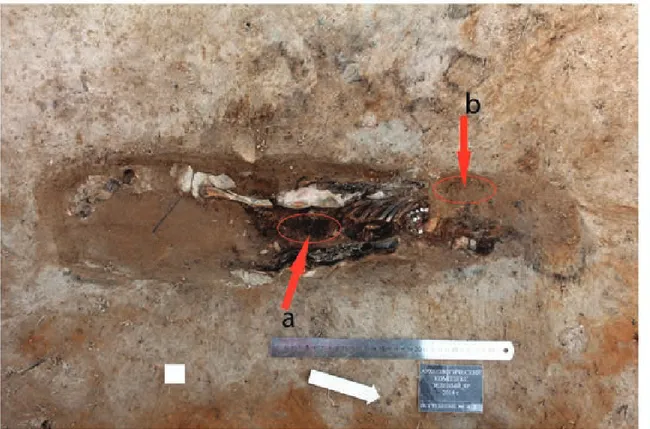
Burial M48 contained remains of an infant with an es-timated age around one year, based on dental eruption and
skeletal development. The body was in supine position
with the head placed at a northwest orientation. The in-fant’s arms were stretched along the body with the palms positioned on the thighs. The neck was bent, so that the chin was pressed to the chest, suggesting that there was
976 Mem Inst Oswaldo Cruz, Rio de Janeiro, Vol. 110(8), December 2015
some support made from perishable material was placed under the head. The infant was wrapped in a fur garment adorned with copper plates, which was further wrapped
in a hide with fur and a layer of birch bark (Gusev 2015).
Since the infant’s sacrum, the os coxae and lumbar
vertebra were completely decomposed, the sample was
collected from the abdominal region (Fig. 2). The sample
weighed about 30 g and was vacuum-packed in the field. In addition, a 50 g control sample was taken from the vicinity
of infant’s head at the same depth as the abdominal sample.
In the laboratory, 3% sodium hydroxide solution was added to the dry sample (Fry 1985, Dufour & Le Bailly 2013). In an hour and a half, the liquid was sifted through a sieve, filled into a plastic test tube, and centrifuged for 7 min (1,500 revolutions per min); distilled water was added to achieve neutral pH level. Upon completing this
cleaning procedure, we added a rich sodium nitrate so-lution (1.38-1.40 g/cm3) to the residue. Sample separa-tion was performed in the same centrifugal tubes. After multiple centrifuge cycles with 0.5% Na3PO4 aqueous
solution and glycerin at a temperature of 22 degrees, we
gathered the supernatant fraction. Having just a few or -ganic particles at hand, we still managed to prepare 20 microslides, following the recommended standard
meth-ods (Reinhard et al. 1986, Araújo et al. 1998). Micro
-scopic examination was conducted using AxioSkop 40 and MicMed 2 var.2. microscopes under 80X and 400X magnification. Measurements were obtained using Ax -ioVision 4.6 and Scope Photo 3.0 software.
RESULTS
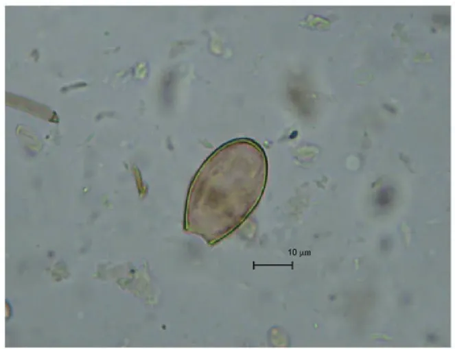
The microscopic examination of the slides revealed four helminth eggs of an oval shape and of light yellow
colour. The operculums of eggs were absent. Some eggs had slight shoulders at the area of operculum attachment.
At the pole opposite to operculum, there was a knob. The eggs measured 34.25-32.39 μm in length and 24.5-18.01 μm in width. Based on their morphology and size, these
eggs belong to the trematodes group of O. felineus (Fig. 3). The control sample was free of eggs. Epidemiolog-ic records indEpidemiolog-icate a high incidence of opisthorchiasis among the modern-day populations of the area where
the Zeleniy Yar burial ground is located (Istomin et al.
2003), corroborating our interpretation.
DISCUSSION
Differential diagnosis - In order to identify species
affiliation of the eggs, we performed a differential diag-nosis. The eggs found in the samples from the burial may
belong to several different genera of helminths. Morpho
-logical characteristics such as an oval shape, light yellow colour, size, and knob have to be further analysed with the aid of differential diagnosis which involves compar -ing helminth eggs of the cestodes Diphyllobothrium sp.
and of the trematodes Clonorchis sinensis, Opisthorchis viverrini, Metorchis bilis.
Diphyllobothriasis is widespread throughout West
-ern Siberia. The Cisuralian area of Yamalo-Nenets
Autonomous Okrug (with Zeleniy Yar burial ground
there) has the highest incidence of this helminth infec-tion among humans in the region. Some records show 340.3 cases per 100,000 people (Rospotrebnadzor State
Reports 2011). In the Ural Mountain area and Eastern
Siberia, D. latum is the most common fish tape worm
infecting humans. In Western Siberia Diphyllobothrium dendriticum and Diphyllobothrium ditremum are also common in humans, although the epidemiological im-pact of the latter on the local human populations is
mini-mal (Yastrebov 2013).
Having the same colour, elliptic shape, and knob, Diphyllobothrium sp. eggs look superficially similar to the eggs of Opisthorchis. Nevertheless, we excluded the
tapeworm eggs as the infection source, because of their bigger size. Typical Diphyllobothrium eggs measure
58-75 μm x 40-50 μm (Ash & Orihel 2007), considerably larger than the 24.5-18.01 μm eggs reported here.
Trematodes, C. sinensis and O. viverrini, also
pro-duce eggs that are morphologically very similar to the observed eggs (Kaewkes et al. 1991, Zhou et al. 2007).
Their size falls within the same range as that of the eggs
we have found (29 μm x 17 μm and 27 μm x 15 μm, re
-spectively). Eggs of trematodes are ovoid-shaped, long
-ish, yellow-brown, and have shoulders at the place of an
operculum attachment and a knob at the opposite egg
pole (Sadun 1955, Liu & Chen 1998). Therefore, exclud -ing C. sinensis and O. viverrini is difficult. However,
the epidemiologic records show that the trematodes C. sinensis and O.viverrini are unlikely to be the source of
infection. Although both helminths are present in South
-east Asia, Korea, and China (Posokhov 2004, Lun et al.
2005, Sripa et al. 2007), they are nonexistent in Western
Siberia (Posokhov 2004). The only known case of clo
-norchiasis in Russia has been documented in the Amur River Basin. Therefore, despite the morphological simi
-larity to the recovered eggs, the trematodes C.sinensis
and O.viverrini are unlikely to be the source of the in-fection, albeit these cannot be completely ruled out.
The eggs present in the M48 sample are also mor-phologically similar to eggs of M. bilis, a helminth from family Opisthorchidae that is present in Western Siberia. Since eggs of O. felineus and M. bilis cannot be differen-tiated by any common medical and parasitological meth-ods, it is likely that some of the diagnosed opisthorchiasis infection in Western Siberia is caused by metorchosis
in-fection or by both (Skrjabin & Petrov 1950, Sidorov 1983, Romashov et al. 2005). However, genus Metorchis is more
978 Mem Inst Oswaldo Cruz, Rio de Janeiro, Vol. 110(8), December 2015
commonly present in the southern territory of Western Siberia, while O. felineus has been recorded in its
north-ern part (Fattakhov 1996). Therefore, final animal hosts
and humans are predominantly infected with opisthor-chiasis in the northern part of Western Siberia, with its
highest rate in the Middle Ob Area, whilst metorchosis
infection rate increases towards the southern regions of
Siberia (Fattakhov 1996). A very low incidence of metor
-chosis in the lower course of the Ob River, where Zeleniy Yar burial ground is located, has been documented. This
low occurrence of metorchosis can be explained by a low frequency of Bethynia tentaculata (family Bithyniidae, genus Bethynia) molluscs that serve as the principal first intermediate host for metorchosis. Although Codiella in-flate (family Bithyniidae, genus Bethynia), the secondary intermediate host of metorchosis, is present in the region, this mollusc has a low susceptibility of metorchosis, lim-iting its propagation(Fattakhov 1996).
A high frequency of opistarchosis infection is re -corded among the modern-day populations of the
Cis-uralian area of Yamalo-Nenets Autonomous Okrug. In 2003, the prevalence rate was 2,585 cases per 100,000 people (Istomin et al. 2003). Among children under the age of 14, opistarchosis was present in 110 individuals
out of 100,000 (Rospotrebnadzor State Reports 2011).
For comparison, the prevalence rate for opistarchosis in the Ob River tributaries is 10-104 cases per 1,000 people (Karpenko et al. 2008). Up to 100% of the indigenous
people, among Khanty, Mansi, and Nenets ethnic
com-munities of the Ob and Irtysh basins in West Siberia are infected with these helminths (Schustov et al. 2002).
Such a high incidence of opistarchosis among
indig-enous people of the reason is caused by the heavy para
-sitic loads found in fish of Ob and Irtysh rivers. In the middle and the lower Ob River Basin, the opistarchosis
infection rate in Leuciscus idus and Leuciscus icuciscus fish is between 80-100%. The invasion degree is 4.5 and 7.5 larvae per gram of muscles, for L. idus and L. Icucis-cus, respectively [Fattakhov (1996) (p. 14)]. Moreover, the high frequency of opistarchosis among the local peo-ple is caused by the consumption of raw, undercooked,
or slightly salted fish (Istomin et al. 2003).
Thus, we may infer that the helminth eggs in the
samples from the Zeleniy Yar burial ground most likely
belonged to the fish tapeworm genus O. felineus. The presence of Opisthorchis eggs in the samples from the burial of an infant under one is noteworthy, as it implies early introduction of solid foods into infant diet. The in-fant could be infected with opisthorchiasis only by con-suming raw or minimally processed contaminated fish. The incubation period of opisthorchiasis is approximate-ly three-four weeks after the initial infection of the
fi-nal host (Vozianova 2000). Consequently, we may infer that the infant from M48 was given raw fish as a nursing
supplement at least one month before his/her death.
The abundance of fish in the rivers of Western Sibe -ria determined the local subsistence strategies at least since the terminal Neolithic (Tupakhina 2013). Large
quantities of fish bones were recovered from the Iron Age Ust-Poluy archaeological site, located in the lower Ob River Basin, suggesting that fish was an important
and possibly the main food source at the time. Similarly,
the medieval Zeleniy Yar archaeological site is also rich in fish deposits (Aleksashenko et al. 2005).
According to ethnographic reports (Bartenev 1896, Dunin-Gorkavich 1910), the indigenous people of the
northern part of Western Siberia consumed large
quanti-ties of raw fish. In 1715, the Russian ethnographer Grego
-ry Novitsky recorded that the population of the Ob River
Basin consumed fish, including fish intestines, in large
amounts (Zuev 1947, Novitsky 1989). People ate consid
-erable quantities of fish liver oil, a local specialty food. Diet included fresh raw fish in the summer and frozen
raw fish in the winter. The same subsistence practice
was described by ethnographer Vasily Zuev in 1771-1772 (Zuev 1947). An XVIII-century historian Gerhard Miller
(1742-1746) wrote about the practice of feeding raw fish
to young children among Nents. Nents children over one year old were given deer oil and chopped fish in order
to get them accustomed to eating raw food (Miller 1999, 2000, Ehlert 2006). Likewise, the Ostyaks (also known as Khanty) fed adult food to children. Furthermore,
Rus-sian historian Constantine Nosilov recorded consumption of several fish species, including Stenodus leucichthys, L. idus, Rutilus rutilus, Lota maculosa, Esox lucius, Perca fluviatilis, Gymnocephalus by Khanty (Nosilov 1937).
Some of these species, such as L. Idus and R. rutilus, could be carriers of O. felineus larvae (WHO 1995), serv -ing as the source of human infection when consumed raw. The Opisthorchis eggs presence in the samples from
the infant’s burial in the Zeleniy Yar burial ground re
-veals a dietary custom of consuming raw or under
-cooked fish during the Middle Ages in Western Siberia. Opisthorchiasis infection observed in the very young in -fant indicates early supplementation of breast milk with minimally processed fish. Ethnographic records from the northern part of Western Siberia corroborate these
findings, revealing numerous cases of feeding raw fish
to their children, which suggests that this dietary prac-tice has considerable antiquity.
ACKNOWLEDGEMENTS
To DSc Adauto Araujo, PhD Vladimir Adaev, PhD Mat
-thieu Le Bailly, for supporting, and to PhD Ekaterina Pechenki
-na and Prof Ron Pinhasi, who improved the quality of our paper.
REFERENCES
Aleksashenko NA, Brusnitsyna AG, Litvinenko MN, Kosintsev PA, Perevalova EV, Razhev DI, Fedorova NV 2005. Zeleniy Yar ar-chaeological complex era Middle Ages in the northern Ob, Ural
Branch of RAS, Salekhard, 368 pp.
Araújo A, Ferreira LF 1996. On hookworms and transpacific contact.
Parasitol Today12: 454-455.
Araújo A, Ferreira LF 2000. Paleoparasitology and the antiquity of human host-parasite relationship. Mem Inst Oswaldo Cruz 95
(Suppl. I): 89-93.
Araújo A, Ferreira LF, Reinhard KJ, Gardner SL 2008. Parasites as probes for prehistoric human migrations? Trends Parasitol 24: 112-115.
Ash LR, Orihel TC 2007. Atlas of human parasitology, 5th ed., Amer -ican Society for Clinical Pathology Press, Chicago, 540 pp.
Bartenev B 1896. In the extreme Northwest Siberia, essays Obdorsk edge, Printing and Lithography MF Paikin, St. Peterburg, 148 pp.
Bonina OM, Fedorov KP 2010. Local foci of opisthorchiidoses in ba -sin of Novosibirsk reservoir. Sib Vestnik SelKhoz Nauki4: 62-68. Bouchet F 1997. Intestinal capillariasis in Neolithic inhabitants of
Chalain (Jura, France). Lancet349: 256.
Bouchet F, West D, Lefèvre C, Corbett D 2001. Identification of par -asitoses in a child burial from Adak Island (central Aleutian Is -lands, Alaska). C R Acad Sci III324: 123-127.
Dufour B, Le Bailly M 2013. Testing new parasite egg extraction methods in paleoparasitology and an attempt at quantification.
Int J Paleopathol 3: 199-203.
Dunin-Gorkavich AA 1910. Tobolsk north, Regional Printing House, Tobolsk, 353 pp.
Ehlert AH 2006. North-Western Siberia in the shipping of labor and materials Georg Miller, Volot, Ekaterinburg, 416 pp.
Fattakhov RG 1996. Ecology parasitic systems opistorhid Ob-Irtysh Basin in terms antropopressy, Tyumen, 50 pp.
Fry GF 1985. Analysis of fecal material. In RI Gilbert Jr, J Mielke (eds.), The analysis of prehistoric diets, Academic Press, Orlan -do, p. 127-154.
Gusev AV 2015. Report on the scientific research work “Comprehen-sive archaeological and anthropological studies of medieval bur -ial grounds near the village of Zelyeny Yar. Priuralsky YANAO district in 2014”, Typography Salekhard, Salekhard, 350 pp.
Horne PD, Tuck JA 1996. Archaeoparasitology at a 17th century colo -nial site in Newfoundland. J Parasitol82: 512-515.
Istomin AV, Shushkova TS, Raegulov BM 2003. Hygienic problems of ecology and human health in the Far North, Nauka, Moscow, 388 pp.
Jaeger LH, Taglioretti V, Fugassa MH, Dias O, Neto J, Iñiguez AM 2013. Paleoparasitological results from XVIII century human re -mains from Rio de Janeiro, Brazil. Acta Trop125: 282-286. Kaewkes S, Elkins DB, Sithithaworn P, Haswell-Elkins MR 1991.
Comparative studies on the morphology of the eggs of Opisthor-chis felineus and lecithodendriid trematodes. Southeast Asian J Trop Med Public Health22: 623-630.
Karpenko SV, Chechulin AI, Yurlova NI, Serbina EA, Vodyanitskaya SN, Krivopalov AV, Fedorov KP 2008. Characteristics of opisthor -chiasis foci in southern West Siberia. Contemp Probl Ecol1: 517-521.
Khomich LV, Irikov SI, Aiupova GE 2002. Taz selkups: essays on traditional culture, Filial Izd-va, St. Petersburg, 149 pp.
Le Bailly M, Leuzinger U, Schlichtherle H, Bouchet F 2005. Diphyl -lobothrium: neolithic parasite? J Parasitol 91: 957-959.
Liu YS, Chen M 1998. Biology of Clonorchis sinensis and control of clonorchiasis, Science Press, Beijing, 117 pp.
Lun Z, Gasser RB, Lai D, Li A, Zhu X, Yu X, Fang YY 2005. Clo -norchiasis: a key foodborne zoonosis in China. Lancet Infect Dis 5: 31-41.
Miller GF 1999. The history of Siberia, Vol. 1, Eastern Literature/ Russian Academy of Science, Moscow, 630 pp.
Miller GF 2000. The history of Siberia, Vol. 2, Eastern Literature/ Russian Academy of Science, Moscow, 796 pp.
Mordvinov VA, Yurlova NI, Ogorodova LM, Katokhin AV 2012.
Opisthorchis felineus and Metorchis bilis are the main agents of liver fluke infection of humans in Russia. Parasitol Int61: 25-31.
Nosilov KD 1937. Northern stories, Regional Publishing, Sverdlovsk,
272 pp.
Novitsky GI 1989. Brief description of the Ostyak people. Columba Russian landsat document. Descriptions of the discovery and study of Siberia, the Far East and the North in the XVII-XVIII centuries, Khabarov, Khabarov, 121 pp.
Posokhov PS 2004. Clonorchosis in the Amur River Basin, 1st ed., Far East State Medical University, DGMU, Khabarovsk, 187 pp. Reinhard K 1992a. Parasitology as an interpretive tool in archaeol
-ogy. Am Antiq57: 231-245.
Reinhard K, Mrozowski AS, Orloski KA 1986. Privies, pollen, para -sites, and seeds - a biological nexus in historic archaeology. MAS-CA Journal4: 31-36.
Reinhard KJ 1988. Diet, parasitism and anemia in the prehistoric southwest, PhD Thesis, Department of Anthropology/Texas
A&M University, Texas, 120 pp.
Reinhard KJ 1992b. The impact of diet and parasitism on anemia in the prehistoric west. In P Stuart-McAdam, S Kent (eds.), Diet, de-mography, and disease: changing perspectives of anemia, Trans-action Publishers, New York, p. 219-258.
Reinhard KJ, Araújo A 2008. Archaeoparasitology. In DM Pearshall (ed.), Encyclopedia of archaeology, New York, p. 145-163.
Romashov BV, Romashov VA, Semenov VA, Filimonova LV 2005. Opisthorchiasis in the upper Don Basin (Voronezh Oblast): the liver fluke fauna, ecological, and biological patterns of circula -tion and foci of opisthorchiases, 1st ed., Voronezh State Univer -sity, Voronezh, 210 pp.
Rospotrebnadzor State Reports 2011. On sanitary-epidemiological situation in Yamalo-Nenets AO, Rospotrebnadzor, Moscow, 73 pp. Sadun EH 1955. Studies on Opisthorchis viverrini in Thailand. Am J
Hyg 62: 81-115.
Savinetsky AB, Khrustalev AV 2013. Paleoparasitological investigations in Mongolia, Middle Asia and Russia. Int J Paleopathol3: 176-181. Schustov AV, Kotelkin AT, Sorokin AV, Ternovoi AA, Loktev VB 2002.
The Opisthorchis felineus paramysin; cDNA sequence and charac -terization of its recombinant fragment. Parasitol Res88: 724-730.
Sidorov EG 1983. Natural foci of opisthorchiasis, Nauka Kaz,
Alma-Ata, 240 pp.
Skrjabin KI, Petrov AM 1950. Superfamily Opisthorchoidea Faust 1929. Trematodes of man and animals, Foundation of Trema-todology, Moscow, 496 pp.
Slepchenko SM, Ivanov SN 2015. Paleoparasitological analysis of soil samples from the Kikki-Akki burial ground of the 17th-19th cen -turies in West Siberia, Russia. J Archaeol Sci 2: 467-472.
Sripa B, Kaewkes S, Sithithaworn P, Mairiang E, Laha T, Smout M, Pairojkul C, Bhudhisawasdi V, Tesana S, Thinkamrop B, Betho-ny JM, Loukas A, Brindley PJ 2007. Liver fluke induces cholan -giocarcinoma. PLoS Med4: 1148-1155.
Tsembalyuk SI 2013. Economy and life of the population krasnooz -erskoy culture based on settlement Marai 1 in lower Priishimya. New materials and methods of archaeological research. Proceed-ings of the II International Conference of Young Scientists, Mos -cow, 19-21 March 2013, Institute of Archeology of the Russian Academy of Sciences, Moskow, p. 70-72.
Tuchkova NA, Glushkov SV, Kosheleva EY, Golovnev AV, Baidak AV, Maximova NP 2013. Selkups. Sketches of traditional culture and Selkups, Tomsk Polytechnic University, Selkup, 318 pp.
settle-980 Mem Inst Oswaldo Cruz, Rio de Janeiro, Vol. 110(8), December 2015
ment of an epoch Eneolithic - the Early Bronze century (Priural-sky district) Yamalo-Nenets Autonomous District, Typography Salekhard, Salekhard, 450 pp.
Vizgalov GP, Kardash AV, Kosintsev PA, Lobanova TV 2013. Histori-cal ecology of the population of the north of Western Siberia, AMB
Yekaterinburg, Nefteyugansk, 376 pp.
Vozianova JI 2000. Infectious and parasitic diseases, Vol. 1, Health, Kiev, 903 pp.
WHO - World Health Organization 1995. Control of foodborne trematode infections. Available from: apps.who.int/iris/bit -stream/10665/41544/1/WHO_TRS_849_(part1).pdf.
Yastrebov KV 2013. Ecology, epidemiology and prevalence diphyllo -bothriasis in Siberia and the Far East. Actual aspects of parasitic diseases in the modern period. Proceedings of All-Russian Confer -ence, Tyumen, 25-26 September 2013, FBUN Tyumen Research Institute of Regional Infectious Pathology, Tyumen, p. 205-207.
Yeh H-Y, Prag K, Clamer C, Humbert JB, Mitchell PD 2015. Human intestinal parasites from a Mamluk Period cesspool in the Chris-tian Quarter of Jerusalem: potential indicators for long distance travel in the 15th century AD. Int J Paleopathol 9: 69-75. Yossepowitch O, Gotesman T, Assous M, Marva E, Zimlichman R,
Dan M 2004. Opisthorchiasis from imported raw fishes. Emerg Infect Dis 10: 2122-2126.
Zach VA, Tsembalyuk SI, Siben AN 2011. Parasites in human life: formulation of the problem. Proceedings of the Ecology of An -cient and Traditional Societies, Tyumen, 14-18 November 2011, IPDN SB RAS, Tyumen, p. 107-110.
Zhou XN, Lv S, Yang GJ, Kristensen TK, Bergquist NR, Utzinger J, Malone JB 2007. Spatial epidemiology in zoonotic parasitic diseases: insights gained at the 1st International Symposium on Geospatial Health in Lijiang. Parasit Vectors2: 10.