• Nenhum resultado encontrado

Application of molecular tools for detection of plant viruses

N/A
N/A
Protected

Academic year: 2020

Share "Application of molecular tools for detection of plant viruses"

Copied!
111
0
0

Texto

(1)

Faculdade de Ciências e Tecnologia

APPLICATION OF MOLECULAR TOOLS FOR

DETECTION OF PLANT VIRUSES

Submitted by

FILIPA ESTEVES

Dissertation for obtaining a Master degree in

Molecular and Microbial Biology

Supervised by Prof. Dr. Filomena Fonseca

Faro, Portugal

2013

(2)

Faculdade de Ciências e Tecnologia

APPLICATION OF MOLECULAR TOOLS FOR

DETECTION OF PLANT VIRUSES

Submitted by

FILIPA ESTEVES

Dissertation for obtaining a Master degree in

Molecular and Microbial Biology

(3)

Declaration

I declare this thesis submitted by me for the degree of Magister Scientiae in Molecular

and Microbial Biology at the University of Algarve is result of my research work and its

contents are my responsibility.

De acordo com a alínea iii) do ponto 4.1 do Anexo II a que se refere o ponto 2 do Artigo 18º do Regulamento nº 287/2021

publicado em Diário da Repúbica, a 24 de Julho de 2012: “Declaro ser a autora deste trabalho, que é original e inédito.

Autores e trabalhos consultados estão devidamente citados no texto e constam da listagem de referências incluída.”

___________________________

Filipa Esteves

©Copyright by Filipa Alexandra Oleiro Esteves. All Rights Reserved.

Após publicação do seu conteúdo em revistas da especialidade e de acordo com o ponto 4.2 do Anexo II a que se refere o

ponto 2 do Artigo 18º do Regulamento nº 287/2021 publicado em Diário da Repúbica, a 24 de Julho de 2012: “A

Universidade do Algarve tem o direito, perpétuo e sem limites geográficos, de arquivar e publicitar este trabalho através de

exemplares impressos reproduzidos em papel ou de forma digital, ou por qualquer outro meio conhecido ou que venha a ser

inventado, de o divulgar através de repositórios científicos e de admitir a sua cópia e distribuição com objetivos

educacionais ou de investigação, não comerciais, desde que seja dado crédito ao autor e editor.”

(4)
(5)

Acknowledgments

I would like to thank my supervisor Prof. Dr. Filomena Fonseca for allowing me

to pursuit my interest in science. I would like to thank you for giving me the opportunity

to work with you. I also thank you for the assistance and advice as well for the support

and guidance during the development of this project. In addition, I would like to thank

the recommendations and corrections to this manuscript.

I would like to thank David for his patience and support during this time. Thank

you for your tolerance and love even in those moments when my temper was

particularly challenging.

I also would like to thank Gui for making be stop and take a breath of fresh air

every day during this process.

Finally, I would like to thank my mother for her unconditional love, support and

encouragement through every step of the way. Thank you for always be beside me and

share my path. I thank you for being an extraordinary reliable person through my entire

life and for always believing in me. Without you I could not have made it here.

(6)
(7)

Abstract

Grapevine leafroll disease (GLRD) is one of the most important virus diseases of

grapevines worldwide, causing major economical impact. The disease has a complex

aetiology and currently eleven phloem-limited viruses, termed in general Grapevine

leafroll-associated virus (GLRaVs), have been identified. Two of the GLRaVs,

GLRaV-1 and GLRaV-3, are included in the European certification scheme of propagation

material. However, the flawed notion that GLRaV-3 is more frequent than GLRaV-1

and that all other GLRaVs are possibly not as relevant for GLRD, has until now

precluded the development of specific serological and molecular detection assays and

limited the scope of molecular characterization of the viruses known to be associated

with the disease. Hence, few studies have addressed the phylodynamics of GLRaVs or

even characterized the genetic structure of their natural populations. This generalized

lack of molecular information, in turn underlie the deficient capacity to detect the

viruses.

The phylogenetic analyses were conducted on the basis of the heat shock protein

70 homologue (HSP70h) and the coat protein (CP) genes for GLRaV-1 and the

HSP70h, the heat shock protein 90 homologue (HSP90h) and the CP genes for

GLRaV-5. The data obtained for GLRaV-1 contributed 83 new CP sequences. This information

was combined with previous analysis by other authors and used for the production of

new polyclonal IgG, capable of detecting CP variants from all the phylogroups

observed. Successful testing of this new tool included tissue print immunoblotting

(TPIB) and in situ immunoassay (ISIA).

The data obtained for GLRaV-5, contributed 61 new CP and 28 new HSP90h

gene sequences. Eight phylogenetic groups were identified on the basis of the CP.

Characterization of the genetic structure of the isolates revealed a higher diversity than

previously reported and allowed the identification of dominant virus variants.

For both GLRaV-1 and GLRaV-5, the effect of vegetative propagation on the

virus transmission dynamics was addressed.

(8)
(9)

Resumo

A Doença do Enrolamento da Videira (GLRD) é uma das mais importantes

doenças virais que afectam as videiras a nível mundial, causando um grande impacto

económico. A doença tem uma etiologia complexa e, actualmente, onze vírus

associados ao floema, designados por Grapevine leafroll-associated virus (GLRaVs)

foram identificados. Dois dos GLRaVs, os GLRaV-1 and GLRaV-3, estão incluídos na

lista de vírus de certificação obrigatória em material de propagação vegetativa indicada

pelo esquema de certificação de material de propagação da União Europeia. Contudo, a

ideia imprecisa de que o GLRaV-3 é mais frequente que o GLRaV-1 e que, todos os

outros GLRaVs, possivelmente, não serão tão revelevantes para a GLRD, tem excluído

o desenvolvimento de análises de detecção serológica e molecular e limitado a

caracterização molecular dos vírus associados à doença. Deste modo, poucos estudos

trataram a filodinâmica dos GLRaVs ou caracterizaram a estrutura genética das suas

populações naturais. Por seu turno, a falta generalizada de informação molecular é

subjacente à deficiente capacidade de detecção dos vírus.

Para o GLRaV-1, as análises filogenéticas foram efectuadas com base no gene

que codifica a heat shock protein 70 homologue (HSP70h), bem como no gene que

codifica a proteína da cápside (CP). Para o GLRaV-5, as análises foram realizadas com

base nos genes que codificam a HSP70h, a heat shock protein 90 homologue (HSP90h)

e a CP. Os dados obtidos para o GLRaV-1 contribuíram com 83 novas sequências da

CP. Esta informação, juntamente com análises anteriores relizadas por outros autores,

foi utilizada para produzir um novo IgG policlonal, capaz de detectar as variantes da CP

de todos os filogrupos observados. Esta nova ferramenta de detecção foi testada com

êxito por tissue print immunoblotting (TPIB) e por in situ immunoassay (ISIA).

A informação obtida para o GLRaV-5, contibuiu com 61 e 28 novas sequências

da CP e HSP90h, respectivamente. Oito grupos filogenéticos foram identificados com

base na CP. A caracterização da estrutura genética dos isolados revelou uma maior

diversidade que a anteriormente publicada e permitiu a identificação de variantes virais

dominantes.

O efeito da propagação vegetativa na dinâmica de transmissão do vírus, foi

analisado para o GLRaV-1, bem como para o GLRaV-5.

(10)
(11)

TABLE OF CONTENTS

Declaration___________________________________________________________ 1

Acknowledgments_____________________________________________________ 3

Abstract_____________________________________________________________ 5

Resumo_____________________________________________________________ 7

Table of Contents______________________________________________________ 9

Presentations and Publications____________________________________________ 13

List of abbreviations___________________________________________________ 15

Chapter 1. Introduction and literature review_____________________________ 21

1.1. Viruses in general____________________________________________ 21

1.2. Plant Viruses_______________________________________________ 22

1.3. Economic Importance of Plant Viruses___________________________ 22

1.4. Virus Transmission in Plants___________________________________ 23

1.5. Virus Diseases of Grapevine___________________________________ 24

1.5.1. Leafroll Disease________________________________________ 25

1.5.1.1. Economic Impact_________________________________ 25

1.5.1.2. Symptomatology_________________________________ 25

1.5.1.3. Implicated Visuses_______________________________ 27

1.5.1.4 Cytopayhology___________________________________ 28

1.5.1.5. Epidemiology____________________________________ 29

1.5.1.6. Certification Scheme of Vegetative Propagation Material__ 30

(12)

1.6.1. Visual Detection and Biological Indexing____________________ 31

1.6.2. Serological Tests________________________________________ 32

1.6.3. Molecular Detection and Typyfycation______________________ 34

1.7. Aim of the Work_____________________________________________ 35

Chapter 2. Grapevine Leafroll-associated Virus 1___________________________ 37

2.1. Introduction________________________________________________ 37

2.2. Material and Methods_________________________________________ 39

2.2.1. Sample Collection and Plant Material_______________________ 39

2.2.2. RNA Extraction________________________________________ 41

2.2.3. cDNA Synthesis and PCR________________________________ 42

2.2.4. Cloning and Transformation_______________________________ 43

2.2.5. Single-Strand Conformation Polymorphism (SSCP)____________ 44

2.2.6. Plasmid DNA Extraction and Restriction Analysis_____________ 44

2.2.7. Sequencing and Sequence Data Analysis_____________________ 45

2.2.8. Estimates of Genetic Distances and Selection Pressure__________ 46

2.2.9. Recombination Analysis__________________________________ 46

2.2.10. Antibodies Development________________________________ 46

2.2.11. SDS-PAGE and Western Blot Analysis_____________________ 47

2.2.12. Tissue Print Immunoblotting (TPIB)_______________________ 48

2.2.13. In Situ Immunoassay (ISIA)______________________________ 48

2.2.13.1. ISIA with Polyclonal IgG__________________________ 48

(13)

2.3. Results_____________________________________________________ 50

2.3.1. PCR amplification_______________________________________ 50

2.3.2. Genetic Diversity of GLRaV-1 Isolates______________________ 50

2.3.3. Phylogenetic Analysis of GLRaV-1 Isolates__________________ 53

2.3.4. GLRaV-1 Phylogroups Diversity___________________________ 56

2.3.5. Recombination and Selection Constraints Analysis_____________ 58

2.3.6. Serological Assays______________________________________ 59

2.4. Discussion__________________________________________________ 65

Chapter 3. Grapevine Leafroll-associated Virus 5___________________________ 69

3.1. Introduction________________________________________________ 69

3.2. Material and Methods________________________________________ 71

3.2.1. Sample Collection and Plant Material_______________________ 71

3.2.2. RNA Extraction and cDNA Synthesis______________________ 73

3.2.3. PCR Amplification______________________________________ 73

3.2.4. Cloning and Transformation_______________________________ 74

3.2.5. Single-Strand Conformation Polymorphism (SSCP)____________ 74

3.2.6. Plasmid DNA Extraction and Restriction Analysis _____________75

3.2.7. Sequencing and Sequence Data Analysis_____________________ 75

3.2.8. Estimates of Evolutionary Divergence and Selection Pressure____ 76

3.2.9. Recombination Analysis__________________________________ 76

3.3. Results_____________________________________________________ 77

3.3.1. PCR amplification_______________________________________ 77

(14)

3.3.3. Phylogenetic Analysis of GLRaV-5 Isolates and Related

Ampeloviruses______________________________________________ 80

3.3.4. GLRaV-5 Phylogroups and Related Ampeloviruses Diversity____ 82

3.3.5. Selection Constraints on CP and HSP90h Genes_______________ 85

3.4. Discussion__________________________________________________ 90

Chapter 4. General Discussion and Conclusions___________________________ 95

(15)

Presentations and Publications

Part of the results reported in this thesis have been published and presented at

scientific meetings.

Publications

Chapter 3:

Esteves, F., Teixeira Santos, M., Eiras-Dias, J. E. and Fonseca F. 2012.

Occurrence of grapevine leafroll-associated virus 5 in Portugal: genetic variability and

population structure in field-grown grapevines. Arch. Virol. 157(9): 1747-1745.

DOI:10.1007/s00705-012-1371-2; IF 2.11.

Poster Presentations

Chapter 2:

Esteves, F., Teixeira Santos, M., Rocha, M. L., Eiras-Dias, J. E. and Fonseca F.

2009. Genetic diversity of portuguese isolates of Grapevine Leafroll-associated Virus 1

(GLRaV-1) based on the capsid protein. In: Proceedings of the 16

th

Congress of ICVG,

Dijon, France, pp. 310-311.

Esteves, F., Teixeira Santos, M., Rocha, M. L., Eiras-Dias, J. E. and Fonseca F.

2012. Novel antibodies for ISIA detection of Grapevine leafroll-associated virus 1

(GLRaV-1) based on the variability of the capsid protein gene. In: Proceedings of the

17

th

Congress of ICVG, UCDavis, USA, pp. 59-60.

Chapter 3:

Esteves, F., Teixeira Santos, M., Eiras-Dias, J. E. and Fonseca F. 2012.

Variability of Grapevine leafroll-associated virus 5 (GLRaV-5) in Portuguese field

grown grapevines. In: Proceedings of the 17

th

Congress of ICVG, UCDavis, USA, pp.

198-199.

(16)
(17)

List of abbreviations

AA

amino acid

AlkB

alpha-ketoglutarate-dependent deoxygenase

AMV

Alfafa mosaic virus

AP

alkaline phosphatase

ArMV

Arabis mosaic virus

BCIP

5-bromo-4-chloro-3-indolyl phosphatase

BIC

Bayesian information criterion

bp

base pair

BSA

bovine serum albumin

CAN

Colecção Ampelográfica Nacional

cDNA

complemantary DNA

cm

centimetre

CMV

Cucumber mosaic virus

CP

coat protein

CPm

minor coat protein

CTV

Citrus tristeza virus

DAS-ELISA

double antibody sandwich enzyme-linked immunosorbent assay

dN

non-synonymous substitutions

dNTP

deoxyribonucleotide triphosphate

ds

double-stranded

(18)

DNA

deoxyribonucleic acid

e.g.

exempli gratia, for example

EDTA

ethylenediaminetetraacetic acid

ELISA

enzyme-linked immunosorbent assay

et al.

et alia, with others

FEL

fixed effects likelihood

FITC

fluoresceine isothiocyanate

g

gram

GARD

genetic recombination detection

GFkV

Grapevine fleck virus

GFLV

Grapevine fanleaf virus

GLRaV

Grapevine leafroll-associated virus

GLRD

Grapevine leafroll disease

ha

hectare

HEL

helicase

HSP70 h

heat shock protein 70 homologue

HSP90 h

heat shock protein 90 homologue

ICTV

International Committee on Taxonomy of Viruses

ICTVdb

International Committee on Taxonomy of Viruses Database

ICVG

International Council for the Study of Virus and Virus-like

Diseases of the Grapevine

i.e.

id est, that is

(19)

IgG

Immunoglobulin G

INRB/INIAV

Instituto Nacional de Recursos Biológicos/Instituto Nacional de

Investigação Agrária e Veterinária

IPTG

isopropyl -D-1-thiogalactopyranoside

ISIA

in situ immunoassay

kb

kilobase

kDa

kilodalton

LB

lysogeny broth

L-Pro

papain-like leader protease

M

molar

mA

milliamp

MAb

monoclonal antibody

mg

milligram

min

minute

ml

millilitre

mM

millimolar

mRNA

messenger ribonucleic acid

MT

methyltransfrease

NBT

nitroblue tetrazolium

nm

nanometres

nt

nucleotide

OIV

International Organisation of Vine and Wine

(20)

PAb

policlonal antibody

PBS

phosphate buffered saline

PCR

polymerase chain reaction

PVDF

polyvinylidene difluoride

PVP-40

polyvinylpyrrolidone wt 40,000

PVY

Potato virus Y

R99

Richter 99

RdRp

RNA-dependent RNA polymerase

REL

random effects likelihood

RFLP

restriction fragment length polymorphism

RNA

ribonucleic acid

rpm

revolutions per minute

RT-PCR

reverse transcription polymerase chain reaction

SDS-PAGE

sodium dodecyl sulphate polyacrilamide gel electrophoresis

SLAC

single likelihood ancestor counting

SO4

Selektion Oppenheim 4

S.O.C

super optimal broth with catabolite repression

spp.

species

ss

single-stranded

SSCP

single-strand conformation polymorphism

TAE

tris acetate-EDTA

(21)

TPIB

tissue print immunoblotting

U

units

UTR

untranslated region

V

volt

Vs

versus

v/v

volume/volume

w/v

weight/volume

X-Gal

5-Bromo-4-chloro-3-indolyl β-D-galactopyranoside

µl

microlitre

µm

micrometre

(22)
(23)

Chapter 1. Introduction and literature review

1.1. Viruses in general

Viruses are obligate intracellular parasites. They need the host cellular

machinery to reproduce, since they lack the genetic information for conducting

biological processes necessary to replicate. Virus particles are designated by virions and

contain the genome enclosed by a coat protein known as capsid. Some viruses also have

an envelope, a lipid component covering the virion surface. The viruses’ genome can be

linear or circular and encompass one or more nucleic acid molecules, either

deoxyribonucleic acid (DNA) or ribonucleic acid (RNA), which can be single-stranded

(ss) or double-stranded (ds). Single-stranded genomes may be positive (+) or negative

(-) sense, or ambisense, as they have the same or the complementary nucleotide

sequence as the mRNA, or both, respectively. Viruses with negative sense RNA

genome need to carry a RNA-dependent RNA-polymerase, since most of their hosts

have no mechanism for RNA-dependent RNA polymerization. The majority of virus

genomes are constituted by a single molecule of nucleic acid (monopartite) but in some

cases the genome is segmented in two (bipartite) or more nucleic acid molecules, which

occurs more frequently with RNA viruses. A segmented genome increases the

possibility of genes to combine in different ways, thus increasing the evolution rates.

Virus genomes can have a size from 3500 nt up to 2400000 nt. In the case of

single-stranded RNA, the fragility and high probability of breaking as well as the higher

mutation rates, contribute to limit the size of the viral genomes, which are usually found

varying from 3.5 kb up to 30 kb (Gelderblom, 1996).

(24)

1.2. Plant Viruses

Plant virus species are determined by groups of properties recognized by the

International Committee on Taxonomy of Viruses (ICTV). These properties include the

plant species infected and the symptoms caused by the viruses, the way viral

transmission occurs, the host species that can be used for experimental infection, the

shape of virus particles, its nucleic acid and proteins, the genome composition and

organization and also their serology. At the present there are 83 genera and 21 families

of plant viruses recognized by the ICTV (ICTV, 2011).

Plant viruses’ morphology may be rigid or flexuous rod-shaped (e.g. tobacco

mosaic virus, TMV and potato virus Y, PVY, respectively), isometric (icosahedral) (e.g.

cucumber mosaic virus, CMV) or bacilliform (bullet shape) (e.g. alfafa mosaic virus,

AMV).

Most plant viruses have RNA genomes, the majority of which are single

stranded and positive sense (Matthews, 1991). Besides the plant viruses with

monopartite genomes, like the members of the Closteroviridae family, there are also

plant viruses with bipartite and tripartite genomes, as the case of the Comoviridae and

Bromoviridae families, respectively.

1.3. Economic Importance of Plant Viruses

Viruses are responsible for many of plant diseases that cause great losses in

crops. These diseases can lead to yield and quality decrease in economically important

crops, as a result of its symptoms which vary according to the disease. Generally, plant

virus symptoms include lesions, necrosis (primary infection), leaf colour alteration and

growth abnormalities (secondary or systemic infection). However, most symptoms can

be common to several viruses and not all viruses cause visible symptoms.

Exact and up to date estimates related to damages caused by plant viruses to a

given crop around the world are usually not available, but there are references to losses

of billions of dollars per year in general terms (Gray and Banerjee, 1999). The costs

(25)

implementation and application of sanitary measures that need to be considered in order

to prevent or control outbreaks, such as propagation material certification programs,

eradication programs or vector control.

1.4. Virus Transmission in Plants

Plant viruses can be transmitted between different hosts through several routes,

including seeds, pollen, propagation material, vectors and mechanical transmission.

Last century research suggested that approximately 20% of plant viruses are

transmitted by seed (Matthews, 1991; Mink, 1993). The infection can occur during or

after the fertilization of the embryo. In the first case the virus can invade the ovule or be

transported by the male gamete and in the second case the virus invade the embryo

during its development (Mink, 1993; Maule and Wang, 1996). Transmission by seeds

usually leads to unhealthy new crops.

Viruses transmitted by pollen can infect the gametes of the embryo or the mother

plant through the fecundated flower (Hull, 2009). This way of transmission seems to be

unusual and only a few viruses are transmitted by pollen (Mink, 1993).

Vegetative propagation is a very common horticultural practice, especially with

perennial species and therefore constitutes a very effective way of viruses’ transmission.

Once the parts of a systematically infected plant are used for propagation, normally the

virus will be introduced into the new host (Hull, 2009). Grafting is an example of

vegetative propagation. In this technique tissues from a plant (scion) are inserted on the

roots of another (rootstock) and if one or both is infected and both are susceptible, the

grafted plant will also become infected.

Viruses can also be disseminated from plant to plant by living organisms that

feed of them and carry virus particles from infected plants to healthy ones, acting as

vectors. This is the most common way of spreading of a virus in nature (Andret-Link

and Fuchs, 2005). The vectors of plant viruses include arthropods (insects and

arachnids), nematodes, fungi and plasmodiophorids (Andret-Link and Fuchs, 2005).

Insect vectors of plant viruses include members of the order Hemiptera, such as aphids,

leafhoppers, planthoppers, whiteflies, mealybugs, mirids and some treehoppers, and

also members from other orders like beetles (Coleoptera), thrips (Thysanoptera),

(26)

grasshoppers (Orthoptera) and mites (Acari). Nematodes responsible for plant virus

transmission

belong

to

genera

Longidorus,

Trichodorus,

Paralongidorus,

Paratrichodorus and Xiphinema. Fungi and plasmodiophorids vectors are included in

the orders Plasmodiophorales (Polymyxa spp. and Spongospora spp,) and Chytridales

(Olpidium spp) (Agrios, 2005). Because aphids transmit 55% of plant viruses, they are

the most important vectors. The remaining virus species are either transmitted by

leafhoppers and beetles (11% each), whiteflies (9%), nematodes (7%) fungi and

plasmodiophorids (5%), and thrips, mites, mirids and mealybugs (2%) (Andret-Link and

Fuchs, 2005), or have no known vector.

According to the way of viruses’ transmission by insect vectors, those are

classified as non-persistent, semi-persistent and persistent. Non-persistent viruses

remain in the stylet of the insect after acquisition and transmission can occur within a

few minutes. Semi-persistent viruses are acquired in about fifteen minutes, enter the

vector foregut and can be transmitted for up to two days. Persistent transmission

involves the passage of the viruses throughout the gut into the haemocoel and then to

the salivary glands. Hence, this type of transmission requires a latency or incubation

period between acquisition and inoculation of the virus, which can remain in the vector

for its lifetime. Non- and semi-persistent viruses, that are not able to replicate in the

insect, are known as non-circulative. Persistent viruses are circulative or propagative,

since they can circulate and replicate in the body of the insect (Katis et al., 2007).

1.5. Virus Diseases of Grapevine

Grapevine (Vitis vinifera L.) is one of the oldest and most economically

important cultivated crops. In 2010, the total vineyard cultivated area worldwide was

estimated at 7.6 million hectares and the grape production was estimated to be

67950000 tons (OIV, 2012). Grapevine crops can be affected by several diseases, which

cause huge economic losses, including those from viruses. Up to date, there are about

70 infectious agents, encompassing virus, viroids and pytoplasmas, recognized by the

International Council for the Study of Virus and Virus-like Diseases of the Grapevine

(ICVG, 2003). However, there are four major virus diseases affecting grapevine:

infectious degeneration/decline, rugose wood complex, fleck and leafroll.

(27)

1.5.1. Leafroll Disease

Grapevine leafroll disease (GLRD) was first reported in California (Goheen et

al., 1958) and nowadays is distributed in all grape-producing regions around the world

(Martelli and Boudon-Padieu, 2006).

1.5.1.1. Economic Impact

GLRD economic impact causes average yield losses of 15-20% (Goheen, 1988,

Martelli and Boudon-Padieu, 2006) but can achieve values up to 68% (Walter and

Martelli, 1997). During a vineyard lifetime, GLRD can cause losses ranging from

$25407/ha, considering a 30% yield reduction, to $41000/ha when considered a 50%

yield reduction and a 10% penalty for low fruit quality (Atallah et al., 2012).

Comparable data is lacking in European countries.

1.5.1.2. Symptomatology

GLRD is latent in American Vitis species and their hybrids used as rootstocks

but symptomatic in European V. vinifera (Uyemoto et al., 2009). The disease affects

rooting ability, graft take and plant vigour. The red and white varieties of V. vinifera

show, respectively, a purple-red or yellow discoloration of the leaf blade with green

primary veins that starts to appear at the basal leaves in late spring or early summer and

progress through the shoot, not affecting the tip leaves, until leaf drop. In advanced

stages of the disease the main veins can also lose their green colour. In some cultivars

the disease can cause mesophyll necrosis, noticeable between the veins. Usually, in

addition to chlorosis, leaves show downward rolling of the margins and thickening of

the blade (Weber et al., 1993, Martelli, 1993). Bunches are smaller and mature

irregularly and later than usual, thus affecting fruit ripening. GLRD symptoms also

include reduced sugar content of the berries, low Brix in the fruit juice (one degree Brix

is 1 gram of sucrose in 100 grams of solution and represents the strength of the solution

as percentage by weight (% w/w), reduced wine colour, phloem disruption, reduction of

protein content, changes in the pattern of peroxidase and polyphenoloxidase

(28)

isoenzymes, potassium depletion and composition as well as aromatic profile of the

musts (Martelli and Boudon-Padieu, 2006).

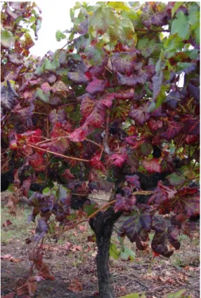
Figure 1.1 Vitis vinifera red wine grape variety

Monvedro with symptoms of leafroll disease. Photo

kindly provided by Dr. Margarida Teixeira Santos.

Figure

1.2

Vitis

vinifera white wine

grape variety Estreito

Macio

with

symptoms of leafroll

disease. Photo kindly

provided

by

Dr.

Margarida

Teixeira

(29)

1.5.1.3. Implicated Viruses

GLRD is one of the most complex viral plant diseases, given the high number of

causal agents associated with it. Leafroll is caused by a complex of viruses, named

Grapevine leafroll-associated viruses (GLRaVs). All GLRaVs are phloem-restricted

filamentous, positive sense single-stranded RNA virus from the family Closteroviridae.

The Closteroviridae family was established in 1998 (Martelli et al., 2000) and

include flexuous filamentous plant virus with either monopartide or bipartide positive

sense single-stranded RNA genomes. The genome members of this family have their

organized into two modules. The first includes a papain-like leader proteinase (L-Pro) at

the 5’ end and two ORFs associated with the replication, one comprising a

methyltransferase (MET) and a helicase (HEL) and the other a dependent

RNA-polymerase (POL) (Dolja et al., 2006). The second module is a quintuple

closterovirus-specific gene block associated with the assembly and movement that codes for a ~6 kDa

hydrophobic protein (p6), a 70 kDa heat shock protein homologue (HSP70h), a

HSP90-like protein of 50-60 kDa, a major coat protein (CP) and a minor CP (CPm) (Dolja et

al., 1994; Dolja et al., 2006; Karasev, 2000).

When it was first established, this family comprised only the genera

Closterovirus and Crinivirus distinguished by having a monopartide and bipartide

genome, respectively. In 2000 it was suggested the creation of a new genus as well the

classification by type of insect vector instead the number of genomic RNAs (Karasev,

2000). The new genus was first named Vinivirus and then Ampelovirus by decision of

the ICTV Study Group on Closteroviruses and Allied Viruses and the revision of the

Closteroviridae family was accepted in 2002 (Martelli et al., 2002).

Until 2009, nine serologically different viruses associated with the leafroll

disease had been identified and named sequentially GLRaV-1, -2, -3, -4, -5, -6, -7, -8,

and -9 according to the order of their discovery. The majority of GLRaVs is classified

as an approved or putative species of the genus Ampelovirus subgroup I (GLRaV-1 and

-3) and subgroup II (GLRaV-4, -5, -6, -9). GLRaV-2 is the only species assigned to the

genus Closterovirus (Martelli et al., 2002). Since 2009 however, GLRaV-8 is no longer

considered a valid species, after its only known sequence (partial CP gene sequence

comprising 273 nt) had been found to be part of the grapevine genome (Bertsch et al.,

2009). GLRaV-7 has so far remained unassigned to any genus. However, new findings

(30)

support the creation of a fourth genus within the Closteroviridae family, provisionally

designated Velarivirus, in which GLRaV-7 would be included (Al Rwahnih et al., 2012,

Martelli et al., 2012). The proposed taxonomic alteration is being examined by the

various bodies of the ICTV and after it should be ratified by the ICTV plenum. Also in

the last years, three other ampeloviruses of subgroup II were reported: GLRaV-10 (or –

Pr), GLRaV-11 (or –De), isolated from the greek grapevine varieties Prevezaniko and

Debina, respectively (Maliogka et al., 2008, Martelli et al., 2012), and GLRaV-Car,

isolated from the V. vinifera cultivar Carnelian (Abou Ghanem-Sabanadzovic et al.,

2010, Martelli et al., 2012).

In advance to the 17

th

Congress of the ICVG (October 2012 at UC Davis,

California) it was proposed to consider GLRaV-4 as a reference species and GLRaV-5,

-6, -9, -Pr, -De and -Car as different strains of GLRaV-4 (Martelli, 2012). However, this

taxonomic approach remains yet to be approved by the ICTV.

1.5.1.4. Cytopathology

Since the works of Rowhani and Golino (1995) and Monis and Bestwick (1996)

an uneven distribution of GLRaVs in the host tissues has been amply documented. This

is related to the fact these viruses are phloem-restricted and spread along the plant

passing through the sieve plates of the sieve tubes, mainly during the growing season.

Given its location, GLRaVs induce alterations in phloem cellular elements, sieve tubes,

companion cells and phloem parenchyma cells, leading to necrotic destruction and

hyperplastic proliferation of sieve tubes (Faoro et al., 1992). These viruses are

characterized by forming intracellular inclusions in phloem cells. The virus particles

aggregate in single or clustered membranous vesicles, which originate from

proliferation of peripheral membrane of mitochondria resealed to cytoplasm, as occurs

with GLRaV-1, -3 and -5, or from vesiculation of the endoplamastic reticulum, as the

case of GLRaV-2 and GLRaV-7 (Martelli and Boudon-Padieu, 2006).

(31)

1.5.1.5. Epidemiology

All GLRaVs are transmitted by grafting (from the rootstock to the scion or vice

versa) and vegetative propagation, both being responsible for medium and long distance

dissemination of the disease (Martelli, 1993). Mechanical transmission of leafroll

through infected pruning equipment or harvesters has not been reported. Transmission

by seed, although already verified for other grapevine viruses, is considered as not

occurring in the case of leafroll associated viruses. GLRaV-2 is the only species with

mechanically transmissible variants to herbaceous hosts, albeit inefficiently

(Goszczynsky et al., 1996).

Until the 1980s, leafroll disease was thought to be spread only through infected

material propagation (Golino, 1993). However, in the 1960s the possibility of leafroll

transmission by vectors had already been referred (Hewitt, 1968). In 1983, for the first

time, grapevine viruses were shown to be transmissible by mealybugs (Rosciglione et

al., 1983). Since then, growers and researchers verified that GLRD spreads within

vineyards over the years, forming aggregates in the distribution patterns of infected

plants, thus suggesting insect transmission (Engelbrecht and Kasdorf, 1990b; Habili et

al., 1995). In 1990, Engelbrecht and Kasdorf, reported the first evidence of GLRaVs

transmission by insects. Subsequently, several reports of transmission of GLRaVs on

the field (Engelbrecht and Kasdorf, 1990a), in laboratory (Rosciglione and Gugerli,

1989) or greenhouses (Tanne et al., 1989) by mealybugs and soft scales have been

shown.

Mealybugs and soft scales are small, phloem-feeding insects from the order

Hemiptera and families Pseudococcidae and Coccidae, respectively. These insects can

cause severe damage in plants either by weakening them when feeding of its nutrients,

or by excreting honeydew suitable for the proliferation of sooty mould fungi which

reduces photosynthesis, or by transmitting viruses while they feed. GLRaVs seem to be

transmitted by these insects in a semipersistent manner (Tsai et al., 2008).

Although transmission of GLRaVs by vectors is still poorly understood, there

are reports of GLRaV-1, -3, -4, -5 and -9 being vectored by mealybug and soft scale

species. GLRaV-1 has been showed to be transmitted by the mealybugs Heliococcus

bohemicus and Phenacoccus aceris and the soft scales Parthenolecanium corni,

Neopulvinaria innumerabilis and Pulvinaria vitis (Belli et al., 1994; Fortusini et al.,

(32)

Heliococcus bohemicus, Phenacoccus aceris, Planococcus citri, P. ficus, Pseudococcus

affinis, Ps. calceolariae, Ps. comstocki, Ps. longispinus, Ps. maritimus, Ps. viburni, and

the soft scales Neopulvinaria innumerabilis and Pulvinaria vitis (Belli et al., 1994;

Cabaleiro and Segura, 1997; Engelbrecht and Kasdorf, 1990a; Garau et al., 1995;

Golino et al., 2002; Petersen and Charles, 1997; Rosciglione et al., 1983; Sforza et al.,

2003). Up to date, transmission of GLRaV-4, -5 and -9 only has been demonstrated

experimentally. GLRaV-4 has been shown to be transmitted by Planococcus ficus (Tsai

et al., 2010), GLRaV-5 by Planococcus ficus (Mahfoudhi et al., 2009; Tsai et al., 2010),

Pseudococcus longispinus (Golino et al., 2002; Sim et al., 2003) and Ceroplastes rusci

(Mahfoudhi et al., 2009) and GLRaV-9 by Planococcus ficus (Tsai et al., 2010) and

Pseudococcus longispinus (Sim et al., 2003; Tsai et al., 2010). GLRaV-2 has no known

vector, but several members of the genus Closterovirus are transmitted by aphids.

1.5.1.6. Certification Scheme of Vegetative Propagation Material

It was already referred that introduction and indiscriminate use of infected

propagation material is one of the most important route of grapevine virus

dissemination throughout the world. To use certified material is one effective way to

prevent and control those agents responsible for the losses in crops yield and quality.

With that purpose certification schemes for grapevine material produced with intent to

be propagated or sold were established worldwide. To be considered as certified

planting material, plants have to be rigorously tested and found free of specified

pathogens. Afterwards plants have to be maintained and propagated for one or several

stages under strict and different approved conditions to ensure health standards and

avoid recontaminations. Because of the uneven distribution of GLRaVs, the

standardized protocol recommends each sample should include four to six branches,

randomly collected from different sides of the vine to be tested (Rowhani and Golino,

1995). Also, although GLRaVs antigens are usually detected throughout the year

(except early in the growing season), samples near the bottom portion of actively

growing stems and petioles usually have the highest concentration of virus (Monis and

Bestwick, 1996), reinforcing that multiple samples should be taken from this tissues to

unambiguously detect GLRaVs.

(33)

In Europe, a certification scheme has been implemented by the Council

Directive 68/193/CEE (9 April 1968, last amended by Directive 2002/11/EC) on the

marketing of material for the vegetative propagation of the vine. According to this

directive, propagation material should be free of GFLV and ArMV from the complex of

infectious degeneration, GLRaV-1 and GLRaV-3 from the grapevine leafroll disease

and GFkV (only for rootstocks). Finland and Sweden were released from the obligation

to apply the Council Directive 68/193/EEC as stated in the Commission Decision

2005/931/EC (21 December 2005).

1.6. Diagnostic Techniques of Plant Viruses

Diagnostic tools for detection of plant viruses have evolved through the years.

Plant viruses can be detected by several diagnostic methods, ranging from visual

observation to molecular detection. Primary detection can be done by symptoms

inspection or using electron microscopy to visualize virus particles. Routine diagnosis

may be accomplished through serological and molecular detection. Further

characterization of isolated virus strains can be accomplished through biological

indexing on indicator grapevine cultivars.

1.6.1. Visual Detection and Biological Indexing

A viral infection is possible to identify in the field by recognizing some typical

visible symptoms. However, this detection method does not identify the specific

infectious agent within a complex aetiology. In fact, the majority of symptoms are not

exclusive of one virus and some viruses may not induce visible symptoms. Moreover,

plants can express a variety of symptoms, when infected by different strains of a single

virus, or similar symptoms caused by different viruses. Thus, posterior identification of

the disease viral agent, by other methods, is always required.

Electron microscopy allows direct observation of the virus particles by

examining a crude plant extract. The particles morphology may be used to identify the

family or genus to which the viral agent belongs to, but not the virus species or strain. In

(34)

addition, this technique may allow viruses that are present in low concentrations to pass

unnoticed.

Biological indexing is a diagnostic tool used in detection and characterization of

causal agents of grapevine virus diseases (Martelli, 1993). It is a technique based in the

observation of the presence or absence of symptoms in herbaceous or woody indicator

plants after their inoculation (herbaceous) or grafting (woody) with a plant that is being

tested (Moore et al., 1948). For each viral disease of the grapevine a suitable indicator

plant has been south and identified. Herbaceous indexing is developed in greenhouses

and involves inoculation through mechanical transmission by scrubbing a crude extract

sample of the plant being tested on the leaves of indicator plants that are sensitive and

systematically develop an array of specifically described symptoms. Woody or field

indexing involves bud-grafting the indicator plants with material from the grapevine

being tested. While herbaceous indexing takes several weeks, woody indexing takes at

least two growing seasons. Even though biological indexing requires labour and time it

is still very much used, and is the technique applied in certification schemes of

propagation material. In the case of grapevine, it is also used for selection of clones to

be commercialized. Nevertheless, given that only the disease symptoms are observed,

this technique allows the detection of a disease but does not identify the causal agent.

1.6.2. Serological Tests

Serological methods are based on reactions between polyclonal (PAb) or

monoclonal (MAb) antibodies and certain antigenic structures of grapevine viruses.

Given the high specificity of the antibody-antigen reaction, these can be developed to a

highly selective degree.

Enzyme-linked Immunosorbent Assay (ELISA) and its Double Antibody

Sandwich form (DAS-ELISA) are the most commonly used serological tests (Engvall

and Perlman, 1971). In the later the antibodies are first immobilized onto the microplate

wells surface (primary antibody) and then a crude extract of the plant being tested is

placed into each well in order to allow the binding of the virus proteins to antibodies.

The viral particles can be detected with enzyme-labelled antibodies (second antibody),

(35)

that convert an unstained substrate into a coloured product, which absorbance can be

measured with a spectrophotometer.

ELISA and DAS-ELISA allow for simultaneously testing of many plants for one

virus and are often favoured for being fast, relatively inexpensive and not technically

challenging. However, they require the existence of antibodies for each virus of interest,

suitable to detect all different strains, without reacting with plant proteins. Often some

commercial antisera are raised against a well-known, widespread virus strain, which

depending on the diversity of the virus, may lead to false negatives by overlooking

plants infected with less common strains. False positives are also a possibility especially

when using polyclonal antibodies rose against a plant sample unsuspectedly infected

with more than one virus. In addition, because a crude plant extract is used, which may

show absorbance at the same peak as the ELISA chromogenic substrates, the technique

lacks in sensitivity, and may fail to detect viruses present in low concentrations.

Western Blotting is a technique which first involves the separation of plant

proteins from an extract by SDS-PAGE. Next the proteins are transferred to a nylon,

nitrocellulose or polyvinylidene difluoride (PVDF) membrane, and the presence of viral

proteins is detected with enzyme-labelled antibodies (Towbin et al., 1979).

Tissue Print Immunoblotting (TPIB) is another diagnostic technique that uses

anti-virus IgG. It consists in pressing the surfaces of freshly cut plant tissues (leaves,

veins, petioles, roots) onto a nylon or nitrocellulose membrane. Virus particles adsorb to

the membranes and are afterwards detected with enzyme-labelled antibodies that are

able to convert a stainless soluble substrate into a stained insoluble product (Cassab and

Varner, 1987). This procedure is simple, fast and inexpensive, allowing testing

hundreds of samples per day or even collecting samples in the field to be processed later

on.

The In Situ Immunoassay (ISIA) technique, uses extremely thin plant tissue

sections (e.g. from veins or petioles), obtained from fresh tissues with a cryostat or with

a microtome after resin inclusion, immobilized onto a microscope slide. Antibodies

labelled with enzymes or fluorescent dyes identify the virus presence in each section by

producing a coloured signal in the precise location of virus particles (Lin et al., 2000),

visualized with a microscope.

(36)

1.6.3. Molecular Detection and Typification

Molecular testing for plant diseases routine diagnostic has become increasingly

popular. These obviously require a priori knowledge of the genome or target gene

sequences. When RNA viruses are being tested for, it is necessary to synthesize cDNA

by a Reverse Transcriptase prior to the amplification of dsDNA (PCR). Hence

RT-PCR and RT-PCR (Saiki et al., 1988) are the most widely used molecular tools for detecting

RNA and DNA plant viruses, respectively. These methods involve the selective

amplification of a specific virus genome fragment, and enable detection even when the

virus is present in very small amounts, with much higher sensitivity that the majority of

the serological techniques. Therefore a molecular result tends to be considered as the

ultimate evidence of the presence or absence of the virus. Thus, sampling for this

purpose is a very critical step in the molecular detection procedure. Representative

samples collected at the right phonological stage are vital in determining the validity of

the results. Even when an adequate sampling protocol is adopted, however,

sample-handling conditions are paramount to obtain reliable results, especially for RNA viruses.

In fact the quality of the extraction step is one of the major pitfalls of using a molecular

detection method. Subsequent technical aspects of obtaining dsDNA from an RNA or

DNA target involve the choice of a DNA polymerase capable of 'proofreading' such as

the Pfu DNA polymerase, which possesses 3' to 5' exonuclease proofreading activity,

and corrects nucleotide-misincorporation errors. Pfu DNA polymerase-generated PCR

fragments are thus blunt-ended and expected to contain fewer errors than Taq-generated

PCR inserts. As a result, Pfu is more commonly used for molecular cloning of PCR

fragments.

Multiplex RT-PCR allows the detection of several viruses in a single PCR

reaction, using multiple primer sets to amplify different length amplicons. It requires

annealing temperatures adjustment but allows obtaining results in a single run.

RT-PCR or PCR followed by Restriction Fragment Length Polymorphism

(RFLP) is a typification method that can be used to identify different variants of the

viruses present in a given sample. It requires previous cloning and sequencing of each

virus variant to determine the respective restriction profile with a chosen array of

endonucleases.

(37)

The use of Real-Time PCR (Syvanen et al., 1988) can allow for simultaneous

detection and typification of specific sequences. This technique is based in the

measurement of fluorescence given by a reporter molecule, which anneals specifically

to the target sequence, increasing as the reaction proceeds. Real-Time PCR is more

sensitive and rapid than traditional PCR, since it does not require end-point analysis

with visualization of final amplification products in agarose gels nor further typification

protocols.

Microarray analysis is a method that allows the identification of several different

nucleotide sequences at the same time (Schena et al., 1995). These target sequences are

labelled with fluorescent dyes and hybridized to probe sequences placed on a solid

surface. In the end, specific laser scanners and image analysis software are used to

determine the occurrence of probe-target hybridization. Theoretically, this technique

permits the detection of several virus sequences in a unique plant; however its

application in plant virology is still very limited.

1.7. Aim of the Work

The most effective way to control grapevine diseases is to prevent its appearance

by strict observation of certification schemes, i.e. propagation and commercialization of

virus-free plant material. To this end, it is extremely important to have efficient

detection and diagnostic tools that distinctively identify each virus associated with a

given disease. In order to develop these tools, which will increasingly rely on molecular

techniques, it is necessary to gather information on way of transmission, genome

arrangement and population genetic structure of the viruses involved. In agreement with

the European directives, propagation material for viticulture industry must be free of

ArMV, GFLV, GFkV, GLRaV-1 and GLRaV-3. The complex aetiology of leafroll

disease and its economic impact

justify the investment not only in the molecular

characterization of GLRaV-1 and -3 but also of all of the known grapevine

leafroll-associated viruses. In fact few studies have tackled the phylodynamics of GLRaV-1 and

-3, and the other GLRaVs have been considered more as a scientific curiosity in terms

of evolution of phylogenetic lineages. Reasons for the later may well be linked to the

(38)

fact that, although some molecular evidence for other GLRaVs had been previously

reported, their complete genome sequences were obtained only in the last few years.

This situation has hitherto precluded the set up of adequate molecular detection assays

and the subsequent analysis of the genetic structure of natural populations. A

widespread notion that GLRaV-3 is far more frequent than GLRaV-1 and that all other

GLRaVs are much less common and of less importance to the GLRD scenario has

resulted.

Given the present state of knowledge, this work aims to contribute to improve

the understanding and diagnosis of leafroll disease and implicated viruses by focusing

on two grapevine leafroll-associated viruses - GLRaV-1 and GLRaV-5, in Portuguese

grapevine cultivars. Although a quarantine virus, sparse molecular information has been

gathered on GLRaV-1 especially regarding the CP gene, in which routine detection

methods are based. GLRaV-5 in turn, even though not currently listed in the European

certification scheme, has been increasingly reported over the last three years in several

grapevine-growing countries. This might indicate that GLRaV-5 is emerging or that it

has been overlooked due to lack of implementation of a specific routine detection assay.

In this scenario, this work will describe the genetic diversity and population genetic

structure of both viruses and, in the case of GLRaV-1, use the collected information will

be used to develop antibodies able to detect the array of coat protein variants known for

this virus.

(39)

Chapter 2. Grapevine Leafroll-associated Virus 1

2.1. Introduction

GLRaV-1 has a positive sense single-stranded RNA genome of approximately

19.5 kb organized into 10 ORFs (Fig. 2.1). ORF1a encodes a helicase (HEL) that

possibly is part of a larger protein at 5’ -end of the genome. ORF1b encodes a

RNA-dependent RNA polymerase (RdRp), which overlaps with ORF1a, followed by a

non-coding region of 793 nt and ORF2 that encodes for a small endoplasmatic reticulum

associated hydrophobic protein (p7). The products of ORF3 and ORF4 are, respectively,

a heat shock protein 70 homologue, HSP70h, and a protein with few similarities to the

HSP90h of closteroviruses. Next are the ORFs which encode in the 5’-3’ direction, the

coat protein (CP), two minor copies of CP (CPm1 and CPm2) and two other proteins

(p22 and p24) of unknown function (Fazeli and Rezaian, 2000). Unlike other GLRaVs,

GLRaV-1 has two diverged copies of the CP. Therefore, the five-gene module

characteristic of closteroviruses is present in GLRaV-1.

Figure 2.1 Schematic representation of the GLRaV-1 genome organization. Boxes represent the

10 ORFs: MT-methyltransfrease; HEL-helicase; RdRp-RNA-dependent RNA polymerase; p7-

7 kDa endoplasmatic reticulum associated hydrophobic protein; HSP70h-heat shock protein 70

homologue; HSP90h-heat shock protein 90 homologue; CP-coat protein; CPm1 and

CPm2-minor copies of CP; p22-22 kDa protein; p24-24 kDa protein. The above scale is graduated in

kb.

Even thought GLRaV-1 is included in certification schemes of grapevine

planting material, very little information is available regarding its molecular variability.

Fazeli et al. (2000, 2001) were the first to sequence the genome (coding regions) of

(40)

GLRaV-1 and to analyze the nucleotide sequences of 10 open reading frames, revealing

the presence of higher degree of sequence variation in ORFs 3, 6 and 7 encoding a

homologue of heat shock protein 70 and two diverged copies of the coat protein (CPm1

and CPm2), respectively. However, up to date, only two studies on the genetic structure

of natural populations of GLRaV-1 were published. One characterized a 540 nt

fragment of the HSP70h gene (Komínek et al., 2005) from grape cultivars of Czech

Republic and the other analyzed the genetic diversity of partial sequences from the

HSP70h, CP, CPm2 and p24 genes (Alabi et al., 2011), in American grapevines from

the states of California, Washington and New York. In fact, only 8 complete sequences

(969 nt) of the coat protein gene are currently available at GenBank in a total of 41

sequences retrieved from 28 isolates collected from 10 countries. Noticeably, further

characterization of the molecular diversity and ecology of the virus from other

grapevine growing regions is necessary. In order to obtain data suitable to improve the

serological and molecular detection methods, a better description of the diversity of the

coat protein, the target for routine serological tests, is badly needed. With that purpose

a total of 20 field isolates of GLRaV-1 from 13 grapevine varieties were analyzed in

this work. Eighty-three new sequences of the complete CP gene were obtained, and

combined with the information at GenBank. A dataset of 120 CP sequences was

assembled and used to develop new antibodies for GLRaV-1 diagnostic.

(41)

2.2. Material and Methods

2.2.1. Sample Collection and Plant Material

Selection of plants to be analyzed in this study was resultant from an ongoing

screening of GLRD symptomatic varieties, using cross information from both

serological and molecular detection assays.

Sixteen of the twenty Grapevine leafroll-associated virus 1 isolates analyzed in

this work were collected from V. vinifera grapevines maintained in the National

Ampelographic Collection (CAN) from the INRB/INIAV situated at Dois Portos,

Portugal (Table 2.1). This collection was established in the field 30 years ago (Teixeira

Santos et al., 2009b), and a “mother plant” from each grapevine variety was multiplied

by grafting onto certified rootstock material (SO4, clone 73). Hence, each variety is at

present represented by seven clone plants. The plants from the collection are maintained

in phytosanitary controlled conditions, free from nematodes and mealybugs, and are

regularly tested for the presence of GLRaV-1, -2, -3, -5, -6 and -7 among other

grapevine viruses by DAS-ELISA using commercially available antisera or by RT-PCR.

The other four isolates were collected from grapevines established in identical

phytosanitary conditions at Quinta do Marquês (INRB/INIAV), in Oeiras, Portugal.

Three of these four isolates were grafted onto certified rootstock material (R99) and are

maintained in the field. The fourth originated from an ungrafted grapevine cultivar

maintained in the greenhouse. In the isolates accession, the numbers in brackets refer to

the number of the variety’s clone plant from which the isolate was obtained.

Samples consisted of different plant tissue, either cortical scrapings from

dormant canes collected in the fall after harvesting season, or petioles and veins from

leaves collected in late spring or early summer, when symptoms are fully visible. To

account the possibility of uneven virus distribution throughout the plant, randomly

collected samples from a minimum of five dormant canes or leaves from six branches

were combined.

(42)

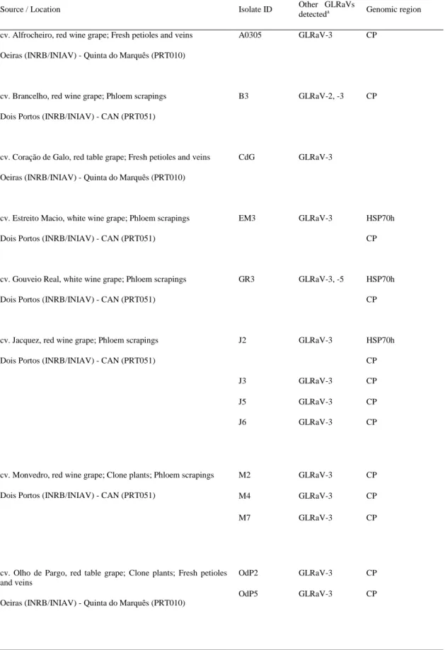
Table 2.1 Details of Grapevine leafroll-associated virus 1 isolates obtained in this study

Source / Location

Isolate ID

Other GLRaVs

detected

a

Genomic region

cv. Alfrocheiro, red wine grape; Fresh petioles and veins

Oeiras (INRB/INIAV) - Quinta do Marquês (PRT010)

A0305

GLRaV-3

CP

cv. Brancelho, red wine grape; Phloem scrapings

Dois Portos (INRB/INIAV) - CAN (PRT051)

B3

GLRaV-2, -3

CP

cv. Coração de Galo, red table grape; Fresh petioles and veins

Oeiras (INRB/INIAV) - Quinta do Marquês (PRT010)

CdG

GLRaV-3

cv. Estreito Macio, white wine grape; Phloem scrapings

Dois Portos (INRB/INIAV) - CAN (PRT051)

EM3

GLRaV-3

HSP70h

CP

cv. Gouveio Real, white wine grape; Phloem scrapings

Dois Portos (INRB/INIAV) - CAN (PRT051)

GR3

GLRaV-3, -5

HSP70h

CP

cv. Jacquez, red wine grape; Phloem scrapings

Dois Portos (INRB/INIAV) - CAN (PRT051)

J2

GLRaV-3

HSP70h

CP

J3

GLRaV-3

CP

J5

GLRaV-3

CP

J6

GLRaV-3

CP

cv. Monvedro, red wine grape; Clone plants; Phloem scrapings

Dois Portos (INRB/INIAV) - CAN (PRT051)

M2

GLRaV-3

CP

M4

GLRaV-3

CP

M7

GLRaV-3

CP

cv. Olho de Pargo, red table grape; Clone plants; Fresh petioles

and veins

Oeiras (INRB/INIAV) - Quinta do Marquês (PRT010)

OdP2

GLRaV-3

CP

(43)

Source / Location

Isolate ID

Other GLRaVs

detected

a

Genomic region

cv. Pical-Polho, red wine grape; Phloem scrapings

Dois Portos (INRB/INIAV) - CAN (PRT051)

PP1

GLRaV-3

CP

cv. São Saúl, red wine grape; Phloem scrapings

Dois Portos (INRB/INIAV) - CAN (PRT051)

SS3

GLRaV-2, -3

HSP70h

CP

cv. Sousão Vinhos Verdes, red wine grape; Phloem scrapings

Dois Portos (INRB/INIAV) - CAN (PRT051)

SVV1

GLRaV-3

HSP70h

CP

cv. Tinta Lameira, red wine grape; Phloem scrapings

Dois Portos (INRB/INIAV) - CAN (PRT051)

TL3

GLRaV-3

HSP70h

CP

cv. Vinhão Douro, red wine grape; Clone plants; Phloem

scrapings

Dois Portos (INRB/INIAV) - CAN (PRT051)

VD4

GLRaV-3

CP

VD7

GLRaV-3

CP

a

Molecular detection with CP gene virus-specific primers after cDNA synthesis (Esteves et al., 2009a,

Esteves et al., 2009b, , Esteves et al., 2012, Teixeira Santos et al., 2009a).

2.2.2. RNA Extraction

Total RNA was extracted from samples with the E.Z.N.A.

TM

Plant RNA Mini

Kit (Omega Bio-Tek, USA) according to manufacturer’s Protocol II for difficult

samples with a few modifications as described by MacKenzie et al., 1997. Plant

material (0.25 g in the case of cortical scrapings and 0.7 g in the case of petioles and

veins) was macerated in a cooled sterile mortar with liquid nitrogen. The powder was

homogenised with RNA extraction buffer [4 M guanidine thiocyanate, 0.2 M sodium

acetate, pH 5.0, 25 mM EDTA, 2.5% (w/v) PVP-40 and 1% (v/v)

-mercaptoethanol

(added just before use)], which was added in a 1:10 or 1:3 proportion (w/v), when using

cortical scrapings or petioles/veins, respectively. The sample homogenate (circa 1.4 ml)

was transferred to a 1.5 ml microcentrifuge tube and mixed with 100 µl of 20% (w/v)

sodium lauryl sarkosyl. After incubation at 70 ºC for 10 min with sporadic agitation,

samples were centrifuged at 10,000 x g for 10 min at room temperature. From this point

on, manufacturer’s instructions were followed.

Imagem

Figure  1.1  Vitis  vinifera  red  wine  grape  variety  Monvedro with symptoms of leafroll disease
Figure 2.1 Schematic representation of the GLRaV-1 genome organization. Boxes represent the  10  ORFs:  MT-methyltransfrease;  HEL-helicase;  RdRp-RNA-dependent  RNA  polymerase;  p7-  7 kDa endoplasmatic reticulum associated hydrophobic protein; HSP70h-he
Table 2.1 Details of Grapevine leafroll-associated virus 1 isolates obtained in this study
Table 2.2 Gene specific primers for Grapevine leafroll-associated virus 1 used in this work
+7

Referências

Documentos relacionados

Figure 29 – Theoretical results for the FWM signal without absorption for three intensities of the input lasers using the complete solution (blue) and only the direct pathway

Foi utilizado o conceito Lean Canvas (Maurya, 2012) como modelo de negócios de forma a expor o projecto, e também as metodologias Lean para eliminar recursos e actividades

The probability of attending school four our group of interest in this region increased by 6.5 percentage points after the expansion of the Bolsa Família program in 2007 and

No Comité de Embaixadores foram adiantados os primeiro contornos de algumas propostas que poderiam começar a concretizar-se: o enquadramento sistemático na CPLP

ções às escolas para a implantação de Programas de Saú- de. Deve possuir conhecimentos referentes à Educação da Saúde e estar <Jpto a desenvolver uções

The objective of this study was to model, analyze and compare the structure of spatial dependence, as well as the spatial variability of the water depths applied by a

The nurses agreed with the universal access to technology, which should also consider the results of their use and the professional opinion based on what is best for the