Ovicidal activity of succinic acid isolated from sisal waste
(Agave sisalana) against gastrointestinal nematodes of goats
Atividade ovicida do ácido succínico isolado do resíduo de sisal (Agave sisalana) sobre nematoides gastrintestinais de caprinos
Nathália Silva de Souza Santos1 Jener David Gonçalves Santos2 Francianne Oliveira Santos1 Taiane Menezes Serra1 Hélimar Gonçalves de Lima1 Mariana Borges Botura2
Alexsandro Branco2 Maria José Moreira Batatinha1*
ISSNe 1678-4596
INTRODUCTION
Parasitic infections caused by gastrointestinal nematodes in goats remain as a global challenge (JABBAR et al., 2006; NABUKENYA et al., 2014). These infections are responsible
for a significant economic impact due to weight loss, reduced milk production and delayed growth
(ROEBER et al., 2013). Development of nematode resistance to drugs commercially available encouraged the search for products of plant origin (FERREIRA et al., 2013). Such products can provide potential alternatives to the use of synthetic nematicides because they degrade to non-toxic products and cause
fewer side effects to non-target organisms and within
the broader environment.
Succinic acid (SA) is a product of the metabolism of plants and micro-organisms (WANG
et al., 2011) and has shown biological activities, such
as anxiolytic (CHEN, 2003; VOLCHEGORSKII et al., 2015), and the induction for IL-8 production in
inflammatory processes (GRAHAM et. al., 2013).
CHUNGSAMARNYART & JANSAWAN (2001) reported the action of SA, isolated from the fruit of
Tamarindus indicus (tamarind), on engorged females
of Rhipicephalus (Boophilus) microplus. In this
sense, previous studies of short-chain organic acids revealed nematicide action (SANTOS et al., 2007; NGUYEN et al., 2013).
Agave sisalana (sisal) is of a great
economic interest because it is a source of fiber in
semi-arid areas. Brazil is the largest producer and 1Escola de Medicina Veterinária, Universidade Federal da Bahia (UFBA), 40170-110, Salvador, BA, Brasil. E-mail: mjmb@ufba.br. *Corresponding author.
2Universidade Estadual de Feira de Santana (UEFS), Feira de Santana, BA, Brasil.
ABSTRACT: This study was conducted to evaluate the in vitro anthelmintic activity of the succinic acid (SA) isolated from sisal waste against gastrointestinal nematodes of goats, using the egg hatching and larvae motility assays. In addition, potential cytotoxicity of SA on Vero cell
cultures was investigated by means of MTT (3-4,5-dimethylthiazol-2-yl, 2,5diphenyltetrazolium bromide) test. The SA induced a significant
inhibition of egg hatching (P<0.05) at all concentrations tested (60 to 250µg mL-1), and the concentrations to inhibit 50% (EC
50) and 90%
(EC90) values (mean ± standard deviation) were 90.3±2.8 and 130.6±3.5µg mL-1, respectively. The SA has not shown larvicidal activity. The
SA was less toxic to the Vero cells, with the mean percentage of cell viability equal to 85±6.2% at the concentration of 130µg mL-1. The results suggested that SA has potential anthelmintic effect; although, more research is needed to confirm its activity in vivo.
Key words: Agave sisalana, sisal waste, succinic acid, anthelmintic, goats.
RESUMO: O objetivo deste estudo foi avaliar a atividade anti-helmíntica in vitrodo ácido succínico (AS) isolado do resíduo de sisal sobre nematódeos gastrointestinais de caprinos, utilizando os ensaios de inibição da eclosão de ovos e motilidade larval. Além disso, a citotoxicidade do AS em culturas de células Vero foi investigada empregando-se o teste de MTT (brometo de 3-4,5-dimetiltiazol-2-ilo, brometo
de 2,5-difeniltetrazólio). O AS promoveu redução significativa no percentual de eclosão de ovos (P<0,05) em todas as concentrações testadas
(60 a 250μg mL-1). As médias (± desvio padrão) da CE
50 e CE90 (Concentração efetiva 50% e 90%) sobre ovos foram de 90.3±2.8 e 130±3.5μg
mL-1, respectivamente. O AS não apresentou atividade larvicida. O AS foi menos tóxico para as células Vero, com média do percentual de viabilidade celular igual a 85±6.2% na concentração de 130μg mL-1. Os resultados sugerem que o AS tem potencial efeito anti-helmíntico,
embora sejam necessários a realização de estudos in vivo para confirmar seu uso terapêutico.
Palavras-chave: Agave sisalana, resíduos de sisal, ácido succínico, anti-helmíntico, caprinos.
exporter of sisal fibers worldwide (IBGE, 2013), where 4% of the sisal leaves produce fiber and the remaining material (waste) is discarded (SHARMA
and VARSHNEY, 2012). Our research group has investigated the biological activity of different
extracts and fractions obtained from sisal waste
against nematodes of goats. In the context, the
flavonoid and saponin fractions from ethyl acetate extract showed ovicidal and larvicidal effects,
respectively (SILVEIRA et al., 2012; BOTURA et. al., 2013; SANTOS et al., 2015). In a continuous
study, we described the evaluation of the in vitro
anthelmintic and cytotoxicity activity of the SA isolated from the same extract on eggs and larval stage (L3) of nematodes of goats, and on African green monkey´s kidney cell line (Vero).
MATERIALS AND METHODS
Materials
Ethyl acetate, ethanol and methanol
(analytical grade) from VETEC were used. Analytical thin-layer chromatography (TLC) was performed on commercial aluminum plates coated with silica gel
(0.025mm) (Merck, Darmstadt, Germany). Spots
were visualized by spraying with 1M H2SO4 and
heating to 100°C. Silica gel (Kielselgel 60, 70-230
mesh) was used for open-column chromatography.
Carbon-13 Nuclear Magnetic Resonance (13C NMR)
spectra were obtained using a Varian Gemini 300
equipment.
Plant material
The Agave sisalana waste was collected
after the process of decortication of the leaves, on a sisal farm located in Valente, in the state of Bahia (S 11°24’53.4”), in May 2012.
Obtaining of succinic acid
The obtaining of succinic acid from
sisal waste was performed using the methodology described by SANTOS et al. (2015). Briefly, the ethyl acetate extract was subjected to open-column chromatography packed with silica gel to yield the succinic acid in fractions eluted with EtOAc (100%) and EtOAc/MeOH (8:2). Crystals obtained were
identified with 1H NMR (Proton Nuclear Magnetic
Resonance) and 13C NMR (Carbon Nuclear Magnetic
Resonance).
Anthelmintic activity
All early-life stages of trichostrongylids
used in this study were obtained from goats
naturally infected and kept at the School of Veterinary Medicine, Federal University of
Bahia. The generic identification of the nematode population was determined according to UENO
and GONÇALVES (1998). The feces cultures of
those animals indicated the presence of 86% of
Haemonchus spp., 10% Oesophagostomum spp.
and 4% Trichostrongylus spp.
Egg hatching assay (EHA)
Eggs were isolated from feces of goats naturally infected with gastrointestinal
nematodes as described by HUBERT and
KERBOEUF (1992). The bioassay was performed following COLES et al. (1992). Egg suspension was distributed in 96-well plates (100 fresh
eggs 100µL-1/well) and mixed with the same
volume of the SA dissolved in distilled water at
different concentrations (60; 90; 130; 180 and 250µg mL-1). Negative and positive controls
were distilled water and thiabendazole (25µg
mL-1), respectively. After a 48-hour incubation at
25°C, egg hatching was blocked by the addition
of Lugol’s iodine solution. Number of eggs
and larvae L1 per well was counted. Inhibition percentage of egg hatching was determined using the following ratio: [Number of eggs/(number of
eggs + number of L1 larvae)] x 100.
Larval motility assay
For the larval motility assay, a suspension of infective larvae (L3) was distributed in 24-well
plates (50 larvae/100µL/well) and added with the
succinic acid (1,000µg mL-1) in the same volume
(100µL) (FERREIRA et al., 2013). Both a negative
control with distilled water and a positive control
with levamisole (250µg mL-1) were also prepared.
The results were expressed as the percentage of
mobile larvae.
Cytotoxicity assay
The commercial Vero cell line (Vero - ATCC® CCL-81TM) was obtained from African
Green Monkey´s (Cercopithecus aethiops)
kidney and maintained in RPMI (Roswell Park
Memorial Institute) medium supplemented
with penicillin G (100UI mL-1), streptomycin
(100mg/mL) and 10% fetal equine serum. Cells were cultured at 37ºC in a humidified 5%
CO2 incubator.
In the moment of the experiments,
cells were placed in 96-well plates at a density
prior to treatment. Thereafter, cells were treated with succinic acid diluted in RPMI medium (60;
90; 130; 180 and 250µg mL-1). Negative control
group was treated only with RPMI. Plates were
kept in an incubator for an additional 24h and then
the assessment of cell viability was performed by
means of the 3-(4,5-dimethylthiazol-2-yl)-2,5-diphenyltetrazolium bromide (MTT) test according to HANSEN et al. (1989).
After the treatment period, the
culture medium was removed and MTT (1mg
mL-1, 100µL) was equally added to each well.
After three hours of incubation, lysis buffer
containing 20% sodium dodecyl sulfate (SDS) and 50% dimethylformamide (DMF) was added,
maintaining the plates incubated for another
12h. Optical absorbance was measured using a wavelength (405-600nm) plate reader. Results were expressed as percentages of viability of
treated groups related to the control group.
Statistical analysis
Results are expressed as mean ±
standard deviation (S.D.). The data were analyzed using an ANOVA and were compared using Tukey´s test (5%). For each biological assay, three independent experiments were performed, with five repetitions for each concentration and
controls. The EC50 and EC90 for the ovicidal tests
were individually calculated for each experiment
using a non-linear regression analysis All
statistical analyses were performed with the
GraphPrism version 5.0.
RESULTS AND DISCUSSION
The succinic acid (SA) was isolated from the ethyl acetate extract from sisal waste in accordance with SANTOS et al. (2015) and its chemical characterization was made using NMR
data. The 13C NMR (Figure 1) showed only three
signals attributed to the two methylenic carbons and two carbons of the carboxylic acid.
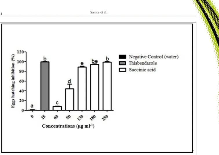
The SA inhibited egg hatching in a concentration-dependent manner. The mean percentage inhibition of egg hatching ranged from
15.2 to 97.2% (Figure 2). The mean and standard
deviations of EC50 and EC90 of the three experiments
were 90.3+2.8 and 130.6+3.5µg mL-1, respectively.
In the larval motility assay, the mean percentage of
mobile larvae observed in the group treated with SA (82±7.7%) did not differ statistically (P>0.05) from the negative control (98.2±1.5%).
The SA was effective in preventing
the development of the eggs. According to VERCRUYSSE et al. (2001), a synthetic product
is effective when it promotes anthelmintic activity
above 90%. The mean value reported for the EC90
(130.6μg mL-1) was, respectively, equal and higher to
those reported by BOTURA et al. (2013) for the ethyl
acetate extract (130μg mL-1) and for flavonoid fraction
(70µg mL-1) obtained in this same sisal residue
extract. The authors attribute this ovicidal activity
to the presence of homoisoflavonoids detected in the chemical analysis of flavonoid fraction (BOTURA et
al., 2013). Bearing in mind that the succinic acid used
in this study was also obtained from the ethyl acetate
extract from the sisal liquid residue, the results suggested a possible synergistic or additive action of
the SA with these flavonoids.
Ovicidal activities of isolated organic acids from plants have been reported. Research results performed by SANTOS et al. (2007) have demonstrated the action of oleanolic acid obtained from the Rheedia gardneriana fruit on the egg hatching of Meloidogyne incognita, yet with low
percentage of hatching inhibition (63.5%) after ten
days of treatment using the concentration of 800µg
mL-1, which was six times greater than the mean of
EC90 (130.6µg mL-1) reported in this study.
The succinic acid had no effect on larval motility at the concentration used. BOTURA et al. (2013) reported moderate larvicidal activity of the
saponin fraction from the sisal waste (efficacy of 64.1%). These results suggested the participation of
one more chemical component in the anthelmintic activity of A. sisalana.
The cytotoxicity effects of SA on Vero
cells were reported in figure 3. The treatment with SA
(90 to 250µg mL-1) induced a significant reduction
in the percentage of cell viability compared to the
negative control (P<0.05), with mean of percentage from 90±5.13 to 78.4±5.1%, respectively. According
to the ISO 10993-5 (2009), a substance is considered
toxic (MTT test) when it showed percentages of over 30% of non-viable cells. Thus, no sign of cytotoxicity was observed after the exposition with SA. This result suggested low potential for toxicity of succinic acid at a concentration in which it has pronounced
anthelmintic effect.
CONCLUSION
The succinic acid from sisal waste
(Agave sisalana) showed an ovicidal activity
against gastrointestinal nematodes of goats and low
potential for toxicity on Vero cell cultures, suggesting the participation and promising potential of this constituent on the anthelmintic activity reported
for Agave sisalana; although, it did not show
effectiveness on the larvae of these parasites. Further research, including in vivo studies, are required in order to assess the antiparasitic potential of this acid.
Figure 2 - Inhibition percentage (mean ± S.D) of egg hatching of gastrointestinal nematode eggs of goats treated with succinic acid (SA)
ACKNOWLEDGMENTS
We would like to thank the Conselho Nacional de Desenvolvimento Científico e Tecnológico (CNPq), Universidade
Estadual de Feira de Santana (UEFS) and Universidade Federal
da Bahia (UFBA) for the financial support. We also thank teacher
Abilio Borghi for the grammar review of the manuscript.
BIOETHICS AND BIOSSECURITY COMMITTEE APPROVAL
We, authors of the article entitled “Ovicidal
activity of succinic acid isolated from sisal waste (Agave sisalana) against gastrointestinal nematodes of goats” declare, for all due purposes, that the project that gave rise to the present data t has not been submitted for evaluation to the Ethics Committee of the Universidade Federal da
Bahia / Escola de Medicina Veterinária e Zootecnia, but we are aware of the content of the Brazilian resolutions of the
Conselho Nacional de Controle de Experimentação Animal
(CONCEA) <http://www.mct.gov.br/index.php/content/
view/310553.html> if it involves animals. Thus, the authors
assume full responsibility for the data presented and are available for possible questions, should they be required by the competent authorities.
REFERENCES
BOTURA, M.B. et al. In vitro ovicidal and larvicidal activity of Agave sisalana Perr. (sisal) on gastrointestinal nematodes of goats. Veterinary Parasitology, v.192, p.211-217, 2013. Available from: <http://www.sciencedirect.com/science/article/pii/ S0304401712005596>. Accessed: Mar. 10, 2014. doi: 10.1016/j. vetpar.2012.10.012.
CHEN, S.W. Anxiolytic-like effect of succinic acid in mice. Life Sciences, v.73, p.3257-3264, 2003. Available from: <http://www. sciencedirect.com/science/article/pii/S0024320503008105>. Accessed: Mar. 10, 2014. doi: 10.1016/j.lfs.2003.06.017.
CHUNGSAMARNYART, N.; JANSAWAN, W. Effect of
Tamarindus indicus L. against the Boophilus microplus. Kasetsart Journal (Natural Science), v.35, p.34-39, 2001. Available from:
<http://kasetsartjournal.ku.ac.th/abstractShow.aspx?param=YXJ0
aWNsZUlEPTEwOTl8bWVkaWFJRD05MjU=>. Accessed: Apr.
28, 2014.
COLES, G.C. et al. World association for the advancement of veterinary parasitology (WAAVP) methods for the detection of anthelmintic resistance in nematodes of veterinary importance. Veterinary Parasitology, v.44, p.35-44, 1992. Available from: <http://www.sciencedirect.com/science/
article/pii/030440179290141U>. Accessed: Apr. 12, 2014. doi: 10.1016/0304-4017(92)90-141-U.
FERREIRA, L.E. et al. In vitro anthelmintic activity of aqueous leaf extract of Annona muricata L. (Annonaceae) against Haemonchus contortus from sheep. Experimental Parasitology, v.134, p.327-332, 2013. Available from: <http://www.sciencedirect.com/ science/article/pii/S0014489413001094>. Accessed: June 23, 2014. doi: 10.1016/j.exppara.2013.03.032.
GRAHAM, L.S. et al. Effects of succinic acid and other microbial fermentation products on HIV expression in macrophages. BioResearch Open Access, v.2, p.385-391, 2013. Available from: <http://online. liebertpub.com/doi/pdfplus/10.1089/biores.2013.0013>. Accessed: Oct. 27, 2014. doi: 10.1089/biores.2013.0013.
HANSEN, M.B. et al. Re-examination, and further development
of a precise and rapid dye method for measuring cell growth/
cell kill. Journal Immunological Methods, v.119, p.203-210,
1989. Available from: <http://www.sciencedirect.com/science/ article/pii/0022175989903979>. Accessed: Aug. 13, 2014. doi:
10.1016/0022-1759(89)90397-9.
HUBERT, J.; KERBOEUF, D. A micro larval development assay for the detection of anthelmintic resistance in sheep nematodes.
Veterinary Record, v.130, p.442-446, 1992. Available from: <http://veterinaryrecord.bmj.com/content/130/20/442.abstract>. Accessed: Oct. 23, 2014. doi: 10.1136/vr.130.20.442.
INSTITUTO BRASILEIRO DE GEOGRAFIA E ESTATÍSTICA.
Produção da pecuária municipal. 2013. Online. Available
from: <http://www.ibge.gov.br/˂3ftp://ftp.ibge.gov.br/Producao_ Pecuaria/Producao_da_Pecuaria_Municipal/2013/ppm2013.pdf>.
Accessed: June 08, 2015.
INTERNATIONAL STANDARD, ISO 10993-5. Biological evaluation of medical devices - part 5: Tests for in vitro
cytotoxicity. Berlin, Germany, 2009.
JABBAR, A. et al. Anthelmintic resistance: the state of play revisited. Life Sciences, v.79, p.2413-2431, 2006. Available from: <http://www.sciencedirect.com/science/article/pii/ S0024320506006436>. Accessed: June 01, 2015. doi: 10.1016/j. lfs.2006.08.010.
NABUKENYA, I. et al. Anthelmintic resistance in gastrointestinal nematodes in goats and evaluation of FAMACHA diagnostic marker in Uganda. Veterinary Parasitology, v.205, p.666-675, 2014. Available from: <http://www.sciencedirect.com/science/ article/pii/S0304401714004063>. Accessed: Nov. 24, 2014. doi: 10.1016/j.vetpar.2014.07.019.
NGUYEN, D.M.C. et al. Nematicidal activity of
3,4-dihydroxybenzoic acid purified from Terminalia nigrovenulosa
bark against Meloidogyne incognita. Microbial Pathogenesis, v.59-60, p.52-59, 2013. Available from: <http://www.sciencedirect.
com/science/article/pii/S088240101300051X>. Accessed: Apr. 30,
2015. doi: 10.1016/j.micpath.2013.04.005.
ROEBER, F. et al. Impact of gastrointestinal parasitic nematodes of sheep, and the role of advanced molecular tools for exploring epidemiology and drug resistance – an Australian perspective. Parasites & Vectors, v.6, p.1-13, 2013. Available from: <http://parasitesandvectors.biomedcentral.com/
articles/10.1186/1756-3305-6-153>. Accessed: Apr. 2015. doi:
10.1186/1756-3305-6-153.
SANTOS, M.H. et al. Efeito de constituintes químicos isolados da casca do fruto de Rheedia gardneriana sobre a eclosão de juvenis de Meloidogyne incognita Raça 3. Latin American Journal Pharmacy, v.26, p.711-714, 2007. Available from: <http://
www.latamjpharm.org/resumenes/26/5/LAJOP_26_5_1_10.pdf>.
Accessed: Feb. 14, 2015.
SANTOS, J.D.G. et al. Chemicals from Agave sisalana Biomass:
Isolation and Identification. International Journal of Molecular
Science, v.16, n.4, p.8761-8771, 2015. Available from: <http://
www.mdpi.com/1422-0067/16/4/8761/htm>. Accessed: Nov. 27,
2014. doi: 10.3390/ijms16048761.
SHARMA, S.; VARSHNEY, V.K. Chemical analysis of Agave sisalana juice for its possible utilization. Acta Chimica & Pharmaceutica Indica, v.2, p. 60-66, 2012, Available from:
<http://www.tsijournals.com/chemical-sciences/chemical-analysis-of-agave-sisalana-juice-for-its-possible-utilization.pdf>.
Accessed: Mar. 14, 2015.
SILVEIRA, R.S. et al. Action of sisal (Agave sisalana, Perrine) extract in the in vitro development of sheep and goat gastrointestinal nematodes. Experimental Parasitology, v.131, p.162-168, 2012.
Available from: <http://www.sciencedirect.com/science/article/pii/
S0014489412001129>. Accessed: Feb. 13, 2015. doi: 10.1016/j.
exppara.2012.03.018.
UENO, H.; GONÇALVES, P.C. Manual para diagnóstico das helmintoses de ruminantes. Tokyo: Japan International Cooperation Agency, 1998. 147p.
VERCRUYSSE, J. et al. International harmonisation of
anthelmintic efficacy guidelines. Veterinary Parasitology, v.96,
p.171-193, 2001. Available from: <http://www.sciencedirect.com/ science/article/pii/S030440170000443X>. Accessed: Feb. 12, 2015. doi: 10.1016/S0304-4017(00)00443-X.
VOLCHEGORSKII, I.A. et al. Comparative analysis of the anxiolytic effects of 3-Hydroxypyridine and succinic acid derivatives. Bulletin of Experimental Biology and Medicine, v.158, p.756-761, 2015. Available from: <http://link.springer.com/
article/10.1007/s10517-015-2855-3>. Accessed: Mar. 10, 2015.
doi: 10.1007/s10517-015-2855-3.