This is an Open Access article distributed under the terms of the Creative Commons Attribution License, which permits unrestricted use, distribution, and reproduction in any medium, provided the original work is properly cited.
Antiproliferation effects and antioxidant activity of two new
Lactobacillus
strains
Efeitos de antiproliferação e atividade antioxidante de duas cepas novas de Lactobacillus
Abstract
The microorganisms most commonly used as probiotics are lactic acid bacteria, especially those of the genus Lactobacillus. In the present study, two Lactobacillus strains, L. pentosus ITA23 and L. acidipiscis ITA44, previously isolated from mulberry silage, were characterized for their antiproliferative and antioxidant activities. The antiproliferative effects of the strains were investigated using the MTT assay with breast cancer (MDA-MB-231), liver cancer (HepG2) and normal liver (Chang) cell lines. The strains were tested for their antioxidant activity using the FRAP and ABTS methods. The results showed that the two Lactobacillus strains had good antiproliferative effects against both cancer cell lines tested, while their effects on the normal cells were weak. Based on the results of the antioxidant tests, the intact cells and cell-free extracts of the two Lactobacillus strains showed more than 135 and less than 50 µg trolox/ml of antioxidant activity, respectively. Lactobacillus pentosus ITA23 and L. acidipiscis ITA44 can be considered as potential probiotic candidates for humans because of their antioxidant activity and antiproliferation effects against cancer cells.
Keywords: Probiotic; Lactobacillus; Antiproliferation; Antioxidant.
Resumo
Os micro-organismos mais comumente usados como probióticos são as bactérias lácticas, especialmente aquelas do genus Lactobacillus. No presente estudo, duas cepas de Lactobacillus, L. pentosus ITA23 e L. acidipiscis ITA44, previamente isoladas da silagem de amora, foram caracterizados por suas atividades antiproliferativas e antioxidantes. Os efeitos antiproliferativos das cepas foram investigados usando-se o ensaio MTT, com linhas de células cancerígenas de mama (MDA-MB-231) e do fígado (HepG2), e com células normais do fígado (Chang). As atividades antioxidantes das cepas foram testadas utilizando-se os métodos de FRAP e ABTS. Os resultados demonstraram que as duas cepas de Lactobacillus tinham bons efeitos antiproliferativos contra as duas linhas de células cancerígenas, enquanto seus efeitos nas células normais eram fracos. Baseando-se nos resultados dos testes antioxidantes, as células intactas e os extratos livres de células das duas cepas de Lactobacillus demonstraram mais que 135 e menos que 50 µg trolox/mL de atividade antioxidante, respectivamente. Lactobacillus pentosus ITA23 e Lactobacillus acidipiscis ITA44 podem ser considerados como potenciais candidatos probióticos para humanos, devido à sua atividade antioxidante e aos efeitos antiproliferativos contra células cancerígenas.
Palavras-chave: Probiótico; Lactobacillus; Antiproliferação; Antioxidante.
Parisa Shokryazdan1,2, Mohammad Faseleh Jahromi1,2, Fatemeh Bashokouh3, Zulkifli Idrus1, Juan Boo Liang1*
1 Universiti Putra Malaysia, Institute of Tropical Agriculture, Serdang/Selangor - Malaysia
2 Agriculture Biotechnology Research Institute of Iran (ABRII), East and North-East Branch, Mashhad - Iran 3 Universiti Teknologi MARA, Faculty of Medicine, Shah Alam/Selangor - Malaysia
*Corresponding Author
Juan Boo Liang, Universiti Putra Malaysia, Institute of Tropical Agriculture, 43400 UPM, Serdang/Selangor - Malaysia, e-mail: [email protected]
Cite as: Antiproliferation effects and antioxidant activity of two new Lactobacillus strains. Braz. J. Food Technol., v. 21, e2016064, 2018.
1 Introduction
Probiotic is defined as “[...] live microorganisms which, when administered in adequate amounts, confer a health benefit on the host [...]” (FAO, 2001, p. 50). Lactic acid bacteria, including the genus Lactobacillus, are the most commonly used probiotics in the food industry because of their beneficial functional effects and their reputation as “generally recognized as safe” (GRAS) and environmentally friendly bacteria (SAARELA et al., 2000; SALMINEN et al., 1998; KLAENHAMMER; KULLEN, 1999). Probiotics have become highly recognized as supplements for human consumption because of their beneficial effects on the health and well-being (OOI; LIONG, 2010). For example, interference with pathogens and the prevention of infections, immune system stimulation and immunomodulation, anti-carcinogenic and antioxidant activities, reduction of gastrointestinal disorders such as diarrhoea, constipation and the irritable bowel syndrome, alleviation of allergic and lactose intolerance symptoms, reduction in serum cholesterol, blood pressure and risk of gestational diabetes, and the production of beneficial compounds, such as vitamins, short-chain fatty acids (SCFAs) and conjugated linoleic acid are some of their important beneficial effects (LOLLO et al., 2013, 2015; SHOKRYAZDAN et al., 2017). Among these effects, the antiproliferative or cytotoxic effect of probiotic strains against cancer cells is a very important and relatively recent aspect. This probiotic property is important because cancer is considered as the major worldwide cause of morbidity (WHO, 2009). Another recently discovered important functional property of a probiotic strain is its antioxidant activity. Many reactive oxygen species (ROS) are continuously formed in food systems and in the human body (AHOTUPA et al., 1996), and the antioxidant activity of probiotic strains could be a helpful option to encounter the adverse effects of ROS. Therefore, probiotic strains with strong antiproliferative effects and antioxidant activity can be considered as important agents in the food industry for producing functional foods.
Although many different probiotics have already entered the food market, novel strains with more functional probiotic properties than the existing ones are the object of new research due to the growing demand for “healthy” foods in the food industry. In the present study, two previously isolated and identified Lactobacillus strains from the author’s laboratory were used to investigate their antioxidant activity, and also their antiproliferation effects against two different carcinoma cell lines and one normal cell line as the control, to investigate the selective killing effects of the two Lactobacillus strains.
2 Materials and methods
2.1 Lactobacillus strains and sample preparation
Two Lactobacillus strains, namely L. pentosus ITA23 and L. acidipiscis ITA44, previously isolated and identified (SHOKRYAZDAN et al., 2015) from locally prepared mulberry (Morus alba) silage, were used as test strains. De Man-Rogosa-Sharpe (MRS) broth (Merck, Germany) was used as the growth medium for the Lactobacillus strains. The strains were grown at 37 °C in anaerobic jars (Oxoid, UK) containing gaspack (AnaeroGen, Oxoid, UK). Each Lactobacillus strain was sub-cultured three times before the experiment, and the overnight culture (final concentration of 1×10⁷ CFU/ml) of
each strain was centrifuged at 4000 × g for 5 min at 4 °C. The supernatants were collected and filter sterilized through a 0.22 µm pore-size filter (Pall, USA). Filter sterilized supernatants were used as treatments in the antiproliferation assay. The cell pellets were washed three times with deionized water, and freeze dried for use in the antioxidant study. Intact cells [10 mg of each freeze-dried cell sample, re-suspended in 1 ml of PBS (pH 7.2)] and intracellular cell-free extracts were used for the investigation of the antioxidant activity of the two Lactobacillus strains. To prepare the intracellular cell-free extract, 10 mg of each freeze-dried cell sample was re-suspended in 1 ml of deionized water, sonicated on ice for 5 min, centrifuged at 7000 × g for 10 min at 4 °C, and the supernatants collected.
2.2 Antiproliferation assay
2.2.1 Cell culture
Breast cancer (MDA-MB-231), liver cancer (HepG2) and normal liver (Chang) cell lines were obtained from the American Type Culture Collection (ATCC). The cell lines were routinely grown to 80% confluent in Roswell Park Memorial Institute medium (RPMI) supplemented with 10% (v/v) foetal bovine serum (FBS, inactivated at 56 °C for 30 min) and 1% (v/v) penicillin–streptomycin (10,000 IU/ml and 10,000 µg/ml). The cells were incubated in a CO2 incubator at 37 °C in 5% CO2 for maintenance and during the experiment.
2.2.2 MTT assay
For the MTT assay, filter sterilized supernatants from overnight cultures of each Lactobacillus strain were used as the treatment. All the chemicals used in the experiment were from Sigma, USA. The MTT assay followed the method of Haza et al. (2004) with some modifications. Each cell line was placed in a 100 µl/well in 96 well plates (1×10⁶ cells/plate). After incubation at 37 °C for 24 h,
cells. The plates were then incubated for 24, 48 and 72 h. After incubation, 20 µl of MTT (0.5 mg/ml in phosphate buffer saline) was added to each well and the plates incubated for 3 h in the dark. The mixture was then discarded from the wells and 100 µl DMSO (dimethyl sulfoxide) added to the monolayers of the cells in order to solubilize the formazan precipitates. After 10 min of incubation at room temperature, the absorbance was measured at 570 nm using a micro-plate reader.
The percentage of cell viability was calculated using Equation 1:
( ) (= 570 − 570 )× 570
Cell viability % A sample A blank 100 / A control (1)
Where, A570, absorbance at 570 nm; sample, monolayer of each cell line plus different concentrations of treatment in RPMI medium; blank, different concentrations of treatment in RPMI medium; control, monolayer of each cell line plus RPMI medium.
2.2.3 Antioxidant assay
2.2.3.1 FRAP
The ferric reducing ability of plasma “FRAP” assay devised by Benzie and Strain (1996) with some minor modifications was used to investigate the total antioxidant activity of the Lactobacillus strains. An amount of 200 µl of FRAP reagent [300 mM acetate buffer (pH 3.6, containing 6.4 ml 2 M sodium acetate and 93.6 ml 2 M acetic acid), 10 mM 2,4,6-tri (2-pyridyl)-1,3,5-triazine (TPTZ) in 40 mM HCl, and 20 mM ferric chloride in a ratio of 10:1:1 (v:v:v)] was added to 50 µl of intact cells or intracellular cell-free extract of each Lactobacillus strain. After incubation of the mixture for 30 min at room temperature in the dark, the absorbance was measured at 593 nm using a spectrophotometer (Barnstead International, USA).
2.2.3.2 ABTS
The ABTS radical cation decolourization assay described by Tsai et al. (2011), with some modifications, was used to determine the free radical scavenging activity of the Lactobacillus strains. The ABTS radical cation (ABTS°+) was prepared by reacting 7.4 mM ABTS
and 2.6 mM potassium persulfate in equal quantities (1:1, v:v) for 12 h at room temperature in the dark, and diluting the mixture in methanol to obtain an absorbance of 1.1 ± 02 at 734 nm using a spectrophotometer (Barnstead International, USA). An amount of 200 µl of ABTS°+ solution
was added to 50 µl of intact cells or intracellular cell-free extract of each Lactobacillus strain. After incubation at room temperature for 5 min in the dark, the absorbance was measured at 734 nm.
For both the FRAP and ABTS methods, the antioxidant activity was expressed as µg Trolox/ml sample solution
(Trolox Equivalent Antioxidant Capacity), obtained using a standard curve prepared with Trolox.
2.2.3.3 Statistical analysis
The assays were carried out in two independent experiments, each in triplicate. The data were analysed as a completely randomized design (CRD) using the general linear model (GLM) procedure of SAS 9.2 (STATSOFT, 2008), with a model that included treatment effects and experimental error. All multiple comparisons amongst the means were carried out using Duncan’s new multiple range test (α = 0.05).
3 Results and discussion
In the present study, two Lactobacillus strains were investigated for their antiproliferative (cytotoxic) effects against two cancer cell lines, and the antioxidant activities of the strains were also investigated, both using in vitro methods.
Since the selective induction of cancer cell death is a crucial factor for every anticancer agent (DAMIA; BROGGINI, 2004), in this study, the Chang cell line was used as a non-cancerous cell line to determine whether the two Lactobacillus strains were able to selectively kill the cancer cells without having adverse effects on the normal cells. The MTT assay was used to investigate the antiproliferation effects of two Lactobacillus strains. In the MTT assay, viable cells are able to reduce MTT [3-(4,5-Dimethylthiazol-2-yl)-2,5-diphenyltetrazolium bromide, a yellow tetrazole] to a purple formazan product. After dissolution of the formazan product with a dissolution solution, the absorbance was recorded at an appropriate wavelength.
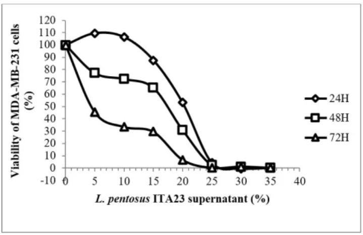
Figures 1, 2 and 3 show the results of the MTT assay for L. pentosus ITA23 against MDA-MB-231, HepG2 and the Chang cell lines, respectively. The supernatant of this strain exhibited good cytotoxic effects against both cancer
Figure 1. Cytotoxic effects of the L. pentosus ITA23 supernatants
cell lines and was able to greatly inhibit their proliferation. At the three incubation times (24, 48 and 72 h of incubation), 27% of L. pentosus ITA23 supernatant was enough to completely kill the MDA-MB-231 cells (Figure 1). However, for the HepG2 cell line, the amount required was 30% at 24h and 25% at 48 and 72 h of incubation (Figure 2). On the other hand, the supernatant of L. pentosus ITA23 showed a lower antiproliferative effect against the normal cell line (Chang) in comparison with the cancer cell lines, since even the highest concentration tested (70%) for the longest incubation time (72 h), did not kill all the normal cells, only 73% of them (Figure 3).
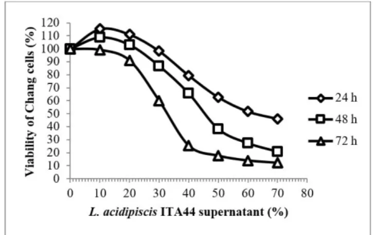
The results of the MTT assay for L. acidipiscis ITA44 against the MDA-MB-231, HepG2 and Chang cell lines are presented in Figures 4, 5 and 6, respectively. L. acidipiscis ITA44, like L. pentosus ITA23, exhibited strong cytotoxic effects against both cancer cell lines. For both the MDA-MB-231 and HepG2 cell lines, 20% of L. acidipiscis ITA44 supernatant revealed 0% viability of the cancer cells after 24 and 48 h of incubation, and after 72 h of incubation, just 16% of the supernatant was enough to kill all the cancer cells. However,
for the normal cells, the antiproliferative effect of the L. acidipiscis ITA44 supernatant was weaker than its effect on the cancer cells. Like the L. pentosus ITA23 supernatant, the L. acidipiscis ITA44 supernatant did not kill all the normal cells even at the highest concentration (70%) and the longest incubation time (72 h).
Figure 2. Cytotoxic effects of the L. pentosus ITA23 supernatants
against the HepG2 cell line. The results are mean values from two independent experiments, each with three replicates.
Figure 3. Cytotoxic effects of the L. pentosus ITA23 supernatants
against the Chang cell line. The results are mean values from two independent experiments, each with three replicates.
Figure 4. Cytotoxic effects of the L. acidipiscis ITA44 supernatants
against the MDA-MB-231 cell line. The results are mean values from two independent experiments, each with three replicates.
Figure 5. Cytotoxic effects of the L. acidipiscis ITA44 supernatants
against the HepG2 cell line. The results are mean values from two independent experiments, each with three replicates.
Figure 6. Cytotoxic effects of the L. acidipiscis ITA44 supernatants
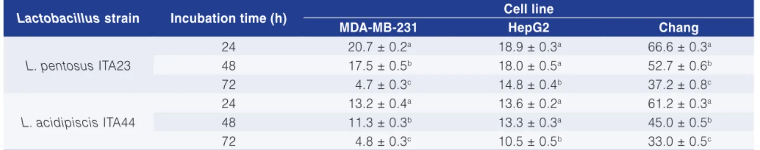
Table 1 shows the results of the MTT assay in the form of the IC50, which is the concentration of the Lactobacillus supernatant that causes a 50% reduction in cell viability. A lower value for the IC50 means greater efficacy of the Lactobacillus supernatant to inhibit proliferation of the cancer cells and vice versa. With regards to the IC50 values, it is clear that the supernatants of both L. pentosus ITA23 and L. acidipiscis ITA44 exhibited stronger antiproliferation effects against the cancer cell lines than against the normal cell line, because the IC50s of the Lactobacillus supernatants on the normal cells were about three to eight times greater than those on the cancer cells. This means that both L. pentosus ITA23 and L. acidipiscis ITA44 were able to selectively kill the cancer cells.
The results of the MTT assay in the form of IC50 (Table 1) also showed that, as expected, longer incubation periods caused more inhibition of proliferation than shorter incubation periods for all the cell lines tested. For example, in the case of L. pentosus ITA23, the IC50s of the supernatants on the MDA-MB-231 and Chang cells were 20.7 and 66.8% for 24 h, 17.5 and 52.7% for 48 h, and 4.7 and 37.2% for 72 h of incubation, respectively, showing that, for each cell line, a longer incubation time led to significantly (p < 0.01) smaller IC50 values than the shorter incubation times. For the HepG2 cell line, an incubation period of 72 h also showed greater (p < 0.01) effectiveness on the inhibition of proliferation of the cells than 24 and 48 h, since the corresponding IC50s were 18.9, 18.0 and 14.8% after 24, 48 and 72 h of incubation, respectively. In the case of L. acidipiscis ITA44, the IC50s values of the supernatants on MDA-MB-231 cells were significantly (p < 0.01) different from each other (13.2, 11.3 and 4.8% for 24, 48 and 72 h of incubation, respectively). The same trend of significant (p < 0.01) differences was observed for the Chang cells (61.2, 45.0 and 33.0% for 24, 48 and 72 h of incubation, respectively). The IC50s values for the supernatants on the HepG2 after 72 h of incubation (10.5%) were also significantly (p < 0.01) higher than those after 24 and 48 h (13.6 and 13.3%, respectively).
Antiproliferative effects of bacterial strains on different cancer cell lines have been reported before
(HAZA et al., 2004; TUO et al., 2010; BIFFI et al., 1997; THIRABUNYANON et al., 2009; CHOI et al., 2006). For example, Choi et al. (2006) investigated seven strains of L. acidophilus, L. casei, L. brevis and L. rhamnosus for their antiproliferative effects on HeLa, MCF7, U-87, HepG2, U2OS and PANC-1 cancer cell lines. They used the heat-killed cells as the treatments in their experiments and found that, of all the strains tested, L. acidophilus 606 (with 21-28% survival of cancer cells as compared to the control) and L. casei ATCC 393 (with 15-20% survival of cancer cells as compared to the control) exhibited the greatest inhibition effects on most of the cancer cells tested. In another study, Tuo et al. (2010) tested heat-killed bacterial cells, genomic DNA, and the cell walls of seven wild strains of L. rhamnosus and L. paracasei, isolated from traditional fermented foods, for their cytotoxic effects on the HT-29 cancer cell line. All the three fractions of the seven Lactobacillus strains exhibited direct antiproliferative activities against HT-29 cells. Of all the strains tested in their study, L. coryniformis ssp. torquens T3L exerted marked antiproliferative activities against the HT-29 cell line, with maximum inhibition rates of 30, 44.9, and 35.9% by its heat-killed bacterial cells, cell wall and genomic DNA, respectively. Haghshenas et al. (2014) also used some metabolites secreted by Enterococcus strains in the MTT assay against different cancer (MCF-7, HeLa, HT29, and AGS) and normal (HUVEC) human cell lines. They found that the metabolites tested had antiproliferative affects against cancer cells, but did not exhibit cell toxicity on the normal cell line. In another study, Desrouillères et al. (2015) used specific probiotic fermented milk components and cell walls extracted from a biomass containing L. acidophilus CL1285, L. casei LBC80R, and L. rhamnosus CLR2 on cancer-induced rats. Based on the results obtained, they concluded that the probiotic bacteria and their metabolites released during the fermentation process could prevent colorectal carcinogenesis. Saxami et al. (2016) also reported a significant reduction in the proliferation of cancer cells by using both heated and non-heated conditioned media of L. pentosus B281 and L. plantarum B282. The results for the antiproliferative activity of the two Lactobacillus
Table 1. IC50s (%) of the Lactobacillus strain supernatants on the different cell lines.
Lactobacillus strain Incubation time (h) Cell line
MDA-MB-231 HepG2 Chang
L. pentosus ITA23
24 20.7 ± 0.2a 18.9 ± 0.3a 66.6 ± 0.3a
48 17.5 ± 0.5b 18.0 ± 0.5a 52.7 ± 0.6b
72 4.7 ± 0.3c 14.8 ± 0.4b 37.2 ± 0.8c
L. acidipiscis ITA44
24 13.2 ± 0.4a 13.6 ± 0.2a 61.2 ± 0.3a
48 11.3 ± 0.3b 13.3 ± 0.3a 45.0 ± 0.5b
72 4.8 ± 0.3c 10.5 ± 0.5b 33.0 ± 0.5c
strains used in the present study are consistent with the above-mentioned studies.
The mechanism of antiproliferative activity of Lactobacillus strains against cancer cells is still unclear. However, it may be attributed to the production of organic acids by the Lactobacillus strains and/or their antioxidant activity. Since some studies have indicated that different antioxidant compounds may have cytotoxic effects on different cancers (KATIYAR; MUKHTAR, 1997; LAMSON; BRIGNALL, 1999; SLAGA; BRACKEN, 1977), Lactobacillus strains which produce antioxidant compounds may have anti-carcinogenic effects against some types of cancer.
In the present study the total antioxidant activity and radical scavenging activity of the Lactobacillus strains was investigated using the FRAP and ABTS methods, respectively. Both intact cells and cell-free extract sections of the Lactobacillus cultures were used for the antioxidant assay, due to the fact that probiotic cells may either stay alive and intact in the intestinal tract of the host, or be lysed and release their intracellular extracts into the intestinal environment.
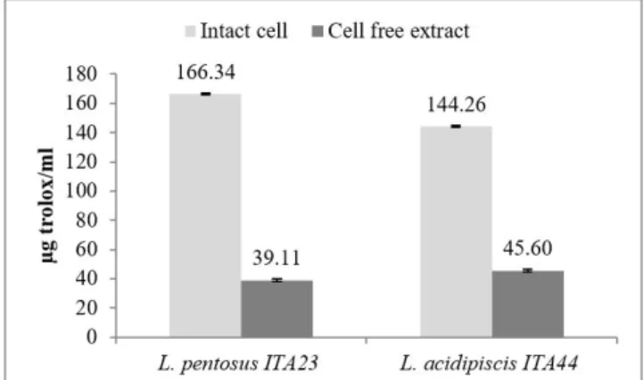
The results revealed that the two Lactobacillus strains tested showed antioxidant activity using both the FRAP and ABTS methods, especially by their intact cells when compared to the intracellular cell-free extracts (Figures 7 and 8). Using the FRAP method (Figure 7), L. pentosus ITA23 showed 135.28 and 23.91 µg trolox/ml antioxidant activity by its intact cells and cell-free extracts, respectively. The other Lactobacillus strain, L. acidipiscis ITA44, also showed similar results of 136.96 and 25.58 µg trolox/ml antioxidant activity by its intact cells and cell-free extracts, respectively. Using the ABTS method (Figure 8), L. pentosus ITA23 and L. acidipiscis ITA44 showed 166.34 and 144.26 µg trolox/ml antioxidant activity by their intact cells, respectively, and 39.11 and 45.6 µg trolox/ml antioxidant activity by their cell-free extracts.
Since the antioxidant activity of lactic acid bacteria is a new approach, there are not too many reports on the antioxidant activity of lactobacilli. In addition, the variation in the expression of the results and experimental conditions led to some difficulties in comparing the results of different experiments. In 2009, Klayraung and Okonogi (2009) investigated the antioxidant activity of two L. fermentum strains, which were isolated from fermented tea-leaves, using both the ABTS and FRAP methods. They reported Trolox Equivalent Antioxidant Capacities of 22.54 and 24.09 µM/ml supernatant for the two L. fermentum strains using the ABTS method, but using the FRAP method, these amounts were 20.63 and 21.26 µM/ml supernatant.
In the present study, the antioxidant activity of the intact cells of each of the Lactobacillus strains was higher than that of the intracellular cell-free extracts for both the
FRAP and ABTS methods. This could be attributed to the peptidoglycan and exopolysaccharide of the Lactobacillus cells, since the antioxidant activity of these fractions of the lactic acid bacteria have already been reported (LIU et al., 2011; CHABOT et al., 2001), although more studies are required to confirm this hypothesis.
In conclusion, both the L. pentosus ITA23 and L. acidipiscis ITA44 strains used in this study, which exhibited 100% killing of the cancer cells by, at the most, 30% of their supernatant, were considered to have remarkable antiproliferative effects against cancer cells. In addition, they showed specific selectivity in killing cancer cells and not normal cells. Furthermore, the two strains tested showed good antioxidant activity, especially by their intact cells. Hence, the two Lactobacillus strains could be considered as good potential probiotics for human consumption, due to their beneficial antioxidant and anticancer effects. Further investigations on their safety have to be carried out before they can be used in the food industry as probiotic strains.
Figure 7. Antioxidant activity of the two Lactobacillus strains
as measured by the FRAP assay. The results are mean values from two independent experiments, each with three replicates.
Figure 8. Antioxidant activity of the two Lactobacillus strains
HAGHSHENAS, B.; NAMI, Y.; ABDULLAH, N.; RADIAH, D.; ROSLI, R.; KHOSROUSHAHI, A. Y. Anti-proliferative effects of Enterococcus strains isolated from fermented dairy products on different cancer cell lines. Journal of Functional Foods, v. 11, p. 363-374, 2014. http://dx.doi.org/10.1016/j.jff.2014.10.002.
HAZA, A. I.; ZABALA, A.; MORALES, P. Protective effect and cytokine production of a Lactobacillus plantarum strain isolated from ewes’ milk cheese. International Dairy Journal, v. 14, n. 1, p. 29-38, 2004. http://dx.doi.org/10.1016/S0958-6946(03)00146-8.
KATIYAR, S. K.; MUKHTAR, H. Tea antioxidants in cancer chemoprevention. Journal of Cellular Biochemistry, v. 67, n. S27, p. 59-67, 1997. PMid:9591194. http://dx.doi.org/10.1002/ (SICI)1097-4644(1997)27+<59::AID-JCB11>3.0.CO;2-G.
KLAENHAMMER, T. R.; KULLEN, M. J. Selection and design of probiotics. International Journal of Food Microbiology, v. 50, n. 1-2, p. 45-57, 1999. PMid:10488843. http://dx.doi.org/10.1016/ S0168-1605(99)00076-8.
KLAYRAUNG, S.; OKONOGI, S. Antibacterial and antioxidant activities of acid and bile resistant strains of Lactobacillus fermentum isolated from miang. Brazilian Journal of Microbiology, v. 40, n. 4, p. 757-766, 2009. PMid:24031422. http://dx.doi.org/10.1590/ S1517-83822009000400005.
LAMSON, D. W.; BRIGNALL, M. S. Antioxidants in cancer therapy; their actions and interactions with oncologic therapies. Alternative Medicine Review, v. 4, n. 5, p. 304-329, 1999. PMid:10559547.
LIU, C. F.; TSENG, K. C.; CHIANG, S. S.; LEE, B. H.; HSU, W. H.; PAN, T. M. Immunomodulatory and antioxidant potential of Lactobacillus exopolysaccharides. Journal of the Science of Food and Agriculture, v. 91, n. 12, p. 2284-2291, 2011. PMid:21560134.
LOLLO, P. C. B.; MOURA, C. S.; MORATO, P. N.; CRUZ, A. G.; CASTRO, W. F.; BETIM, C. B.; NISISHIMA, L.; FARIA, J. A. F.; MARÓSTICA, M.; FERNANDES, C. O.; AMAYA-FARFAN, J. Probiotic yogurt offers higher immune-protection than probiotic whey beverage. Food Research International, v. 54, n. 1, p. 118-124, 2013. http://dx.doi.org/10.1016/j.foodres.2013.06.003.
LOLLO, P. C.; MORATO, P. N.; MOURA, C. S.; ALMADA, C. N.; FELICIO, T. L.; ESMERINO, E. A.; BARROS, M. E.; AMAYA-FARFAN, J.; SANT’ANA, A. S.; RAICES, R. R. S.; SILVA, M. C.; CRUZ, A. G. Hypertension parameters are attenuated by the continuous consumption of probiotic Minas cheese. Food Research International, v. 76, n. Pt 3, p. 611-617, 2015. PMid:28455044. http://dx.doi.org/10.1016/j.foodres.2015.07.015.
OOI, L. G.; LIONG, M. T. Cholesterol-lowering effects of probiotics and prebiotics: a review of in vivo and in vitro findings. International Journal of Molecular Sciences, v. 11, n. 6, p. 2499-2522, 2010. PMid:20640165. http://dx.doi.org/10.3390/ijms11062499.
SAARELA, M.; MOGENSEN, G.; FONDEN, R.; MATTO, J.; MATTILA-SANDHOLM, T. Probiotic bacteria: safety, functional and technological properties. Journal of Biotechnology, v. 84, Acknowledgements
The present work was supported by the Ministry of Higher Education of Malaysia under the LRGS Fasa 1/2012, UPM/700-1/3/LRGS grant. P. Shokryazdan and M.F. Jahromi acknowledge support from Iran’s National Elites Foundation (INEF).
References
AHOTUPA, M.; SAXELIN, M.; KORPELA, R. Antioxidative properties of Lactobacillus GG. Nutrition Today, v. 31, p. 262-265, 1996. Supplement 1. http://dx.doi.org/10.1097/00017285-199611001-00018.
BENZIE, I. F. F.; STRAIN, J. J. The ferric reducing ability of plasma (FRAP) as a measure of “antioxidant power”: the FRAP assay. Analytical Biochemistry, v. 239, n. 1, p. 70-76, 1996. PMid:8660627. http://dx.doi.org/10.1006/abio.1996.0292.
BIFFI, A.; CORADINI, D.; LARSEN, R.; RIVA, L.; DI FRONZO, G. Antiproliferative effect of fermented milk on the growth of a human breast cancer cell line. Nutrition and Cancer, v. 28, n. 1, p. 93-99, 1997. PMid:9200156. http://dx.doi.org/10.1080/01635589709514558.
CHABOT, S.; YU, H. L.; DE LESELEUC, L.; CLOUTIER, D.; VAN CALSTEREN, M. R.; LESSARD, M.; ROY, D.; LACROIX, M.; OTH, D. Exopolysaccharides from Lactobacillus rhamnosus RW-9595M stimulate TNF, IL-6 and IL-12 in human and mouse cultured immunocompetent cells, and IFN-in mouse splenocytes. Dairy Science & Technology, v. 81, n. 6, p. 683-697, 2001. http:// dx.doi.org/10.1051/lait:2001157.
CHOI, S. S.; KIM, Y.; HAN, K. S.; YOU, S.; OH, S.; KIM, S. H. Effects of Lactobacillus strains on cancer cell proliferation and oxidative stress in vitro. Letters in Applied Microbiology, v. 42, n. 5, p. 452-458, 2006. PMid:16620202. http://dx.doi. org/10.1111/j.1472-765X.2006.01913.x.
DAMIA, G.; BROGGINI, M. Improving the selectivity of cancer treatments by interfering with cell response pathways. European Journal of Cancer, v. 40, n. 17, p. 2550-2559, 2004. PMid:15541958. http://dx.doi.org/10.1016/j.ejca.2004.07.020.
DESROUILLÈRES, K.; MILLETTE, M.; VU, K. D.; TOUJA, R.; LACROIX, M. Cancer preventive effects of a specific probiotic fermented milk containing Lactobacillus acidophilus CL1285, L. casei LBC80R and L. rhamnosus CLR2 on male F344 rats treated with 1, 2-dimethylhydrazine. Journal of Functional Foods, v. 17, p. 816-827, 2015. http://dx.doi.org/10.1016/j.jff.2015.06.035.
n. 3, p. 197-215, 2000. PMid:11164262. http://dx.doi.org/10.1016/ S0168-1656(00)00375-8.
SALMINEN, S.; VON WRIGHT, A.; MORELLI, L.; MARTEAU, P.; BRASSART, D.; DE VOS, W. M.; FONDÉN, R.; SAXELIN, M.; COLLINS, K.; MOGENSEN, G.; BIRKELAND, S. E.; MATTILA-SANDHOLM, T. Demonstration of safety of probiotics. International Journal of Food Microbiology, v. 44, n. 1-2, p. 93-106, 1998. PMid:9849787. http://dx.doi.org/10.1016/S0168-1605(98)00128-7.
SAXAMI, G.; KARAPETSAS, A.; LAMPRIANIDOU, E.; KOTSIANIDIS, I.; CHLICHLIA, A.; TASSOU, C.; ZOUMPOURLIS, V.; GALANIS, A. Two potential probiotic lactobacillus strains isolated from olive microbiota exhibit adhesion and anti-proliferative effects in cancer cell lines. Journal of Functional Foods, v. 24, p. 461-471, 2016. http://dx.doi.org/10.1016/j.jff.2016.04.036.
SHOKRYAZDAN, P.; JAHROMI, M. F.; LIANG, J. B.; HO, Y. W. Probiotics: from isolation to application. Journal of the American College of Nutrition. 2017. In press. http://dx.doi.org/10.1080/ 07315724.2017.1337529.
SHOKRYAZDAN, P.; LIANG, J. B.; JAHROMI, M. F.; NORHANI, A. Probiotic potential of lactic acid bacteria isolated from mulberry silage. Journal of Pure and Applied Microbiology, v. 9, n. 2, p. 443-452, 2015.
SLAGA, T. J.; BRACKEN, W. M. The effects of antioxidants on skin tumor initiation and aryl hydrocarbon hydroxylase. Cancer Research, v. 37, n. 6, p. 1631-1635, 1977. PMid:404033.
STATSOFT. Statistic for Windows. SAS OnlineDoc 9.2. Tulsa: StatSoft Inc., 2008.
THIRABUNYANON, M.; BOONPRASOM, P.; NIAMSUP, P. Probiotic potential of lactic acid bacteria isolated from fermented dairy milks on antiproliferation of colon cancer cells. Biotechnology Letters, v. 31, n. 4, p. 571-576, 2009. PMid:19116692. http:// dx.doi.org/10.1007/s10529-008-9902-3.
TSAI, J. C.; HUANG, G. J.; CHIU, T. H.; HUANG, S. S.; HUANG, S. C.; HUANG, T. H.; LAI, S. C.; LEE, C. Y. Antioxidant activities of phenolic components from various plants of Desmodium species. African Journal of Pharmacy and Pharmacology, v. 5, n. 4, p. 468-476, 2011. http://dx.doi.org/10.5897/AJPP11.059.
TUO, Y. F.; ZHANG, L. W.; YI, H. X.; ZHANG, Y. C.; ZHANG, W. Q.; HAN, X.; DU, M.; JIAO, Y. H.; WANG, S. M. Antiproliferative effect of wild Lactobacillus strains isolated from fermented foods on HT-29 cells. Journal of Dairy Science, v. 93, n. 6, p. 2362-2366, 2010. PMid:20494143. http://dx.doi.org/10.3168/jds.2010-3069.