junho de 2015
Joana Filipa Alegria Pereira
Resting-State functional connectivity
in individuals with Williams Syndrome
Universidade do Minho
Dissertação de Mestrado
Mestrado Integrado em Psicologia
Trabalho realizado sob orientação da
Professora Doutora Adriana da Conceição Soares Sampaio
e coorientação do
Professor Doutor José Miguel Montenegro Soares
junho de 2015
Joana Filipa Alegria Pereira
Resting-State functional connectivity
in individuals with Williams Syndrome
Universidade do Minho
DECLARAÇÃO
Nome: Joana Filipa Alegria Pereira
Endereço eletrónico: [email protected]
Número do Cartão de Cidadão: 14220589
Título da dissertação: Resting-State functional connectivity in individuals with Williams Syndrome
Orientadora: Professora Doutora Adriana da Conceição Soares Sampaio Coorientador: Professor Doutor José Miguel Montenegro Soares
Ano de conclusão: 2015
Designação do Mestrado: Mestrado Integrado em Psicologia
É AUTORIZADA A REPRODUÇÃO INTEGRAL DESTA DISSERTAÇÃO APENAS PARA EFEITOS DE INVESTIGAÇÃO, MEDIANTE DECLARAÇÃO ESCRITA DO INTERESSADO, QUE A TAL SE COMPROMETE;
Universidade do Minho, 12/06/2015
ii Index RESUMO ... iv ABSTRACT ... v Introduction ... 6 Williams Syndrome ... 6
Resting State Networks ... 9
The present study ... 12
Method ... 13
Participants ... 13
MRI data acquisition ... 13
Image pre-processing ... 14
Functional connectivity Analysis ... 14
Statistical Analysis ... 15 Results ... 17 RSNs functional connectivity ... 17 Inter-network connectivity ... 19 Discussion ... 19 Future Directions ... 23 References ... 24 Supplementary data ... 41
Index for Tables Table 1. Resting State Seed-Based Region-of-Interest Analysis………..15
Table 2. Resting state networks clusters in WS and TD………...17
Index for Figures Figure 1. One sample t-test results for the RSNs of interest using the pre-defined ROI seed analysis………..16
Figure 2. Intra-RSN group differences found between healthy controls and WS participants………....19
iii Agradecimentos
"Happiness lies in the joy of achievement and the thrill of creative effort."
Franklin D. Roosevelt
Em primeiro lugar, agradeço à minha orientadora Professora Doutora Adriana Sampaio. O seu apoio e incentivo foram cruciais para a concretização deste trabalho.
Ao Professor Doutor José Miguel Soares, agradeço profundamente todo o seu envolvimento e apoio neste projeto.
Ao Pedro Moreira, por toda a ajuda, paciência e apoio ao longo de todo o processo.
Ao Professor Doutor Óscar Gonçalves e à Professora Doutora Joana Coutinho por todas as sugestões e encorajamentos.
À equipa do Laboratório de Neuropsicofisiologia, pela companhia, boa disposição, ajuda e integração ao longo do meu percurso. Obrigada a todos, sem exceção.
A todos aqueles que me acompanharam nesta etapa, um grande obrigado por todo o conforto e companhia.
À minha companheira e amiga, Mélanie. Obrigada pela amizade e apoio ao longo do nosso percurso.
À Rita, um agradecimento muito especial pelo carinho e apoio diários, pelas palavras doces e pela transmissão de confiança e de força, em todos os momentos. Obrigada por me dares sempre a mão ao longo de toda esta caminhada.
À minha avó, que certamente está a sorrir de orgulho.
Por último, aos pilares da minha vida, à minha mãe e ao meu irmão. Agradeço-vos por todo o apoio, amor, paciência e acima de tudo, por acreditarem sempre em mim.
iv Conectividade funcional nas redes neuronais de repouso em indivíduos com Sindrome
de Williams
RESUMO
As redes neuronais de repouso estão associadas a uma variedade de funções cerebrais importantes. Especificamente, padrões de conectividade funcional anormal nestas redes têm sido reportados pela investigação em diversas perturbações psiquiátricas e neurodesenvolvimentais. A natureza da conectividade funcional no Síndrome de Williams, uma perturbação neurodesenvolvimental rara, caracterizada por um perfil variado e enigmático de características cognitivas e comportamentais, permanece pouco explorada. O principal objetivo deste estudo é colmatar esta lacuna, avaliando a conectividade funcional das redes neuronais de repouso em indivíduos com esta perturbação. Foram submetidos a uma sessão de ressonância magnética funcional sete indivíduos com Síndrome de Williams e sete controlo, emparelhados por sexo e idade. Através da análise de regiões de interesse, detectámos conectividade funcional alterada em áreas que pertencem a seis redes diferentes, nomeadamente à default mode network, salience network, sensoriomotor network, visual
networks e à auditory network. O padrão global de hipoconectividade observado nestas redes
poderá estar subjacente ao seu fenótipo cognitivo e comportamental. Em específico, as alterações na default mode network e nas visual networks poderão estar relacionadas com a cognição social anormal e os défices visuo-espaciais que caracterizam estes indivíduos. Este estudo destaca a importância da exploração das redes neuronais de repouso no desenvolvimento.
v Resting-State functional connectivity in individuals with Williams Syndrome
ABSTRACT
Resting state brain networks are associated with a variety of relevant brain functions. Specifically, atypical patterns of functional connectivity in these networks have been reported in several psychiatric and neurodevelopmental disorders. The nature of the brain connectivity in Williams Syndrome, a rare neurodevelopmental disorder, characterized by a variable and enigmatic profile of cognitive and behavioral characteristics, remains unexplored. The main goal of this research is to evaluate the functional connectivity of resting state networks in individuals with Williams Syndrome. Seven Williams Syndrome participants and seven age and gender matching controls underwent a functional Magnetic Resonance Imaging session. Using a region of interest approach, we found altered functional connectivity in areas that correspond to six different networks, namely in the default mode network, dorsal attention network, salience network, sensoriomotor network, visual networks and auditory network. This global pattern of hypoconnectivity in these networks may be underlying their cognitive and behavioral phenotype. Specifically, alterations in the default mode and visual networks may be related with their abnormal social cognition and visual-spatial deficits. This study highlights the importance of studying the resting state networks in development.
6 Introduction
Williams Syndrome
Williams’s syndrome (WS) is a rare neurodevelopmental disorder, firstly identified in 1961 as a clinical entity distinct by Williams, Barratt-Boyes and Lowe (1961). With an expected incidence of 1 in 7,500 to 1 in 20,000 (Stromme, Bjornstad & Ramstad, 2002), this condition is associated to a chromosomal deletion 1-2Mb 7q11.23 band that includes the genes responsible for Elastin and LIM1K, along with a variety of other genes nearby (eg: WSCR1-5; RFC2, FZD9) whose relationship with the cognitive and neurophenotypic changes are not yet completely understood (Osborne et al., 2001). Physically, this population has specific facial features as broad forehead, flat nasal bridge, a snub nose and short, wide mouth with full lips and irregular teeth (Morris & Mervis, 2000). Besides the facial dysmorphology, abnormalities in connective tissue and cardiovascular disease have also been reported, which has been linked to ELN gene deletion (Bellugi, Klima & Wang, 1997; Pober & Dykens, 1996).
WS is characterized by a variable and enigmatic profile of cognitive and behavioral characteristics.These individuals have been described as having mild to moderate intellectual deficits, with several studies showing a mean IQ varying between 50 and 60, with a range of 40-100 (Bellugi, 1997; Bellugi, Mills, Jernigan, Hickok & Galaburda, 1999; Lenhoff, Wang, Greenberg & Levitin & Bellugi, 1998; Reis, Schader, Milne & Stephens, 2003; Sampaio, Férnandez, Henriques, Carracedo, Sousa & Gonçalves, 2009), with these scores remaining stable with increasing age (Howlin, Davies & Udwin, 1998; Searcy, Lincoln, Rose, Klima, Bavar, & Korenberg, 2004).
The majority of the cognitive studies to date describe them as having a fractionate cognitive profile, in which a verbal strength in contrast to significant visuospatial weaknesses has been reported (Bellugi, Bihrle, Jernigan, Trauner & Doherty, 1990; Mervis, Klein-Tasman & Mastin, 2001), including deficits in saccadic eye movement, visuospatial working memory, selective attention, visual constructive and visual motor skills (Atkinson, Anker, Braddick, Nokes, Mason & Braddick, 2001; Farran, Jarrold & Gathercole, 2003; Frangiskakis et al., 1996; Van der Geest et al., 2004). Nevertheless, a within cognitive domain dissociation has been reported with prominent visuospatial construction impairments being complemented by relative strengths in face and object processing (Meyer-Lindenberg, Mervis & Berman, 2006).
7 Regarding personality and behavior phenotypes, individuals with WS have been described as hypersociable (Bellugi et al., 1999) and over friendly. In fact, Levine and Wharton reported that “one of the most striking characteristics of this condition is a unique personality profile that includes a general presentation of exuding happiness” (2000, p.590). However, this is in contrast with evidence showing that individuals with WS are distressed by persistent fears, showing significant behavioral problems as specific phobias (Dykens, 2003). Indeed, a large percentage of WS meets DSM-IV criteria for specific phobia and general anxiety disorder and is also diagnosed with attention deficit/hyperactivity disorder (ADHD) (Martens, Wilson & Reutens, 2008).
Another specific characteristic of this population is their musical skills. Individuals with WS display heightened levels of curiosity and emotional responsivity toward music, an exceptional memory for lyrics as well as proficient capability in composing song lyrics, absolute pitch and facility learning complex rhythms (Lenhoff, 1996). Reports of their musical abilities headed to the conclusion that music is more completely developed than general cognition (Levitin & Bellugi, 1998). Despite this interest, most individuals with WS have hearing loss. In addition, their abnormal auditory perception as odynacusis and auditory
allodynia that produces distress/discomfort in people with WS, also create aversion (Levitin,
Cole, Lincoln & Bellugi, 2005). Odynacusis consist in early awareness of sounds that are either too soft to hear or simply insignificant to others and the auditory allodynia, a fear of specific sounds (Levitin et al., 2005). Other reported neurological abnormalities are their weakness in complex fine motor abilities as well as atypical motor coordination, tone, and gait (Chapman, du Plessis & Pober, 1996; MacDonald & Roy, 1988).
Neuroimaging studies have provided interesting evidence regarding the understanding the neuroanatomical changes that underlie the cognitive and behavioral phenotype of individuals with WS. Morphological, volumetric and functional alterations as well as abnormal asymmetry have been described by several studies in this specific population (Holinger et al., 2005; Sampaio, Sousa, Férnandez, Vasconcelos, Shenton & Gonçalves, 2008). Structural neuroimaging studies have confirmed a reduced overall brain volume (by 11-13%) as well as smaller size of specific sub regions of the cerebral cortex, cerebellar cortex, and subcortical structures in individuals with WS (Eckert et al., 2005; Reiss, et al., 2000). Areas associated with social cognition have also been stated based on their morphological changes, namely the superior temporal gyrus (Sampaio et al., 2008), the amygdala (Capitão et al., 2011) and the corpus callosum (Sampaio et al., 2013), medial
8 prefrontal cortex and anterior cingulate, parietal cortex and precuneus (Gaser et al., 2006; Reiss et al., 2004).
The medial prefrontal cortex has been reported as displaying disproportionate enlargement and increased gyrification when compared with typically developing individuals (Gaser et al., 2006; Reiss et al., 2004). The posterior cingulate cortex and left precuneus in WS, show altered patterns of cortical folding (Gaser et al., 2006). A parietal hypoplasia and increased parietal grey matter volumes in WS have also been described by post-mortem and MRI studies (Galaburda & Bellugi, 2006; Reiss et al., 2000). These structural alterations are paralleled by functional MRI (fMRI) evidence. Specifically, reduced activity in the striatum, dorsolateral prefrontal cortex and dorsal anterior cingulate cortex in a modified Go/No Go task (Mobbs et al, 2007); and between the fusiform face area and amygdala, in social-cognitive tasks (Sarpal et al., 2008). Reduced functional connectivity within frontostriatal pathways have been reported (Meyer-Lindenberg et al., 2004), when compared to control participants. Several studies described also altered patterns of cortical-subcortical connectivity in this syndrome (e.g., Marenco et al., 2007). The abnormal connections observed between prefrontal cortex and the amygdala is believed to contribute to the social phenotype of WS (Meyer-Lindenberg et al., 2004; Meyer-lindenberg et al., 2005).Specifically, the abnormal frontostriatal circuitry combination with altered connectivity between the amygdala and the prefrontal cortex has been evidenced in many studies of WS (Gaser et al., 2006; Gothelf et al., 2008; Meyer-Lindenberg et al., 2004; Munoz et al., 2010).
Interestingly, these are the core regions of a resting state network involved in self-referential and introspective processes, planning the future, perspective taking of the desires, beliefs, and intentions of others - the Default Mode Network (DMN) (Buckner, Andrews Hanna & Schacter, 2008; Raichle, MacLeod, Snyder, Powers, Gusnard & Shulman, 2001). Therefore, structural and functional alterations have been reported in the main hubs of the DMN which are likely to be associated with abnormal social cognition, as understanding the mental states of others (i.s. theory of mind) (Haas & Reiss, 2012).
Consequently, the cognitive and social phenotype of WS are associated with underlying functional and structural brain alterations, particularly in the core regions of one of the most studied resting state networks – the DMN that is composed by specific brain networks involved in social-cognitive (Mars, Neubert, Nooman, Sallet, Toni & Rushworth, 2012). However, the functional connectivity in WS population during the resting state remains unclear.
9 Resting State Networks
The spatial organization of intrinsic activity seems to exceed levels of consciousness, being present under anesthesia (Greicius et al., 2008), and also through the early phases of sleep (Fukunaga et al., 2006; Larson-Prior, Zempel, Nolan, Prior, Snyder & Raichle, 2009).
Awake and sleeping are opposite states, but when someone is awake but not intentionally performing any task, physically or mentally, is called to be in resting state. It's a state where the individual is conscious and prepared to reply promptly to any type of external stimulation or cognitive requirement. In this situation, while the individual is resting and the body is static, the brain instead is actively engaged, exhibiting slow spatiotemporally ordered fluctuations of neuronal activity (Biswal, Zerrin Yetkin, Haughton & Hyde, 1995; Lowe, Mock & Sorenson, 1998). According to Cabral and collaborators (2014), the organization of the resting-state activity is characterized by the existence of temporally correlated activity -or functional connectivity- between different voxels in the brain defining the so-called resting state networks (RSNs). The resting state functional connectivity can be analyzed in two levels: within and between network connectivity.
The overall correspondence between RSN patterns and functionally interacting brain regions proposes that RSN could reflect coherent neuronal signalling within functional systems (Cordes et al. 2000; Leopold, Murayama & Logothetis, 2003; Lowe, Dzemidzic, Lurito, Mathews & Phillips, 2000). Many functional connectivity studies have reported a number of networks that are intensely functionally linked during the rest (Beckmann, Deluca, Devlin & Smith, 2005; Calhoun, Kiehl & Pearlson, 2008; Damoiseaux et al, 2006; Rosazza, Minati, Ghielmetti, Mandelli & Bruzzone, 2011; Smith et al, 2009; Van den Heuvel, Mandl & Hulshoff, 2008; Van Dijk, Hedden, Venkataraman, Evans, Lazar & Buckner, 2010).
Of the several RSNs, the DMN may be readily designated as the most important, because of their cognitive and affective associations. The introspective mental processes, autobiographical memories, the tendency of human minds to wander, to the ability to rethink about the recent past and to imagine future events, and the connection between the internal and external attention while monitoring the world around us seem to be promoted by this network (Buckner et al., 2008; Greicius, Krasnow, Reiss & Menon, 2003; Mason, Norton, Van Horn, Wegner, Grafton & Macrae, 2007). The key regions of the DMN are the precuneus, the posterior cingulate cortex, the medial prefrontal cortex and the bilateral inferior parietal cortex (Raichle et al., 2001). The DMN is also known to interface task performance and emotion (Simpson, Snyder, Gusnard & Raichle, 2001). Mars and collaborators (2012)
10 reported that specific brain networks involved in social-cognitive tasks match the different nodes of the DMN. Thus most of the brain regions of the DMN have been found to superpose with important areas supporting the social cognition, which contrast with a task-based performance, as it is considered a task-negative network.
Differently, in a task-based scenario, the DMN is suppressed and it has been termed the task-negative network inversely correlated with task-positive networks (TPN) (Dutta, Mckie & Deakin, 2014). That is, the greater suppression of the DMN is associated with a improved performance in several cognitive tasks (Mayer, Roebroeck, Maurer & Linden, 2010; Singh & Fawcett, 2008). However, the DMN activity continues to a significant degree during simple sensory tasks, in which satisfactory task performance is conceivable with only minimal attentional assets (Greicius et al., 2003; Wilson, Molnar-Szakacs & Iacoboni, 2008), under conscious sedation (Greicius et al., 2008) as well as during the initial stages of sleep (Horovitz et al., 2008).
Additionally, other resting state networks have also been identified. A task-positive network (TPN) (Fox, Snyder, Vincent, Corbetta, Van Essen & Raichle, 2005) or frontoparietal network (Toro, Fox & Paus, 2008), consists on a set of regions believed to be involved in cognitive control (e.g., Alain, Yu & Grady, 2008; D'Esposito, Detre, Alsop, Shin, Atlas & Grossman, 1995; Dove, Brett, Cusack & Owen, 2006). That is, when someone gets involved in carrying out an externally driven task (e.g. working memory), the activity in task-related regions increase and the default mode activity declines. Specifically, two lateralized parietal-frontal networks have been described, the Dorsal Attention Network (DAN) and the Ventral Attention Netwok (VAN). The DAN includes the posterior parietal cortex and the frontal eye fields, and is believed to participate in orientating visual spatial attention (Corbetta, Kincade, Ollinger, McAvoy & Shulman, 2000; Hopfinger, Buonocore & Mangun, 2000; Weissman, Giesbrecht, Song, Mangun & Woldorff, 2003). More specifically, this network is thought to produce and sustain endogenous signals related to present task goals (Corbetta et al., 2000; Hopfinger et al., 2000), and the activation of sensory (Weissman, Gopalakrishnan, Hazlett & Woldorff, 2005; Woldorff, Hazlett, Fichtenholtz, Weissman, Dale & Song, 2004) and motor (Astafiev, Shulman, Stanley, Snyder, Van Essen & Corbetta, 2003) regions of the brain that are important to achieve those goals, hold task-relevant information online in short-term memory (Pessoa, McKenna, Gutierrez & Ungerleider, 2002), and link stimuli to responses (Rushworth, Johansen-Berg, Göbel & Devlin, 2003). The VAN, including the inferior frontal gyrus, the temporal–parietal junction, and the ventrolateral prefrontal cortex (Corbetta, Patel & Shulman, 2008), is thought to make important contributions to stimulus-driven reorienting
11 of covert visual spatial attention (Corbetta et al., 2000; Serences, Shomstein, Leber, Golav, Egeth & Yantis, 2005). This network is thought to generate an “interrupt signal” when a important stimulus appears at an unanticipated location to help to terminate the present focus of attention, thereby allowing spatial attention to change to a new location (Corbetta et al., 2008).
Functional MRI studies looking at spontaneous (“resting-state”) functional connectivity among brain regions have shown that the dorsal and ventral networks are evidently distinguishable on the basis of their correlation patterns even under task-free conditions (Fox, Corbetta, Snyder, Vincent & Raichle, 2006; He, Chen, Gong & Evans, 2009). So, taken together, the dorsal and ventral networks are two anatomically separated cortical systems with functionally specific nodes supporting specific procedures for attentional control (Vossel, Geng & Fink, 2014). The connections between the dorsal and ventral attention networks are also believed to make important contributions to reorienting covert visual spatial attention (Corbetta et al., 2008).
The Salience Network (SN) is involved in filtering information to support behaviour choice, having an important role in assessing the relevance of internal and external stimuli in order to guide behaviour (Seeley et al., 2007). Menon and Uddin (2010) have suggested that the main role of this network is to allow switching between the default mode and task-related conditions of brain connectivity. It is structurally correlated to the anterior cingulate cortex, the presupplementary motor area, and anterior insulae (Bonnelle et al., 2012; Elton & Gao, 2014; Seeley et al., 2007;).
The Sensorimotor Network (SMN) involves the precentral gyrus, post-central gyrus and the supplementary motor area (Rosazza & Minati, 2011). This network seems to be engaged with regions that anatomically and functionally correspond to motor as well as sensory areas and is associated with the spontaneous fluctuations reflecting the active motor tasks (Rosazza & Minati, 2011).
The auditory network (AN) is essential to process auditory stimuli, such as tone/pitch discrimination, music, and language (Laird et al., 2011). Includes the primary and secondary auditory cortices, and corresponds most strongly to action-execution-speech, cognition-language-speech, and perception-audition paradigm. (Smith et al, 2009). This component involves the superior temporal gyrus, the hesch's gyrus, the insula, bilateral superior temporal gyri, and posterior insular cortex (Rosazza & Minati, 2011).
In the visual domain, three distinct networks have been reported: the first one is characterized by activity in the medial visual areas, namely striate cortex and extra-striate
12 regions typically medial, such as lingual gyrus (Rosazza & Minati, 2011). The second one, the lateral visual network, comprises the middle temporal visual association area at the temporo-occipital junction and is frequently essential in complex (emotional) stimuli (Laird et al., 2011). The third network is associated with the activity in the striate cortex and in occipital visual areas (Rosazza & Minati, 2011). The components that comprise medial and occipital visual networks, are important in simple visual processes, for example, a flickering checkerboard, and higher-order visual stimuli, as the orthography, respectively (Beckmann et al., 2005; Damoiseaux et al., 2006; Laird et al., 2011). Still in the visual domain, another distinction of visual networks can be done: the primary visual network (PVN), compromising the bilateral calcarine regions and lingual regions, and the high-level visual network (HVN), including the bilateral supperior occipital regions and middle occipital regions (Zhang et al., 2015).
Over the last decade, a large number of studies have reported altered resting brain activity in a wide range of neuropsychiatric (Guerrero-Pedraza et al., 2012; Pomarol-Clotet et al., 2008) and neurodevelopmental disorders (Kennedy & Courchesne, 2008a, 2008b; Menon, Leroux, White & Reiss, 2004; Vega, Hohman, Pryweller, Dykens & Thornton-Wells, 2015). These results not only exemplify the importance of balancing resting-state dynamics for an optimal cognitive function, but also offer insights to comprehend the intrinsic mechanisms related (Kringelbach, Green & Aziz, 2011).
So far the only study that examined the development/characterization of functional connectivity on resting state networks in Williams Syndrome is from Vega and collaborators (2015) that studied the resting state functional connectivity in Down syndrome compared to WS and typically developing controls. The major aim of this study was to generate insights into resting state brain function across this two different neurodevelopmental disorders. Their results showed that WS individuals exhibited significantly greater between-network connectivity compared to controls for the frontoparietal-DMN network pair, as well as poorer levels of within-network connectivity in the DMN, ventral attention network and sensoriomotor network.
The present study
Therefore, taking into account abnormal social cognitive functioning in WS together with structural and functional evidence regarding key regions of the DMN, as well as resting state abnormal functional connectivity, we expect that individuals with WS have lower levels
13 of within-network connectivity in the DMN, SMN, DAN, AN, VAN, HVN and PVN when compared with typically developing individuals.
Therefore, the main goal of this research is to evaluate the functional connectivity of resting state networks in individuals with WS and in a typically group (TD) matched on age and sex.
Method Participants
The study included 10 participants with WS [4 males and 6 females, mean age 21.33 (SD = 6.75), age range 16 to 37 years], and 11 typically developing individuals (TD) [6 males and 5 females, mean age 22.00 (SD = 6.65), age range 9-36 years]. No significant age differences were found between the two groups (p > 0.05). After excluding participants due to movement artifacts (n = 3), seven participants with WS and their respective control counterparts were included in the analysis [WS: three males and four females, mean age 23.00; SD = 7.00; TD: three males and four females, mean age 24.14, SD = 6.12]. Participants in the WS group tested positive in fluorescence in situ hybridization (FISH) for deletion of the elastin gene in chromosome 7, and the presence of any sensorial or speech disorder, as well as comorbidity with severe psychopathology not associated with the syndrome were defined as exclusion criteria. The typically developing group was composed of individuals without a history of sensorial, psychiatric, neurological disorder or cognitive impairment.
The study was conducted in accordance with the principles expressed in the Declaration of Helsinki and was approved by the Ethics Committee of Centro Hospitalar do Porto (Portugal). The study goals and tests were explained to all participants and all gave informed written consent.
MRI data acquisition
Participants were scanned on a clinical approved Philips (Philips Achieva, Best, Netherlands) MRI scanner on Centro Hospitalar do Porto. The imaging sessions included one structural T1 - weighted and one functional T2* - weighted acquisition conducted in the same day. For structural analysis, a T1 high-resolution anatomical sequence, 3D SENSitivity Encoding (SENSE) was performed with the following scan parameters: repetition time (TR) = 7.85 s, echo time (TE) = 4.00 ms, 170 sagittal slices with no gap, flip angle (FA) = 8º,
in-14 plane resolution = 1.0 x 1.0 mm2 and slice thickness = 2.0 mm. During the resting-state fMRI acquisition, using gradient echo weighted echo-planar images (EPIs), the participants were instructed to keep their eyes closed and to think about nothing in particular. The imaging parameters were: 100 volumes, TR = 3 s, TE = 40 ms, FA = 90º, in-plane resolution = 3.0 x 3.0 mm2, 45 interleaved slices, slice thickness = 3.2 mm, imaging matrix 72 x 74 and FoV = 235 mm.
Image pre-processing
Before any data processing and analysis, all acquisitions were visually inspected by a certified neuroradiologist and confirmed that they were not affected by critical head motion and that participants had no brain lesions. To achieve signal stabilization and allow participants to adjust to the scanner noise, the first three fMRI volumes (9 seconds) were discarded. Resting state data preprocessing was performed using FMRIB Software Library (FSL v5.0.4, www.fmrib.ox.ac.uk/fsl). Images were firstly corrected for slice timing using the first slice as reference and Fourier phase shift interpolation. To correct for head motion, images were realigned to the mean image with a six-parameter rigid-body spatial transformation and estimation was performed at 0.9 quality, 4 mm separation, 5 mm full-width at half-maximum (FWHM) smoothing kernel and using 2nd degree B-Spline interpolation. Three WS and two control participants were excluded once they exceed head motion higher than 3 mm in translation or 1º in rotation. Images were then spatially normalized with a non-linear transformation to the MNI (Montreal Neurological Institute) standard coordinate system using the FSL template through the combination of the rigid-body co-registration matrix and warp of the nonlinear transformation. Data were then re-sampled to 3x3x3 mm3 using sinc interpolation, smoothed to decrease spatial noise with a 8 mm, FWHM, Gaussian kernel. Images were then temporally band-pass filtered (0.01-0.08 Hz) and grand mean-scaled.
Functional connectivity Analysis
Functional connectivity analysis was performed using a seed-driven approach with FSL. The seed ROIs were defined as 6 mm radius spheres (except 4 mm for SMN) centred on MNI coordinates used to identify the most well-known resting-state networks in the literature. Coordinates for auditory network, default-mode network, dorsal attention network, salience network, left and right ventral attention networks, visual network and sensorimotor network
15 were obtained from previous studies (Table 1). The mean time series from each ROI was entered as predictor in a dual-regression analysis in order to estimate the subject-specific components.
Table 1
Resting State Seed-Based Region-of-Interest Analysis
Network Seed/mask region
Coordinates (MNI) Reference x y z DMN PCC 1 -55 17 Woodward, 2011: http://www.ncbi.nlm.nih.gov/pubmed/214 58238 DAN left IPS/SPL -25 -53 52 right IPS/SPL 25 -57 53 SN
left fronto-insular cortex -32 26 -14 right fronto-insular
cortex 38 22 -10
VAN-L left DLPFC -42 34 20
VAN-R right DLPFC 44 36 20
VN primary visual cortex -2 -82 4
Maneshi, 2012 :
http://www.plosone.org/article/related/inf o%3Adoi%2F10.1371%2Fjournal.pone.0
050359
AN
left primary auditory
cortex 55 -22 9 Schmidt, 2013 :
http://www.ncbi.nlm.nih.gov/pubmed/240 98513
right primary auditory
cortex -41 -27 6
SMN
left motor cortex -36 -25 57 Van Dijk, 2010 :
http://www.ncbi.nlm.nih.gov/pubmed/198 89849
right motor cortex 36 -25 57
MNI coordinates of seed and mask regions for each analysis.Statistical Analysis
For functional analysis (using the SPM8 software), two-tailed independensamples t-test were performed. The comparison of intra-RSN functional connectivity between groups was performed using a random effects analysis on SPM8 (Statistical Parametrical Mapping, version 8, http://www.fil.ion.ucl.ac.uk). One sample t-tests were performed to confirm that RSNs of interest were obtained using the pre-defined ROI (Figure 1). Thereafter, two-sample t-tests were performed and results were considered significant at p < 0.05 corrected for multiple comparisons using a combination of a peak threshold of p < 0.005 with a minimum
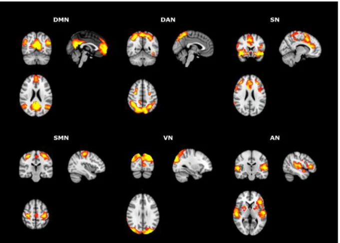
16 cluster size (196 voxels). The minimum cluster size was determined over 1000 Monte Carlo simulations using AlphaSim program distributed with REST software tool (http://resting-fmri.sourceforge.net/) with the following input parameters: individual voxel probability threshold = 0.005, cluster connection radius = 2 mm, gaussian filter width (FWHM) = 10x10x9 mm (smoothness estimated with 3dFWHMx program), number of Monte Carlo simulations = 1000 and mask was set to the corresponding RSN template mask. Anatomical labeling was defined by a combination of visual inspection and Anatomical Automatic Labeling atlas (AAL) (Tzourio-Mazoyer et al., 2002).
Figure 1. One sample t-test results for the RSNs of interest using the pre-defined ROI seed
analysis.
In order to study inter-RSNs functional connectivity, the RSNs' time series from each individual were initially extracted. Thereafter, correlations between pairs of time-courses were estimated using the “glmfit” function on Matlab. The obtained coefficients were transformed to Z-scores using the Fisher's r to z transformation. These values were then used as inputs in two-sample t-tests in order to test for group differences. In order to account for multiple comparisons, results were considered to be significant at the p<.005 level.
17 Results
RSNs functional connectivity
At rest, increased functional connectivity (FC) between the structures comprising the DMN was found in TD in two clusters: the first cluster (347 voxels) included the anterior cingulate and the frontal medial orbitofrontal cortex; the second cluster (281 voxels) included the precuneus, calcarine and cuneus of the left hemisphere. Increased FC between the SN and the supplementar motor area and the right superior frontal gyrus was observed in the TD group (597 voxels). In addition, the same was observed in another cluster (348 voxels) comprising the middle frontal gyrus and precentral gyrus of the right hemisphere. TD individuals displayed increased FC between the components of the DAN and the right inferior frontal gyrus – pars triangularis. Regarding the visual network, it was observed that TD individuals presented increased FC in the middle and superior occipital lobes (cluster size=266 voxels) of the left hemisphere. On the other hand, with respect to the same network, TD group displayed diminished FC in the left inferior and left middle temporal gyrus (454 voxels), the supplemental motor area and the paracentral lobule (703 voxels), and in the left middle frontal gyrus (261 voxels). Significantly increased FC in TD was also found between the AN and the right superior occipital gyrus (198 voxels), as well as increased FC between the SMN and other regions in three clusters: the right middle frontal gyrus and superior frontal gyrus (216 voxels); the left supplementar motor area and the right superior frontal gyrus (308 voxels); and the middle and inferior divisions of the left temporal gyrus and in the left inferior occipital gyrus (451 voxels). The magnitude and graphic representation of these differences are represented on Table 2 and Figure 2.
Table 2
Resting state networks clusters in WS and TD
Condition RSN Regions Peak MNI
coordinates Cluster size (voxels) Maximum Z score Controls > WS
AN Right superior occipital gyrus 30, -76, 18 198 3.41 DAN Right inferior frontal – pars
triangularis 54, 40, 2 206 3.53
18 Right medial orbitofrontal cortex 0, 42, -8 3.30
Left anterior cingulate -4, 40, 8 3.19 Left precuneus -2, -64, 28
281
3.47 Left calcarine region -4, -68, 20 3.09
Left cuneus -14, -60, 24 2.96
PVN Left superior occipital gyrus -82, -84, 24 266 3.69 Left middle occipital gyrus -26, -92. 14 3.52
SN
Right supplementar motor area 0, 6, 52
597 4.26
Right superior frontal gyrus 14, 14, 54 3.79 Right middle frontal gyrus 42, -4, 64
348 3.33
Right precentral gyrus 58, 6, 44 3.17
SMN
Right middle frontal gyrus 30, 38, 36 216 4.46 Right superior frontal gyrus 22, 30, 60
308
3.71 Left supplementar motor area 0, 18, 66 3.12 Right superior frontal gyrus 8, 24, 60 2.81 Left inferior temporal gyrus -58, -52, -14
451
3.19 Left inferior occipital gyrus -52, -68, -12 3.15 Left middle temporal gyrus -54, -42, -8 3.81
Controls < WS PVN
Left middle temporal gyrus -36, 14, -36
454 4.86
Left inferior temporal gyrus -48, 22, -28 3.55 Right supplementar motor area 2, -18, 76
703 4.36
Left paracentral region -10, -24, 68 4.16 Left superior frontal gyrus -18, 48, 16
261
3.72 Left middle frontal gyrus -26, 52, 8 3.71
Group differences (Controls > WS in red) and (Controls < WS in blue) at rest, in brain regions of the AN, DAN, DMN, PVN, SMN and SN maps (two-sample t-tests, corrected for multiple comparisons, p <0.05).
19
Figure 2. Intra-RSN group differences found between healthy controls and WS participants.
Control participants evidenced enhanced FC with the DMN, DAN, SN and SMN. Dissociable effects within the PVN were found, with control individuals presenting augmented FC with occipital structures and WS with frontal and temporal regions. Statistical maps fully corrected for multiple comparisons through the combination of a height threshold of p < 0.005 and a minimum cluster size of 196 voxels. Colorbar represents T-statistic of the corresponding tests (orange bar: Control>WS; blue bar: WS>Control).
Inter-network connectivity
The comparison of FC correlation coefficients between control and WS individuals revealed that no differences in the inter-network connectivity between groups, indicating a similar association between the studied networks.
Discussion
WS is a complex neurodevelopmental disorder that affects multiple cognitive domains and brain systems. A question regarding the nature of the brain connectivity in WS (hypoconnectivity or hyperconnectivity) remains to be unanswered. In this way, we addressed a critical gap in the literature by examining whole-brain intrinsic functional connectivity in
20 this population and identifying specific brain networks that could successfully discriminate the WS group from a TD group in a resting state condition. These results offer a first look into the large-scale brain networks at rest that possibly underlies the cognitive and behavioral symptoms of WS.
Using a region of interest (ROI) approach, we found altered functional connectivity in areas that belong to six different networks, namely in the DMN, DAN, SN, SMN, visual networks and AN. In fact, the PVN was the most altered, indicated by both an increase and decrease in the FC in distinct clusters. In the inter-network connectivity analysis, no significant interaction was found when the groups were compared, indicating a similar connectivity between the studied networks. A reduced FC between structures comprising the DMN was observed in the WS group when compared with the TD group. These findings are consistent with the study of Vega and collaborators (2015) that documented results showing a decreased connectivity within the DMN of participants with WS, when compared with TD individuals and Down syndrome.
The hypoconnectivity observed in the DMN subregions was evident in two major clusters: the anterior hub of the DMN, including the anterior cingulate and the medial orbitofrontal regions; and a posterior hub that included the precuneus, calcarine and cuneus. Regions of the anterior DMN have been connected with social awareness, comprising the capacity to attribute mental state to others (Gusnard, Akbudak, Shulman, & Raichle, 2001; Lane et al., 1998). The anterior hub also has been involved in more general roles in social cognition as theory of mind and monitoring reward, emotional insight and emotional self-control, empathy, and mentalizing abilities (Kringelbach & Rolls, 2004; Li, Mai, & Liu, 2014; Mars et al., 2012; Zhang & Chiang-shan, 2012). These social-cognitive dimensions, as theory of mind and self-referential processing, have also been linked with the posterior hubs of the DMN.
The alterations in the FC of these DMN subregions may underlie the difficulties presented by this population in social awareness and intelligent behavior. The social cognitive profile of WS is characterized by a reduced ability to understand the mental state of others (Santos, Silva, Rosset & Deruelle, 2010), problems in emotional perception and emotional self-control, as in metalizing abilities, empathy and theory of mind. Those behavioural characteristics have been associated with activation of the anterior cingulate and medial orbitofrontal regions (Gusnard et al.,2001; Lane et al., 1998). Consistent with these findings, structural abnormalities of the orbitofrontal cortex have also been described in individuals with WS (Reiss et al. 2004), which may be related with abnormal recognition of emotional
21 faces in this specific population (Mimura et al., 2010). On the left hemisphere, a decreased FC in WS was found in areas that are involved in visuospatial processes and are responsible for visual processing, including execution or preparation of spatially guided behaviors (Astafiev et al., 2003), namely the precuneus, calcarine and cuneus. The alterations in FC observed in these regions may be part of the neurobiological basis underlying one of the most characteristic phenotypic features demonstrated by individuals with WS - their prominent visuo-spatial construction impairments (Atkinson et al., 2001; Farran et al., 2003; Meyer-Lindenberg et al., 2006).
The distinct pattern of FC observed in PVN, namely reduced FC in one cluster and increased FC in other three, meets the visuospatial skills that characterize WS. The PVN is a cluster of regions that plays a central role in spatial cognition. Visual perception and processing of spatial information in relation to several aspects of spatial cognition, namely the orientation, perception and spatial memory, perception, and orientation (Munneke, Heslenfeld & Theeuwes, 2010; Possin, 2010). The perceptual processing in WS is characterized by their strengths in face and object processing, but also by their extended impairments in the visuospatial construction. In this way, the distinct pattern of functional connectivity detected in this network may underlie the distinct visual profile of this specific population. Vega and collaborators (2015) also observed lower FC in the visual domain in WS on resting state, namely in the visual orientation network.
A hypoconnectivity in a cluster of the AN was also found, specifically in the right superior occipital gyrus. This finding along with a decrease in the proportion of the posterior portions of the forebrain areas, comprising the occipital lobes, besides the contribute for the unusual visual profile of the individuals with the WS (Reiss et al., 2000), also may determine the unusual auditory profile of the individuals with the WS featured by attractiveness to music and a variety of broadband noises, the aversion toward certain sounds (auditory allodynia), as well the hyperacusis.
A decreased FC in the inferior frontal gyrus – pars triangularis (IFT), a cluster that belongs to the DAN was found in individuals with WS. Several studies have been linked the frontal lobe activity to the executive functions, a range of processes, such as working memory, inhibitory control and attentional shifting (Friedman, Miyake, Corley, Young, DeFries & Hewitt, 2006; Miyake, Friedman, Emerson, Witzki, Howerter & Wager, 2000). In this way, our results, specifically poorer levels of FC in this region, may underlie the impairments in executive functioning that individuals with WS show, and that have been stated by earlier
22 studies (Menghini, Addona, Costanzo & Vicari, 2010; Porter, Coltheart & Langdon, 2007; Rhodes, Riby, Park, Fraser & Campbell, 2010).
A hypoconnectivity patter was also perceived in the SN of the WS group, mostly observable in the right hemisphere. Abnormal connectivity within SN has also been reported in autism, another neurodevelopmental disorder (Menon & Uddin, 2009). This network plays an important role in recruiting brain regions for sensory information as well in initiating dynamic switches between the executive control networks and the DMN. Thus the reduced FC observed in this network can be linked to the decreased FC detected in the DAN as well as to the hypoconnectivity observed in the DMN. The poor levels of the FC in these three networks could explain many of the behavioral features in WS, such the difficulties in the attention shifting, the difficulty in keep attention in their internal states and in some cases the ADHD that is diagnosed in some patients with WS.
Regarding the SMN a hypoconnectivity was also detected in some areas of this network in WS. Since this component is engaged with motor and sensory areas, the alterations that were found may be related to the abnormal motor capacities manifested by these individuals throughout their development, namely the atypical motor coordination and the weakness in complex fine motor abilities. Our results are consistent with Vega and collaborators (2015) that also detected reduced within-network connectivity in the SMN.
The results of the present study are consistent with our hypotheses. In general, a situation of hypoconnectivity was found in several networks in WS during rest. However, in the PVN besides the reduced FC, was also detected situation of hyperconnectivity. A general situation of hypoconnectivity have been reported in other neurodevelopmental disorders, namely in the autism Spectrum Disorders. These data are supported by the theory of hypoconnectivity in frontal-posterior connectivity in autism (Just et al, 2004, 2007). Although autism and WS present rather distinctive and opposite profiles as regards to the social cognitive profile, both disorders, in general share the same pattern of FC, characterized by poorer levels of functional connectivity in resting state networks, when compared with typically developing individuals.
In conclusion, the present study reveals that functional underconnectivity in WS is not restricted to DMN, but is also prevalent in the other networks during rest, so the lack of coordination among neural centers is general. In this way, this research supports the idea that the WS brain has a specific pattern of functional connectivity even in a resting state.
23 Although the promising data found in our study, our results should be interpreted with caution due to some limitations that should be addressed. The small size of our sample and not having a neuropsychological assessment of the participants are the major restrictions.
Future Directions
Future studies are needed to more closely evaluate the implications of the resting state functional connectivity in WS brain. It would be extremely important to investigate the whole brain architecture of the functional connectivity using other methods, such as graph theory. Besides the neuroimaging, it will be interesting to use neuropsychological assessments about the WS individuals on the study of the FC in this population. It will be also interesting to compare these results with other studies in other neurodevelopmental disorders group, such as ADHD and Fragile X syndrome, as well as, study the FC in a specific age group.
24 References
Alain, C., Yu, H., & Grady, C. (2008). The inferior parietal lobe contributes to auditory spatial working memory and sensorimotor integration. Journal of Cognitive Neuroscience, 20, 285-295.
Astafiev, S. V., Shulman, G. L., Stanley, C. M., Snyder, A. Z., Van Essen, D. C., & Corbetta, M. (2003). Functional organization of human intraparietal and frontal cortex for attending, looking, and pointing. The Journal of Neuroscience, 23(11), 4689-4699. Atkinson, J., Anker, S., Braddick, O., Nokes, L., Mason, A., & Braddick, F. (2001). Visual and
visuospatial development in young children with williams syndrome. Developmental Medicine & Child Neurology, 43(05), 330-337. doi:
http://dx.doi.org/10.1017/S0012162201000615
Beckmann, C. F., DeLuca, M., Devlin, J. T., & Smith, S. M. (2005). Investigations into resting-state connectivity using independent component analysis. Philosophical Transactions of the Royal Society of London B: Biological Sciences, 360(1457), 1001-1013. doi: 10.1098/rstb.2005.1634
Bellugi, U., Bihrle, A., Jernigan, T., Trauner, D., & Doherty, S. (1990). Neuropsychological, neurological, and neuroanatomical profile of williams syndrome. American Journal of Medical Genetics, 37(S6), 115-125. doi: 10.1002/ajmg.1320370621
Bellugi, U., Klima, E. S., & Wang, P. P. (1997). Cognitive and neural development: Clues from genetically based syndromes. In D. Magnusson (Ed.), The lifespan development of individuals: Behavioral, neurobiological, and psychosocial perspectives: A synthesis (pp. 223-243). Cambridge University Press.
25 Bellugi, U., Mills, D., Jernigan, T., Hickok, G., & Galaburda, A. (1999). Linking cognition,
brain structure and brain function in williams syndrome. In H. Tager-Flusberg (Ed.), Neurodevelopmental disorders: Contributions to a new framework from the cognitive neurosciences (pp. 111– 136). Cambridge, MA: MIT Press
Biswal, B., Zerrin Yetkin, F., Haughton, V. M., & Hyde, J. S. (1995). Functional connectivity in the motor cortex of resting human brain using echo‐planar MRI. Magnetic Resonance in Medicine, 34(4), 537-541. doi: 10.1002/mrm.1910340409
Bonnelle, V., Ham, T. E., Leech, R., Kinnunen, K. M., Mehta, M. A., Greenwood, R. J., & Sharp, D. J. (2012). Salience network integrity predicts default mode network function after traumatic brain injury. Proceedings of the National Academy of Sciences, 109(12), 4690-4695. doi: 10.1073/pnas.1113455109
Buckner, R. L., Andrews‐Hanna, J. R., & Schacter, D. L. (2008).The brain's default network. Annals of the New York Academy of Sciences, 1124(1), 1-38. doi:
10.1196/annals.1440.011
Cabral, J., Kringelbach, M. L., & Deco, G. (2014). Exploring the network dynamics underlying brain activity during rest. Prog Neurobiol, 114, 102-131.
http://dx.doi.org/10.1016/j.pneurobio.2013.12.005
Calhoun, V. D., Kiehl, K. A., & Pearlson, G. D. (2008). Modulation of temporally coherent brain networks estimated using ICA at rest and during cognitive tasks. Human Brain Mapping, 29(7), 828-838. doi: 10.1002/hbm.20581
Capitão, L., Sampaio, A., Sampaio, C., Vasconcelos, C., Férnandez, M., Garayzábal, E., Shenton, M. E., & Gonçalves, O. F. (2011). MRI amygdala volume in williams Syndrome. Research in Developmental Disabilities, 32(6), 2767-2772.
26 Chapman, C. A., du Plessis, A., & Pober, B. R. (1996). Neurologic findings in children and
adults with williams syndrome. Journal of Child Neurology, 11(1), 63-65. doi: 10.1177/088307389601100116
Corbetta, M., Kincade, J. M., Ollinger, J. M., McAvoy, M. P., & Shulman, G. L. (2000). Voluntary orienting is dissociated from target detection in human posterior parietal cortex. Nature Neuroscience, 3(3), 292-297. doi: 10.1038/73009
Corbetta, M., Patel, G., & Shulman, G. L. (2008). The reorienting system of the human brain: From environment to theory of mind. Neuron, 58(3), 306-324.
doi:10.1016/j.neuron.2008.04.017
Cordes, D., Haughton, V. M., Arfanakis, K., Wendt, G. J., Turski, P. A., Moritz, C. H., Quigley, M. A., & Meyerand, M. E. (2000). Mapping functionally related regions of brain with functional connectivity MR imaging. American Journal of
Neuroradiology, 21(9), 1636-1644.
Damoiseaux, J. S., Rombouts, S. A. R. B., Barkhof, F., Scheltens, P., Stam, C. J., Smith, S. M., & Beckmann, C. F. (2006). Consistent resting-state networks across healthy
subjects. Proceedings of the National Academy of Sciences, 103(37), 13848-13853. doi: 10.1073/pnas.0601417103
D'Esposito, M., Detre, J. A., Alsop, D. C., Shin, R. K., Atlas, S., & Grossman, M. (1995). The neural basis of the central executive system of working memory. doi: 10.1038/378279a0 Dove, A., Brett, M., Cusack, R., & Owen, A. M. (2006). Dissociable contributions of the
mid-ventrolateral frontal cortex and the medial temporal lobe system to human memory. Neuroimage, 31(4), 1790-1801. doi: 10.1016/j.neuroimage.2006.02.035
Dutta, A., McKie, S., & Deakin, J. W. (2014). Resting state networks in major depressive disorder. Psychiatry Research: Neuroimaging, 224(3), 139-151. doi:
27 Dykens, E. M. (2003). Anxiety, fears, and phobias in persons with williams syndrome.
Developmental Neuropsychology, 23(1-2), 291-316. doi: 10.1080/87565641.2003.9651896
Eckert, M. A., Hu, D., Eliez, S., Bellugi, U., Galaburda, A., Korenberg, J., Mills, D., & Reiss, A. L. (2005). Evidence for superior parietal impairment in williams
syndrome. Neurology, 64(1), 152-153. doi: 10.1212/01.WNL.0000148598.63153.8A Elton, A., & Gao, W. (2014). Divergent task-dependent functional connectivity of executive
control and salience networks. Cortex, 51, 56-66. doi:10.1016/j.cortex.2013.10.012 Farran, E. K., Jarrold, C., & Gathercole, S. E. (2003). Divided attention, selective attention
and drawing: Processing preferences in williams syndrome are dependent on the task administered. Neuropsychologia, 41(6), 676-687. doi:10.1016/S0028-3932(02)00219-1 Fox, M. D., Corbetta, M., Snyder, A. Z., Vincent, J. L., & Raichle, M. E. (2006). Spontaneous
neuronal activity distinguishes human dorsal and ventral attention systems. Proceedings of the National Academy of Sciences, 103(26), 10046-10051. doi:
10.1073/pnas.0604187103
Fox, M. D., Snyder, A. Z., Vincent, J. L., Corbetta, M., Van Essen, D. C., & Raichle, M. E. (2005). The human brain is intrinsically organized into dynamic, anticorrelated
functional networks. Proceedings of the National Academy of Sciences, 102(27), 9673-9678. doi: 10.1073/pnas.0504136102
Frangiskakis, J. M., Ewart, A. K., Morris, C. A., Mervis, C. B., Bertrand, J., Robinson, B. F., Klein, B. P., Ensing, G. J., Everett, L. A., Green, E.D., Pröschel, C., Gutowski, N. J., Noble, M., Atkinson, D. L., Odelberg, S. J., & Keating, M. T. (1996). LIM-kinase1 hemizygosity implicated in impaired visuospatial constructive cognition. Cell, 86(1), 59-69. doi: http://dx.doi.org/10.1016/S0092-8674(00)80077-X
28 Friedman, N. P., Miyake, A., Corley, R. P., Young, S. E., DeFries, J. C., & Hewitt, J. K.
(2006). Not all executive functions are related to intelligence. Psychological Science, 17(2), 172-179. doi: 10.1111/j.1467-9280.2006.01681.x
Fukunaga, M., Horovitz, S. G., van Gelderen, P., de Zwart, J. A., Jansma, J. M., Ikonomidou, V. N., Chu, R., Deckers, R. H. R., Leopold, D. A., & Duyn, J. H. (2006).
Large-amplitude, spatially correlated fluctuations in BOLD fMRI signals during extended rest and early sleep stages. Magnetic Resonance Imaging, 24(8), 979-992. doi:
10.1016/j.mri.2006.04.018
Galaburda, A. M., & Bellugi, U. (2006). V. Multi-level analysis of cortical neuroanatomy in williams syndrome. Journal of Cognitive Neuroscience, 12(1), 74-88. doi:
10.1162/089892900561995
Gaser, C., Luders, E., Thompson, P. M., Lee, A. D., Dutton, R. A., Geaga, J. A., Hayashi, K. M., Bellugi, U., Galaburda, A. M., Korenberg, J. R., Mills, D. L., Toga, A. W., & Reiss, A. L. (2006). Increased local gyrification mapped in williams
syndrome. Neuroimage, 33(1), 46-54. doi:10.1016/j.neuroimage.2006.06.018
Gothelf, D., Searcy, Y. M., Reilly, J., Lai, P. T., Lanre‐Amos, T., Mills, D., Korenberg, J. R., Galaburda, A., Bellugi, U., & Reiss, A. L. 2008). Association between cerebral shape and social use of language in williams syndrome. American Journal of Medical Genetics Part A, 146(21), 2753-2761. doi: 10.1002/ajmg.a.32507
Greicius, M. D., Kiviniemi, V., Tervonen, O., Vainionpää, V., Alahuhta, S., Reiss, A. L., & Menon, V. (2008). Persistent default‐mode network connectivity during light sedation. Human Brain Mapping, 29(7), 839-847. doi: 10.1002/hbm.20537
Greicius, M. D., Krasnow, B., Reiss, A. L., & Menon, V. (2003). Functional connectivity in the resting brain: A network analysis of the default mode hypothesis. Proceedings of the National Academy of Sciences, 100(1), 253-258. doi: 10.1073/pnas.0135058100
29 Guerrero-Pedraza, A., McKenna, P. J., Gomar, J. J., Sarro, S., Salvador, R., Amann, B.,
Carrión, M. I., Landin-Romero, R., Blanch, J., & Pomarol-Clotet, E. (2012). First-episode psychosis is characterized by failure of deactivation but not by hypo-or hyperfrontality. Psychological Medicine, 42(01), 73-84. doi:
http://dx.doi.org/10.1017/S0033291711001073
Gusnard, D. A., Akbudak, E., Shulman, G. L., & Raichle, M. E. (2001). Medial prefrontal cortex and self-referential mental activity: Relation to a default mode of brain function. Proceedings of the National Academy of Sciences, 98(7), 4259-4264. doi: 10.1073/pnas.071043098
Haas, B. W., & Reiss, A. L. (2012). Social brain development in williams syndrome: The current status and directions for future research. Frontiers in Psychology, 3. doi: 10.3389/fpsyg.2012.00186
He, Y., Chen, Z., Gong, G., & Evans, A. (2009). Neuronal networks in alzheimer's disease. The Neuroscientist. doi: 10.1177/1073858409334423
Holinger, D. P., Bellugi, U., Mills, D. L., Korenberg, J. R., Reiss, A. L., Sherman, G. F., & Galaburda, A. M. (2005). Relative sparing of primary auditory cortex in williams syndrome. Brain research, 1037(1), 35-42. doi: 10.1016/j.brainres.2004.11.038 Hopfinger, J. B., Buonocore, M. H., & Mangun, G. R. (2000). The neural mechanisms of
top-down attentional control. Nature neuroscience, 3(3), 284-291. doi: 10.1038/72999 Horovitz, S. G., Fukunaga, M., de Zwart, J. A., van Gelderen, P., Fulton, S. C., Balkin, T. J., &
Duyn, J. H. (2008). Low frequency BOLD fluctuations during resting wakefulness and light sleep: A simultaneous EEG‐fMRI study. Human Brain Mapping, 29(6), 671-682. doi: 10.1002/hbm.20428
30 Howlin, P., Davies, M., & Udwin, O. (1998). Cognitive functioning in adults with williams
syndrome. Journal of Child Psychology and Psychiatry, 39(2), 183-189. doi: 10.1111/1469-7610.00312
Just, M. A., Cherkassky, V. L., Keller, T. A., Kana, R. K., & Minshew, N. J. (2007). Functional and anatomical cortical underconnectivity in autism: Evidence from an FMRI study of an executive function task and corpus callosum morphometry. Cerebral cortex, 17(4), 951-961. doi: 10.1093/cercor/bhl006
Just, M. A., Cherkassky, V. L., Keller, T. A., & Minshew, N. J. (2004). Cortical activation and synchronization during sentence comprehension in high-functioning autism: Evidence of underconnectivity. Brain, 127(8), 1811-1821. doi:
http://dx.doi.org/10.1093/brain/awh199
Kennedy, D. P., & Courchesne, E. (2008a). Functional abnormalities of the default network during self-and other-reflection in autism. Social Cognitive and Affective
Neuroscience, 3(2), 177-190. doi: 10.1093/scan/nsn011
Kennedy, D. P., & Courchesne, E. (2008b). The intrinsic functional organization of the brain is altered in autism. Neuroimage, 39(4), 1877-1885.
doi:10.1016/j.neuroimage.2007.10.052
Kringelbach, M. L., Green, A. L., & Aziz, T. Z. (2011). Balancing the brain: Resting state networks and deep brain stimulation. Frontiers in integrative neuroscience, 5. doi: 10.3389/fnint.2011.00008
Kringelbach, M. L., & Rolls, E. T. (2004). The functional neuroanatomy of the human orbitofrontal cortex: Evidence from neuroimaging and neuropsychology. Progress in neurobiology, 72(5), 341-372. doi: 10.1016/j.pneurobio.2004.03.006
Laird, A. R., Fox, P. M., Eickhoff, S. B., Turner, J. A., Ray, K. L., McKay, D. R., Glahn, C., Beckmann, C. F., Smith, S. M., & Fox, P. T. (2011). Behavioral interpretations of
31 intrinsic connectivity networks. Journal of Cognitive Neuroscience, 23(12), 4022-4037. doi: 10.1162/jocn_a_00077
Lane, R. D., Reiman, E. M., Axelrod, B., Yun, L. S., Holmes, A., & Schwartz, G. E. (1998). Neural correlates of levels of emotional awareness: Evidence of an interaction between emotion and attention in the anterior cingulate cortex. Journal of Cognitive
Neuroscience, 10(4), 525-535. doi: 10.1162/089892998562924
Larson-Prior, L. J., Zempel, J. M., Nolan, T. S., Prior, F. W., Snyder, A. Z., & Raichle, M. E. (2009). Cortical network functional connectivity in the descent to sleep. Proceedings of the National Academy of Sciences, 106(11), 4489-4494. doi: 10.1073/pnas.0900924106 Lenhoff, H. M. (1996). Music and williams syndrome: A status report and goals. In seventh
international professional Williams syndrome conference, Valley Forge, PA.
Lenhoff, H. M., Wang, P. P., Greenberg, F., & Bellugi, U. (1997). Williams syndrome and the brain. Scientific American, 277, 68-73.
Leopold, D. A., Murayama, Y., & Logothetis, N. K. (2003). Very slow activity fluctuations in monkey visual cortex: Implications for functional brain imaging. Cerebral
Cortex, 13(4), 422-433. doi: 10.1093/cercor/13.4.422
Levine, K., & Wharton, R. (2000). Williams syndrome and happiness. American Journal on Mental Retardation, 105(5), 363-371. doi: 10.1352/0895-8017
Levitin, D. J., & Bellugi, U. (1998). Musical abilities in individuals with williams syndrome. Music Perception, 357-389.
Levitin, D. J., Cole, K., Lincoln, A., & Bellugi, U. (2005). Aversion, awareness, and attraction: Investigating claims of hyperacusis in the williams syndrome phenotype. Journal of Child Psychology and Psychiatry, 46(5), 514-523. doi: 10.1111/j.1469-7610.2004.00376.x
32 Li, W., Mai, X., & Liu, C. (2014). The default mode network and social understanding of
others: What do brain connectivity studies tell us. Frontiers in Human Neuroscience, 8. doi: 10.3389/fnhum.2014.00074
Lowe, M. J., Dzemidzic, M., Lurito, J. T., Mathews, V. P., & Phillips, M. D. (2000). Correlations in low-frequency BOLD fluctuations reflect cortico-cortical connections. Neuroimage, 12(5), 582-587. doi: 10.1006/nimg.2000.0654
Lowe, M. J., Mock, B. J., & Sorenson, J. A. (1998). Functional connectivity in single and multislice echoplanar imaging using resting-state fluctuations. Neuroimage, 7(2), 119-132. doi: 10.1006/nimg.1997.0315
MacDonald, G. W., & Roy, D. L. (1988). Williams syndrome: A neuropsychological profile. Journal of Clinical and Experimental Neuropsychology, 10(2), 125-131.
doi:10.1080/01688638808408229
Marenco, S., Siuta, M. A., Kippenhan, J. S., Grodofsky, S., Chang, W. L., Kohn, P., Mervis, C. B., Morris, C. A., Weinberger, D. R., Meyer-Lindenberg, A., Pierpaoli, C., & Berman, K. F. (2007). Genetic contributions to white matter architecture revealed by diffusion tensor imaging in williams syndrome. Proceedings of the National Academy of Sciences, 104(38), 15117-15122. doi: 10.1073/pnas.0704311104
Mars, R. B., Neubert, F. X., Noonan, M. P., Sallet, J., Toni, I., & Rushworth, M. F. (2012). On the relationship between the “default mode network” and the “social brain”. Frontiers in Human Neuroscience, 6. doi: 10.3389/fnhum.2012.00189
Martens, M. A., Wilson, S. J., & Reutens, D. C. (2008). Research Review: Williams syndrome: a critical review of the cognitive, behavioral, and neuroanatomical phenotype. Journal of Child Psychology and Psychiatry, 49(6), 576-608. doi: 10.1111/j.1469-7610.2008.01887.x
33 Mason, M. F., Norton, M. I., Van Horn, J. D., Wegner, D. M., Grafton, S. T., & Macrae, C. N.
(2007). Wandering minds: the default network and stimulus-independent thought. Science, 315(5810), 393-395. doi: 10.1126/science.1131295
Mayer, J. S., Roebroeck, A., Maurer, K., & Linden, D. E. (2010). Specialization in the default mode: Task‐induced brain deactivations dissociate between visual working memory and attention. Human brain mapping, 31(1), 126-139. doi: 10.1002/hbm.20850
Menghini, D., Addona, F., Costanzo, F., & Vicari, S. (2010). Executive functions in
individuals with williams syndrome. Journal of Intellectual Disability Research, 54(5), 418-432. doi: 10.1111/j.1365-2788.2010.01287.x
Menon, V., Leroux, J., White, C. D., & Reiss, A. L. (2004). Frontostriatal deficits in fragile X syndrome: Relation to FMR1 gene expression. Proceedings of the National Academy of Sciences, 101(10), 3615-3620. doi: 10.1073/pnas.0304544101
Menon, V., & Uddin, L. Q. (2010). Saliency, switching, attention and control: A network model of insula function. Brain Structure and Function, 214(5-6), 655-667. doi: 10.1007/s00429-010-0262-0
Mervis, C. B., Klein-Tasman, B. P., & Mastin, M. E. (2001). Adaptive behavior of 4-through 8-year-old children with williams syndrome. Journal Information, 106(1). doi:
10.1352/0895-8017
Meyer-Lindenberg, A., Hariri, A. R., Munoz, K. E., Mervis, C. B., Mattay, V. S., Morris, C. A., & Berman, K. F. (2005). Neural correlates of genetically abnormal social cognition in williams syndrome. Nature neuroscience, 8(8), 991-993. doi: 10.1038/nn1494 Meyer-Lindenberg, A., Kohn, P., Mervis, C. B., Kippenhan, J. S., Olsen, R. K., Morris, C. A.,
& Berman, K. F. (2004). Neural basis of genetically determined visuospatial construction deficit in williams syndrome. Neuron, 43(5), 623-631. doi: 10.1016/j.neuron.2004.08.014
34 Meyer-Lindenberg, A., Mervis, C. B., & Berman, K. F. (2006). Neural mechanisms in
williams syndrome: A unique window to genetic influences on cognition and behaviour. Nature Reviews Neuroscience, 7(5), 380-393. doi: 10.1038/nrn1906
Mimura, M., Hoeft, F., Kato, M., Kobayashi, N., Sheau, K., Piggot, J., Mills, D., Galaburda, A., Korenberg, J. R., Bellugi, U., & Reiss, A. L. (2010). A preliminary study of
orbitofrontal activation and hypersociability in williams syndrome. Journal of Neurodevelopmental Disorders, 2(2), 93-98. doi: 10.1007/s011689-009-9041-8 Miyake, A., Friedman, N. P., Emerson, M. J., Witzki, A. H., Howerter, A., & Wager, T. D.
(2000). The unity and diversity of executive functions and their contributions to complex “frontal lobe” tasks: A latent variable analysis. Cognitive Psychology, 41(1), 49-100. doi: 10.1006/cogp.1999.0734
Mobbs, D., Eckert, M. A., Mills, D., Korenberg, J., Bellugi, U., Galaburda, A. M., & Reiss, A. L. (2007). Frontostriatal dysfunction during response inhibition in williams
syndrome. Biological Psychiatry, 62(3), 256-261. doi: 10.1016/j.biopsych.2006.05.041 Morris, M. D., C. A., & Mervis, D, C. B. (2000). Williams syndrome and related
disorders. Annual review of genomics and human genetics, 1(1), 461-484. doi: 10.1146/annurev.genom.1.1.461
Munneke, J., Heslenfeld, D. J., & Theeuwes, J. (2010). Spatial working memory effects in early visual cortex. Brain and Cognition, 72(3), 368-377. doi:
10.1016/j.bandc.2009.11.001
Muñoz, K. E., Meyer-Lindenberg, A., Hariri, A. R., Mervis, C. B., Mattay, V. S., Morris, C. A., & Berman, K. F. (2010). Abnormalities in neural processing of emotional stimuli in williams syndrome vary according to social vs. non-social content. Neuroimage, 50(1), 340-346. doi: 10.1016/j.neuroimage.2009.11.069