1(Trabalho 082-12). Recebido em: 31-01-2012. Aceito para publicação em: 25-07-2012.
2Eng. Agro., Doutorando em Fitotecnia (Bolsista Fapemig) pela Universidade Federal de Viçosa, Viçosa - MG, 36570-000. E-mail: [email protected]
3Eng. Agro., D.Sc. Universidade Federal de Viçosa, Departamento de Fitotecnia, Viçosa - MG, 36570-000. E-mails: [email protected], [email protected]
4Eng. Agro., D.Sc. Universidade Federal de Viçosa, Departamento de Estatística, Viçosa - MG, 36570-000. E-mail: [email protected]
SALT STRESS CHANGE CHLOROPHYLL
FLUORESCENCE IN MANGO
1CICERO CARTAXO DE LUCENA2, DALMO LOPES DE SIQUEIRA3, HERMÍNIA EMILIA PRIETO MARTINEZ3, PAULO ROBERTO CECON4
ABSTRACT -This study evaluated the tolerance of mango cultivars ‘Haden’, ‘Palmer’, ‘Tommy Atkins’
and ‘Uba’ grafted on rootstock ‘Imbú’ to salt stress using chlorophyll fluorescence. Plants were grown in
modified Hoagland solution containing 0, 15, 30, and 45 mmol L-1 NaCl. At 97 days the parameters of the
chlorophyll fluorescence (F0, Fm, Fv, F0/Fm, Fv/Fm, Fv’/Fm’, ΦPSII = [(Fm’-Fs)/(Fm’)], D = (1- Fv’/Fm’) and
ETR = (ΦPSII×PPF×0,84×0,5) were determined. At 100 days, the leaf emission and leaf area, toxicity
and leaf abscission indexes were determined. In all cultivars evaluated, in different degree, there were de
-creases in photochemical efficiency of photosystem II, enhanced concentrations from 15 mmol L-1 NaCl.
The decreases in the potential quantum yield of photosystem II (Fv/Fm) were 27.9, 18.7, 20.5, and 27.4%, for
cultivars ‘Haden’, ‘Palmer’, ‘Tommy Atkins’, and ‘Uba’, respectively, when grown in 45 mmol L-1 NaCl. It
was found decreases in leaf emission and mean leaf area in all cultivars from 15 mmol L-1 NaCl. There were
increases in leaf toxicity of 33.0, 67.5, 41.6 and 80.8% and in leaf abscission of 71.8, 29.2, 32.5, and 67.9%
for the cultivars ‘Haden’, ‘Palmer’, ‘Tommy Atkins’, and ‘Uba’ respectively, when grown in 45 mmol L-1
NaCl. Leaf toxicity and leaf abscission were not observed in 15 mmol L-1 NaCl. The decrease in F
v/Fm ratio
were accompanied by decreasing in leaf emission and increased leaf toxicity index, showing, therefore, the potential of chlorophyll fluorescence in the early detection of salt stress in mango tree.
Index terms: Mangifera indica L., photosystem II, vegetative growth, salt stress.
ESTRESSE SALINO ALTERA A FLUORESCÊNCIA DA
CLOROFILA EM MANGUEIRA
RESUMO - Este trabalho teve como objetivo avaliar o efeito do estresse salino sobre a eficiência fotoquímica
do fotossistema II (PSII) nas cultivares de manga ‘Haden’, ‘Palmer’, ‘Tommy Atkins’ e ‘Ubá’ enxertadas
sobre o porta-enxerto ‘Imbu’. Foi utilizada solução nutritiva de Hoagland modificada contendo 0; 15; 30e
45 mmol L-1 NaCl. Aos 97 dias após a exposição ao estresse salino, foram avaliados os parâmetros da
fluorescência da clorofila (F0, Fm, Fv, F0/Fm, Fv/Fm, Fv’/Fm’, ΦPSII = [(Fm’-Fs)/(Fm’)], D = (1- Fv’/Fm’) e ETR
= (ΦPSII×PPF×0,84×0,5). Aos 100 dias, foram avaliados a emissão foliar, a área média de folhas (cm2),
o índice de toxidez nas folhas e o índice de abscisão foliar. Em todas as cultivares, em graus diferenciados, ocorreram decréscimo na eficiência fotoquímica do fotossistema II, na emissão de folhas, e aumento nos
índices de toxidez e abscisão foliar, intensificados nas concentrações a partir de 15 mmol L-1 NaCl. As
plantas cultivadas em 45 mmol L-1 NaCl apresentaram decréscimos na razão F
v/Fm de 27,9; 18,7; 20,5 e
27,4%, incremento no índice de toxidez foliar de 33,0; 67,5; 41,6 e 80,8% e no índice de abscisão foliar de 71,8; 29,2; 32,5 e 67,9% para as cultivares ‘Haden’, ‘Palmer’, ‘Tommy Atkins’ e ‘Uba’, respectivamente.
Os decréscimos na razão Fv/Fm foram acompanhados de redução na emissão de folhas e aumento no índice
de toxidez foliar, mostrando, portanto, o potencial da fluorescência da clorofila na detecção precoce de
estresse salino em mangueira.
INTRODUCTION
The mango (Mangifera indica L.) under
irri-gated conditions is widely cultivated in the semi-arid northeast of Brazil, where many have problems of soil salinity (AUDRY; SUASSUNA, 1995, HECK
et al., 2003). Environmental factors such as salinity,
which affect plant growth, have been investigated
using measurements of quantum efficiency of photosystem II (PS II) (HAVAUX et al., 1988).
This Fv/Fm ratio expresses the quantum yield of
photochemical processes of photosystem, namely
the relative efficiency of light energy capture by PS II (BAKER, 2008).
Environmental conditions that provide the concentration of intracellular Na+ ions, K+ and Cl-, leads to irreversible inactivation of photosystem I (PS I) and PS II. This inactivation may also occur in
the electron transport respiratory chain (ALLAKH
-VERDIEV et al., 2000). The use of fluorescence parameters allow to assess the reduction in electron
transport disorder diagnosed by the emission of heat
in the form of infrared radiation or by fluorescence.
This methodology is based on the kinetics of light
absorbed by antenna pigments and the excitation
energy transferred to the reaction centers of
photo-system I and II (KRAUSE; WEIS, 1991).
According to Schreiber et al. (1998), the
relationship between Fm (all reduced plastoquinone)
and F0 (all oxidized plastoquinone) is approximately
5 to 6 in healthy leaves and adapted to shade. Under optimal conditions for the plant, the proportion of
radiant energy emitted as fluorescence is reduced. However, under stressful conditions, the chlorophyll fluorescence changes, (ALLAKHVERDIEV et al.,
2000). Thus, in vivo fluorescence of chlorophyll pro
-vides an early indication of photosynthetic dysfunc-tion and can be used as a test to locate possible sites
of lesions induced by salinity within the chloroplasts (SMILLIE; NOTT, 1982).
Thus, the analysis of the performance of photosystem II in physiological parameters that is
important for the diagnosis of stress in plants, which show stress responses in a short time. This study
aimed to evaluate the effect of salt stress on the
photochemical efficiency of photosystem II (PS II)
from mango cultivars ‘Haden’, ‘Palmer’, ‘Tommy Atkins’ and ‘Uba’.
MATERIALS AND METHODS
The experiment was conducted in the green
-house of the Plant Science Department, at the Federal University of Viçosa, in Viçosa-MG, from March 26th to August 8th, 2008. Hydroponic system was used
static, aerated with Hoagland solution modified with
the following concentrations: N (13.0 mmol L-1), P
(1.0 mmol L-1), K (4.0 mmol L-1), S (2.0 mmol L-1), Ca (5.0 mmol L-1), Mg (2.0 mmol L-1) e B (25.0 µmol L-1), Mn (2.0 µmol L-1), Zn (2.0 µmol L-1), Cu (0.5 µmol L-1), Mo (0.5 µmol L-1) and Fe (80 µmol L-1).
Seedlings were grafted in mango trees on the rootstock ‘Imbu’ with approximately 18 months from
the nursery. The experiment was conducted in a facto
-rial (4 x 4) in randomized blocks, with five replicates
and one plant each. The factorial design consisted of four concentrations of NaCl (0, 15, 30, and 45 mmol L-1) and four mango cultivars (‘Haden’, ‘Palmer’, ‘Tommy Atkins’, and ‘Uba’). The EC (dS.m-1) for treatments 0, 15, 30 and 45 mmol L-1 NaCl were 1.26, 2.46, 4.04 and 5.68 dS.m-1, respectively.
Plants were grown in a volume of seven dm3
of nutrient solution, the volume being restored to its
initial value with deionized water on alternate days. The pH was adjusted to 5.5 ± 0.2 with an acid solution
(0.1 mol L-1 HNO
3) and/or basic solution (0.1 mol L-1 KOH), also on alternate days. The electrical con
-ductivity (EC) of the solution was monitored weekly, and performed the renewal of the solution when it was observed a depletion of 20% of the initial value
of the electrical conductivity of the control (0 mmol
L-1 NaCl). Analyses of chlorophyll fluorescence were
performed at 97 days after exposure to salt stress with the aid of model modulated fluorometer PEA (Plant Efficiency Analyzer, Hansatech Instruments Limited, UK). The induction kinetics of chlorophyll
fluorescence followed the pattern described by Ro
-hácek (2002). At the end of the experiment 100 days after exposure to salt stress, plants were collected by
evaluating the leaf numbers, the average leaf area (cm2) model LICOR LI-3100 (LI-COR, Inc., Lincoln,
Nebraska, EUA1987), the rate of leaf abscission (%)
obtained by the ratio (LNb/LNe × 100), where LNb:
the leaf numbers at the beginning of the experiment,
and LNe: is the leaf numbers at the end of the experi
-ment, the index of toxicity in leaves (%) obtained by
ratio (LAd/LAt × 100), where LAd: the damaged leaf area and LAt: is the total leaf area, using all the leaves
of each plant, obtained with the aid of image analysis system (Win Days Delta-Device, UK). The results were subjected to analysis of variance and regression at 5% probability using the “Systems for Genetic
RESULTS AND DISCUSSION
Leaf emission, abscission and toxicity indexes
There was a decrease in the leaf numbers, in all cultivars with increasing concentrations of NaCl
(p≤0.05). In plants grown at lower concentration (15
mmol L-1 NaCl) the decreased estimate average of
the leaf numbers, was 25.0 ± 2.0% for all cultivars.
However, at higher concentration (45 mmol L-1 NaCl)
the decrease in the leaf numbers was around 75.0%
in the all cultivars (Figure 1A).
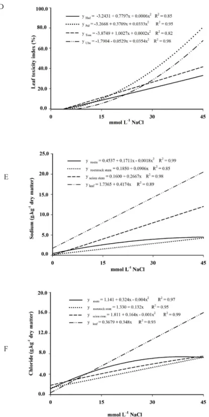
The low emission of shoots results in a de
-crease in leaf area, causing an excessive in-crease in
the Na + and Cl- levels in developed leaves (data not
shown) and trigger the process of senescence (Fig. 1C) and necrotic leaves (Fig. 1D), which alter the photosynthetic process (data not shown), reducing
the amount of photoassimilates important for plant
growth (MUNNS, 2002).
Besides reducing the emission of leaves, the
average leaf area, was lower with increasing salinity
in all cultivars. The decrease in average leaf area (Figure 1B) probably occurred due to the reduction
of meristematic activity, and mainly due to lower potential wall, initially caused by water and osmotic stress, leading to lower occurrence of cell turgor and consequently a lower cell expansion (RADIĆ
et al., 2005).
With the increase of the period of plant ex
-posure to stress, there was likely an ionic imbalance due to the excessive content of Na+ and Cl+ in tissues (Figures 1E and 1F). These results suggest that there
may have been limited ability to compartmentaliza
-tion of ions in the vacuole with consequent loss of membrane integrity, followed by the appearance of foliar symptoms of phytotoxicity (Figure 1D).
The leaf abscission was increased from a
concentration of 15 mmol L-1 NaCl (p≤0.05). In 15 mmol L-1 NaCl the rate of leaf abscission was similar to control plants (Figure 1C). The cultivar ‘Tommy
Atkins’ showed lower rates of leaf abscission, whereas the cultivars ‘Haden’ and ‘Uba’ have higher
rates of leaf abscission at concentrations of 15 mmol L-1 NaCl. These results show that the salinity toler -ance depends on the combination scion/rootstock,
recorded by the index of abscission around 70% in the cultivars ‘Haden’ and ‘Uba’, while cultivars ‘Palmer’ and ‘Tommy Atkins’ index showed around 30.0%,
both at the highest concentration of salts. Bañuls e Primo-Millo (1995) also observed different rates of leaf abscission depending on the rootstock in citrus cultivars subjected to salt stress.
Although it was observed reduction of leaf
emission (Figure 1A) and average leaf area (Figure
1B), only after the concentration of 30 mmol L-1
NaCl phytotoxicity symptoms in the leaves became apparent and were observed in all cultivars analyzed
(Figure 1D).
Initial fluorescence (F0)
The cultivars showed increases in initial fluorescence (F0) with increasing salinity (p≤0.05).
However, in 15 mmol L-1 NaCl with all cultivars the
photosynthetic apparatus appears to be little affected by salinity (Figure 2A). Hipkins and Baker (1986)
define F0 as a benchmark for determining the fluo
-rescence of the other variables. However, the F0 is
not always a constant; its value may increase if the PSII reaction center is compromised or if the transfer
of excitation energy from the antenna to the reac
-tion centers is impaired (SCHREIBER et al., 1998). Thus, the increase in F0 observed in mango cultivars
studied may be associated with damage to the pho
-tosynthetic apparatus, such as inactivation partially reversible or even irreversible reaction centers of
PSII (YAMANE et al., 1997), when these plants were subjected to higher concentrations of NaCl.
Maximal fluorescence (Fm)
The cultivars ‘Palmer’ and ‘Tommy Atkins’
had higher maximum fluorescence (Fm) at all NaCl
concentrations, while ‘Haden’ and ‘Uba’ had lower
mean values of Fm (Figure 2B). However, the great
-est mean maximum fluorescence (Fm) observed in
‘Palmer’ cannot be related to the greater efficiency of
energy capture and converted into chemical energy in step biochemistry of photosynthesis, as seen in the
actual efficiency of photosystem II (Figure 3B) and
the rate of electron transport (Figure 3D) was ob
-served in this cultivar when submitted to salt stress.
According to Silva et al. (2008) the highest values of Fm and Fv/Fm in ‘Palmer’ can be associated with
a higher number of active reaction centers of PSII (RC/CS0) as well as to higher levels of chlorophyll observed in this cultivar, since the increase of these
relations did not reflect a significant increase in en
-ergy absorption, indicating a low capacity to transfer
energy from the light-collecting systems (antenna). Thus, it appears in Figure 2B that the re-duction of Fm can be associated with increased
non-photochemical dissipation as heat (Figure 3C),
associated with the xanthophyll cycle (MÜLLER
et al., 2001). The reduction of electron acceptors such as NADP+, and energy requirements as ATP,
depending on enzyme-sensitive Na + and Cl- are
undermining the activity cycle of carbon fixation
(ABDEL-LATIF, 2008) can also be a reason for the
less than the rate of reduction, thus leading to a reduction of Fm with increasing salinity.
Quantum yield baseline (F
0/Fm)
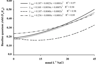
The quantum yield baseline (F0/Fm) was
increased due to the high concentrations of NaCl in the nutrient solution (Figure 2D). The higher
F0/Fm indicates that the initial rate of reduction of
the plastoquinone (Qa) was higher than the rate of plastoquinone reoxidation by b (Qb) and the activity of photosystem I (PSI) when plants were exposed
to higher concentrations of NaCl. Rohácek (2002) points to the increase relation F0/Fm as stress indica-tive suggesting normal values as standard, observed
between 0.14 and 0.20.
Potential quantum yield (F
v/Fm)
The increase in salt concentration provided
the decline in maximum quantum yield (Fv/Fm).
Decrease of efficiency of the capture of light energy
in all cultivars (Fig. 2C). All cultivars in the absence of stress (control) had Fv/Fm ratio within the range
established by Bolhàr-Nordenkampf and Orquist (1993). These authors have established the value
of Fv/Fm ratio between 0.750 and 0.850 in healthy
plants in the absence of stress (biotic or abiotic).
Effective quantum yield (F
v’/Fm’)
The increase in salt concentration brought the decline of effective quantum yield (Fv’/Fm’) (p≤0.05). At higher concentrations of NaCl (45 mmol L-1) F
v’/Fm’ showed significant decline, and
we observed 36.0% reduction in ‘Haden’, 25.3% in ‘Palmer’, and 34.8% in ‘Uba’, especially the cultivar
‘Tommy Atkins’ which showed a smaller reduc
-tion (18.7%) (Figure 3A). The term Fv’/Fm’ (Genty
parameter) represents the conversion efficiency of
the energy of electrons by open reaction centers of photosystem II (PSII) into chemical energy (SCH-REIBER et al., 1994). The effective quantum yield best represents the variations in quantum yield of photosynthesis than the ratio Fv/Fm (potential
quantum yield) and can be used together with pho
-tochemical quenching (qP) to estimate the rate of
electron transport is the photosynthetic photon flux (PPF) on incident photosynthetic tissue is known
(GENTY et al., 1989).
Photosystem II Efficiency (ΦPSII)
The efficiency of photosystem II (ΦPSII)
showed decreases with increasing the salt concen
-tration in the nutrient solution (p≤0.05). According to Schreiber et al. (1998), 1 mol of photons causes
excitation 1 μmol electrons of the chlorophyll in
this condition, it appears that the efficiency of photosystem II (ΦPSII) represents the proportion
of these electrons that are used during photochemi-cal reduction of NADP +. The cultivars ‘Palmer’ and
‘Uba’ showed the greatest reduction in ΦPSII, while
‘Haden’ and ‘Tommy Atkins’ showed tolerance up
to a concentration of 15 mmol L-1 NaCl (Figure 3B).
Thermal Dissipation (D)
The dissipation (quenching) of light energy focused on the photosynthetic apparatus to heat
in-creased with increasing NaCl in the nutrient solution
(p≤0.05). The increase in the dissipation of light en-ergy in the form of heat (D) reached average values
of 75% in the cultivar ‘Haden’ and the other 30%
when cultured in 45 mmol L-1 NaCl (Figure 3C).
Ac-cording to Schreiber et al. (1998), during application of saturating light pulse and after the tissue has been kept in the dark, the quantum yield of photochemi-cal process (Ph) reduces to zero and the emission of
fluorescence and heat dissipation become maximum
(Fm and Dm ). In plants under stress, the increase in non-photochemical in the form of thermal energy (D)
is correlated with increasing proportion xanthophyll/ chlorophyll and the rapid conversion of violaxanthin
to zeaxanthin in the presence of light, featuring pho
-toinhibition (Demmig-Adams; Adams, 1992). Electron Transport Rate (ETR)
The mango cultivars analyzed showed a re
-duction in transport rate (ETR) with increasing NaCl concentration (p≤0.05). However, the reduction in ETR occurred differentially among cultivars (Figure 3D). In 45 mmol L-1 NaCl, only the cultivar ‘Tommy
Atkins’ showed less reduction (29.8%) of ETR, with the other cultivars showing an average reduction of
50% in the ETR. The electron transport reduction
mediated by PSII and PSI in isolated thylakoid mem-brane of Synechococcussp. subjected to salt stress was
reported by Allakhverdiev et al. (2000). The effect of
ionic caused by the influx of sodium ions through the channels K+/Na+ increases the concentration of salt in the cytosol and causes the dissociation of plastocyanin
or cytochrome c553 complex PSI causing decrease
in the rate of electron transport mediated by PSI and
continua... A
B
FIGURE 1 – (A) Number of emitted leaves (leaf.tree-1), (B) mean leaf area (cm2.leaf-1), (C) leaf abscission
index (%), (D) leaf toxicity index (leaf necrosis area or chlorosis/leaf total area), (E) sodium
content (g.kg-1 dry matter) and (F) chloride content (g.kg-1 dry matter) in mango tree cultivars ‘Haden’ (Had), ‘Palmer’ (Pal), ‘Tommy Atkins’ (Tom) and ‘Ubá’ (Uba) grafted on rootstock
‘Imbú’, grown in nutrient solution and submitted to salt stress.
D
E
continua... A
B
FIGURE 2– (A) Initial fluorescence (F0), (B) maximal fluorescence (Fm), (C) potential quantum yield (Fv/Fm), (D) baseline quantum yield (F0/Fm) in mango tree cultivars ‘Haden’ (Had), ‘Palmer’
(Pal), ‘Tommy Atkins’ (Tom) and ‘Ubá’ (Uba) grafted on rootstock ‘Imbú’, grown in nutrient
solution and submitted to salt stress.
D
continua... A
C
D
FIGURE 3 – (A) Effective quantum yield (Fv’/Fm’), (B) photosystem II efficiency (ΦPSII), (C) thermal dissipation (D), (D) electron transport rate (ETR) in mango tree cultivars ‘Haden’ (Had),
‘Palmer’ (Pal), ‘Tommy Atkins’ (Tom) and ‘Ubá’ (Uba) grafted on rootstock ‘Imbú’, grown
in nutrient solution and submitted to salt stress.
CONCLUSIONS
The mango cultivars analyzed showed re-ductioninleaf andtheaverage areaof leaveswhen
exposedtoconcentrationsgreater than15mmolL-1
NaCl. There were novisible symptomsoffoliar
phy-totoxicityat a concentration of15mmolL-1
NaCl.
All chlorophyll fluorescenceparameters evaluated
were altered in thepresenceofNaClin the nutrient
solution. The decreaseinphotochemical efficiency of photosystem II, in differing degrees, occurred in allcultivarsandwasenhancedat concentrations above 15mmolL-1
NaCl.
REFERENCES
ABDEL-LATIF, A. Phosphoenolpyruvate carboxyl
-ase activity of wheat and maize seedlings subjected to
salt stress. Australian Journal of Basic and Applied Sciences, Punjab, v. 2, n. 1, p. 37-41, 2008.
ALLAKHVERDIEV, S. I.; MURATA, N. Salt stress
ALLAKHVERDIEV, S. I.; SAKAMOTO, A.; NISHIYAMA, Y.; INABA, M.; MURATA, N. Ionic
and osmotic effects of NaCl-induced inactivation of Photosystems I and II in Synechococcus sp. Plant Physiology, Rockville, v. 123, n. 3, p. 1047-1056. 2000.
AUDRY, P.; SUASSUNA, J. A salinidade das águas
disponíveis para a pequena irrigação no sertão do Nordeste: caracterização, variação sazonal, limita -ção de uso. Recife: CNPq, 1995. 128 p.
BAKER, N. R. Chlorophyll Fluorescence: A probe
of photosynthesis in vivo. Annual Review of Plant Physiology, Boca Raton, v. 59, p. 89-113, 2008.
BAÑULS, J.; PRIMO-MILLO, E. Effects of salinity
on some Citrus scion-rootstock combinations. An-nals of Botany, Oxford, v. 76, n. 1, p. 97-102. 1995.
BOLHÀR-NORDENKAMPF, H. R.; OQUIST, G. Chlorophyll fluorescence as a tool in photosynthesis research. In: HALL, D. O.; SCURLOCK, J. M. O.; BOLHÀR-NORDENKAMPF, H. R.; LEEGOOD,
R. C.; LONG, S. P. (Ed.). Photosynthesis and
production in a changing environment: a field
and laboratory manual. London: Chapman & Hall, 1993. p. 193-206.
DEMMIG-ADAMS, B.; ADAMS III, W. W. Pho
-toprotection and other response of plants to high light stress. Annual Review Plant Physiology and Plant Molecular Biology, Palo Alto, v. 43, n. 1, p. 599-626. 1992.
GENTY, B.; BRIANTAIS, J. M.; BAKER, N. The
relationship between quantum yield of photosyn
-thetic electron transport and quenching of chlorophyll
fluorescence. Biochimica et Biophysica Acta,
Am-sterdam, v. 990, n. 1, p. 87-92. 1989.
HAVAUX, M.; ERNEZ, M.; LANNOYE, R. Cor
-relation between heat tolerance and drought toler
-ance in cereals demonstrated by rapid chlorophyll
fluorescence tests. Journal of Plant Physiology,
Leipzig, v. 133, n. 5, p. 555-560. 1988.
HECK, R. J.; TIESSEN, H.; SALCEDO, I. H.; SAN
-TOS, M. C.Soil Chemical Changes under Irrigated Mango Production in the Central São Francisco River
Valley, Brazil. Journal of Environmental Quality,
Riverside, v. 32, n. 4, p. 1414-1421. 2003.
HIPKINS, M. F.; BAKER, N. R. Spectroscopy. In:
HIPKINS, M. F.; BAKER, N. R. (Eds.)
Photosyn-thesis-energy transduction: a pratical approuch.
Oxford: IRL Press, 1986. p. 51-101.
KRAUSE, H.; WEIS, E. Chlorophyll Fluorescence
and Photosynthesis: The Basics. Annual Review of Plant Physiology and Plant Molecular Biology, Palo Alto, v. 42, n. 1, p. 313-349. 1991.
LI-COR. LI-3100 area meter instruction manual. Nebraska: Li Cor, 1987. p.33. (Publication, 7903-20).
MÜLLER, P.; LI, X. P.; NIYOGI, K. K.
Non-Pho-tochemical Quenching. A Response to Excess Light Energy. Plant Physiology, Rockville, v.125, n. 4, p. 1558-1566. 2001.
MUNNS, R. Comparative physiology of salt and
water stress. Plant, Cell and Environment, Oxford,
v. 25, n. 2, p. 239-250, 2002.
RADIĆ, S.; PROLIĆ, M.; PAVLICA, M.; PEV
-ALEK-KOZLINA, B. Cytogenetic effects of osmotic
stress on the root meristem cells of Centaurea ragu-sina L. Environmental and Experimental Botany,
Oxford, v. 54, n. 3, p. 213-218, 2005.
ROHÁCEK, K. Chlorophyll fluorescence param
-eters: the definitions, photosynthetic meaning, and
mutual relationships. Photosynthetica, Prague, v. 40, n. 1, p. 13-29, 2002.
SCHREIBER, U.; BILGER, W.; NEUBAUER, C.
Chlorophyll fluorescence as a nonintrusive indica
-tor for rapid assessment of in vivo photosynthesis.
In: SCHULZE, E. D.; CALDWELL, M. M. (Ed.).
Ecophysiology of photosynthesis. Berlin: Springer, 1994. p. 49-70.
SCHREIBER, U.; BILGER, W.; HORMANN, H.;
NEUBAUER, C. Chlorophyll fluorescence as a di
-agnostic tool: basics and some aspects of practical relevance. In: RAGHAVENDRA, A. S. (Ed.). Pho-tosynthesis: a comprehensive treatise. Cambridge: Cambridge University Press, 1998. p. 320-336.
SILVA, M. M.; ZAMPERLINI, G. P.; COSTA, A. N.; COSTA, A. F.; CAETANO, L. C. S.; SILVA, D.
SMILLIE, R. M.; NOTT, R. Salt tolerance in crop
plants monitored by chlorophyll fluorescence in
vivo . Plant Physiology, Rockville, v. 70, n. 4, p. 1049-1054, 1982.
UNIVERSIDADE FEDERAL DE VIÇOSA. Sis-tema de análises estatísticas e genéticas - SAEG. Versão 8.0. Viçosa: Ed. UFV, 2000.
YAMANE, Y.; KASHINO, Y.; KOILE, H.; SATOH,
K. Increase in the fluorescence F0 level reversible