Corresponding author: Dr. Maxwel Adriano Abegg. ORCID: 0000-0002-0328-1122 e-mail: [email protected]
Received 20 April 2018 Accepted 4 July 2018
Short Communication
Journal of the Brazilian Society of Tropical Medicine Vol.:52:e-20180152: 2019 doi: 10.1590/0037-8682-0152-2018
Anti-
Candida
and anti-quorum sensing activity of airborne
microorganisms detected by a rapid method
Andria Lopes Cruz
[1], Fabiana Silva de Souza
[1]and Maxwel Adriano Abegg
[1],[2][1]. Curso de Farmácia, Instituto de Ciências Exatas e Tecnologia, Universidade Federal do Amazonas, Itacoatiara, AM, Brasil. [2]. Laboratório de Micologia, Programa de Pós-Graduação Stricto Sensu em Ciência e Tecnologia para Recursos Amazônicos,
Universidade Federal do Amazonas, Itacoatiara, AM, Brasil.
Abstract
Introduction: Introducing new antibiotics to the clinic is critical. Methods: We adapted a plate method described by Kawaguchi and coworkers in 20131 for detecting inhibitory airborne microorganisms. Results: We obtained 51 microbial colonies antagonist
to Chromobacterium violaceum, purified and retested them, and of these, 39 (76.5%) were confirmed. They comprised 24
bacteria, 13 fungi, and 2 yeasts. Among the fungi, eight (61.5%) produced active extracts. Among the bacterial, yeast, and fungal strains, 17 (44.7%) and 12 (31.6%) were active against Candida albicans and Candida parapsilosis, respectively. Conclusions:
The proposed screening method is a rapid strategy for discovering potential antibiotic producers.
Keywords:Candida sp. Chromobacterium violaceum. Quorum sensing. Antimicrobials.
According to Lewis2, the rise and spread of antimicrobial
resistance presents a unique challenge to both science and medicine. Resistance to antimicrobials has led to the selection
of a list of six priority pathogens (two gram-positive and four gram-negative) termed ESKAPE (Enterococcus faecium,
Staphylococcus aureus, Klebsiella pneumoniae, Acinetobacter baumannii, Pseudomonas aeruginosa, and Enterobacter
species), which require rapid development of new antibiotics (reviewed in3). In case of fungal infections, invasive candidiasis
is increasingly caused by non-albicansCandida intrinsically resistant to antifungals4.
The crisis in antimicrobial discovery, has prompted experts to call for the revival of natural product drug discovery2.
However, the enormous background of known and/or nuisance compounds presents a serious barrier to discovery2. Despite this,
recent results like those presented by Maffioli et al.5 bring hope
to the search for new leads from microbial-extract screening. An alternative to looking for drugs that kill bacteria is to search for molecules that target virulence factors with minimal to no effect on growth6.
Bacteria coordinate gene expression as a function of cell density in a communication mechanism named quorum sensing
(QS)6. Therefore, bacteria are capable of performing tasks as
a group, including migration to more favorable environments, biofilm formation, virulence gene expression, bacteriocin and antibiotic production, bioluminescence, and pigment production, among others6,7. Considering the importance of QS, the search for inhibitors against this mechanism has become a research target8.
In QS, signaling molecules are usually called autoinducers and QS inhibition can be accomplished by interfering with the synthesis, secretion and degradation of these molecules, as well as by hampering their recognition by receptor proteins such as LuxR type homologs6,7. Nowadays, despite promising results9 (also reviewed in depth in6), according to Defoirdt6, it will probably take many more years before these agents can be used in the clinic.
In 2013, Kawaguchi and coworkers1 proposed a supposedly
more efficient and faster method to select candidate fungal
producers of anti-Candida albicans antibiotics. In this study, we adapted this method using the strain Chromobacterium violaceum
ATCC 12472, which naturally produces a violet pigment (violacein), to screen for antibiosis, anti-quorum sensing
2/5
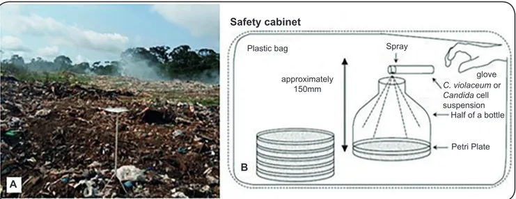
FIGURE 1: Example of a collection site for airborne microorganisms in the municipal dump of Itacoatiara, Amazonas. Three Luria-Bertani Petri dishes supplemented with 5 g/L sodium chloride were fully opened and exposed for 15 minutes at a height of 1.2 m from the soil (A)(see materials and methods). In (B), method for the inoculation of Chromobacterium violaceum or Candida sp. cell suspensions in Petri dishes with previously collected air microorganisms. A cell suspension of approximately 60 µL was sprayed on the air collection plates kept in a plastic bag installed in a biological safety cabinet. Adapted from Kawagushi et al.1.
collect air samples in the municipality of Itacoatiara, Amazonas, Brazil.
The air sampling of microorganisms was preferably performed in places with high circulation of people, such as a university campus and municipal fairs, particularly in places
with selling grain products, such as flour. Considering the
high amount of fungi, the air in the municipal dump was also sampled. Collection was also carried out in forest environments around the municipality of Itacoatiara. The twelve locations of air sampling and their geographic coordinates were as follows:
Private Land in Itacoatiara (S 03o04’31.0’’; W 058o27’38.4’’),
Canaçari District 1 (S 03o03’56.6’’; W 058o27’13.1’’), Canaçari
District 2 (S 03o03’40.6’’; W 058o26’33.4’’), Canaçari District
3 (S 03o03’09.5’’; W 058o26’01.1’’), University Restaurant
(S 03o08’34.746’’; W 058o25’55.258’’), Parking Lot of the
University Campus (S 03o8’35.678’’; W 58o25’52.231’’);
University Campus Autoclave Room (S 03o8’33.439’’;
W 58o25’53.976’’); University Campus Cleaning Room
(S 03o8’33.626’’; W 58o25’53.368’’), University Campus Tree
1 (S 03o8’35.678’’; W 58o25’52.231’’), Itacoatiara Municipal
Dump (S 03o8’50.744’’; W 58o25’39.727’’), University Campus
Tree 2 (S 03o8’33.77’’; W 58o25’52.35’’), and Municipal Flour
Market (S 03o8’33.77’’; W 58o25’52.35’’).
To obtain anemophilous (airborne) microorganisms, three Petri dishes with a modified Luria Bertani (LB) agar
supplemented with 5 g/L NaCl instead of 10 g/L were used.
This modification was made considering a single observation where we noticed a more diverse growth of filamentous fungi
with 5 g/L and no apparent alterations in violacein production by C. violaceum (data not shown). The plates were opened for
15 min with the lid off and faced downwards, at a height of
1.2 m from the soil, using a flat support; presence of walls was
considered and an attempt was made to collect samples across
the sampled area. After exposure, the plates were transported to the laboratory and incubated at 30°C for up to three days,
with 12-hour dark/light cycles (in order to better simulate day and night conditions), to form visible colonies. In parallel, a
C. violaceum cell suspension was prepared from overnight grown cells and adjusted to OD600 = 0.15, and approximately
60 µL was sprayed on the plates using an atomizer (the spray was applied 3 to 4 times in order to achieve this volume) after the
three days of growth, as described by Kawaguchi1, and shown
in Figure 1. Precautions were taken such that the suspension completely covered the plate. After 15 minutes of drying at
room temperature by opening the plates in a laminar flow
bio-safety cabinet, the plates were incubated in inverted position at
28°C for 24-36h. Inhibition of QS was indicated by an opaque
halo around the fungal colonies and classic antibiosis was indicated by a clear halo7.
Potential inhibitory microorganisms that formed a halo of inhibition were repeatedly subcultured on potato dextrose agar
(PDA) plates to obtain pure cultures and these were used to
repeat the test against C. violaceum (Figure 2).
The anemophilous microorganisms were first characterized
as filamentous fungi, yeasts, or bacteria based on their
macroscopic and microscopic morphology. For the filamentous fungal genus identification, the evaluated macromorphological
characteristics included colony size, texture, relief, and pigmentation. Micromorphological analyses were performed using the micro-cultivation technique in PDA to identify the
vegetative structures and especially the specific reproductive
structures of the genus, as described by Watanabe11.
To obtain filamentous fungal extracts, the 13 isolated fungi were grown on PDA for three days. After that, six plugs
of 6 mm in diameter were cut out of the agar and added to
30 mL of Czapeck-Dox broth and six plugs were added to
approximately 150mm
Plastic bag Spray
glove C. violaceum or Candida cell suspension
Half of a bottle
Petri Plate
Safety cabinet
FIGURE 2: Plates illustrating the results of direct tests with the rapid method of airborne microorganisms against Chromobacterium violaceum
applied by spraying. The red circles demonstrate colonies with inhibitory activity in A, B, and C. D, E, and F, demonstrate the inhibitory activity against
C. violaceum by microorganisms isolated from colonies from the rapid method plates. In G, a halo of inhibition against C. albicans and in H, a halo of inhibition against C. parapsilosis is observed in the plug diffusion test.
A B C
D E F
G H
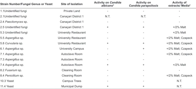
TABLE 1: Inhibitory activity of anemophilous fungi isolated by a rapid screening method against Candida sp. and Chromobacterium violaceum.
Strain Number/Fungal Genus or Yeast Site of Isolation Activity on albicansCandida * Candida parapsilosisActivity on extracts/ MediaActivity of ≠
1.1Unidentified fungi Private Land - -
-2.1Unidentified fungi Canaçari District 1 N.T. N.T.
-2.4 Paecilomyces sp. Canaçari District 1 - -
-2.5 Unidentified fungi Canaçari District 1 + + +/2% Malt
5.3 Unidentified fungi University Restaurant - - +/2% Malt
5.5 Aspergillus sp. University Restaurant - - +/2% Malt; Czapeck
5.8 Curvularia sp. University Restaurant + + +/2% Malt; Czapeck
6.1 Aspergillus sp. University Campus - + +/2% Malt; Czapeck
7.1 Aspergillus sp. Autoclave Room - - +/2% Malt; Czapeck
7.3 Aspergillus sp. Autoclave Room - -
-7.4 Aspergillus sp. Autoclave Room + + +/2% Malt
8.2 Fusarium sp. Cleaning Room - -
-8.4 Penicillium sp. Cleaning Room - - +/2% Malt; Czapeck
10.3 Yeast Campus Trees - - N.T.
11.4 Yeast Municipal Dump + + N.T.
Legend: *activity determined by a plug diffusion assay (see methods); ≠activity against Chromobacterium violaceum ATCC 12472 determined by a hole test in LB agar (see methods). N.T.: not tested (the fungal colonies did not develop sufficiently to perform the test).
30 mL of 2% malt extract broth. The fungi were then grown
at ambient temperature for three days in a shaker at 150 rpm. The culture was mixed with 15 mL of absolute ethanol and mixed for one day. The extracts were dried in aseptic conditions
and resuspended to a final concentration of 10 mg/mL with
sterile distilled water. The extracts were then tested against
C. violaceum using the agar well diffusion test applying 50 µL
of extract to each hole12. The tests were performed in triplicate
with sterile distilled water as the negative control (Table 1).
The tests of antagonism for the fungi and yeasts against
Candida albicans ATCC 18804 and Candida parapsilosis
ATCC 22019 were performed using the agar plug diffusion
method. According to Balouiri et al.12, this method is often used
to highlight the antagonism between microorganisms. For this, the fungi and were previously grown on PDA plates on yeast
extract-peptone-dextrose media (YPD), respectively, for three days and the bacteria were grown on LB media for 36 hours; 6 mm plugs were aseptically cut with a sterile cork borer and transferred to YPD plates previously inoculated by the pour plate technique along with 100 µL suspension of fresh Candida sp. cells at OD600 = 0.15. Measurements such as inhibition halos were performed after 24–48h of incubation at 30°C.
By opening three plates in twelve different locations, 51 microbial colonies that manifested antagonism against
C. violaceum were obtained in the proposed rapid screening method. These colonies were isolated in pure culture and
retested against the same strain, and 39 cultures were found
to manifest halos of inhibition whereas 12 did not show any
inhibition (Figure 2), resulting in false positives. Strains 7.3 (Aspergillus sp.) and 7.4 (also an Aspergillus strain) produced
the largest halos of inhibition. Of the 39, the halos were turbid in five cases, and showed the presence of microorganisms
when observed through the microscope, suggesting an anti-QS
activity at first.
4/5
above. For eight filamentous fungal strains, the extract of at least
one medium was active against C. violaceum (Table 1). It was
observed that in most cases, strains with active extracts in one of the media also showed activity in the other media tested. Despite the apparent conidiation of the fungi in PDA by means of the reference used11, we could not attribute four fungi to any
given genus with an acceptable level of confidence (Table 1).
In the plug diffusion test against Candida sp., among 13
fungi strains and two yeasts, four (33.3%) were active against
C. albicans, and five (41.7%) against C. parapsilosis by
exhibiting a clear halo of inhibition (Figure 2). Interestingly, in
most cases, strains active against C. albicans were also active against C. parapsilosis. Of the 24 bacterial strains isolated with activity against C. violaceum, 13 strains (3.1, 4.4, 5.6,
5.7, 9.2, 10.1, 10.2, 10.5, 11.2, 12.1, 12.3, 12.4, and 12.5) (54.2 %) showed activity against C. albicans and seven strains (5.6,
9.2, 10.1, 10.4, 12.1, 12.2, and 12.4) (29.2 %) showed activity
against C. parapsilosis on the agar plug diffusion test.
Although strains active against C. violaceum could be further tested against other bacteria, we chose to examine their activity in altering Candida growth considering the importance of Candida resistance4, and a previous observation that a
QS molecule (3-oxo-C12 homoserine lactone) could inhibit
C. albicans filamentation13.
There is some evidence in literature that activity against
C. violaceum (a gram-negative species) can extend to other
bacterial species, including gram-negative species, as shown by carvacrol14 and other compounds9. We recognize that in future
research, active strains and extracts must be further screened against additional bacterial species and a panel of ESKAPE species3 and of pathogenic Candida species4.
The apparent QS inhibitory activity of five strains that
manifested turbid halos in the plate test employing C. violaceum
ATCC 12472, can only be considered as a preliminary result, as colorless transparent halos can be difficult to discern from
QS inhibition, which forms a turbid colorless halo7. In this
case, strain extracts have to be further tested in a confirmatory
violacein inhibition assay7.
In QS inhibitory assays, an important limitation is that the QS-regulated phenotypes are often co-dependent on other factors and/or dependent on the metabolic activity of cells. Therefore, evidence of QS disintegration is not always strong and many compounds reported to inhibit QS have proven to be ineffective when studied in greater depth. To avoid this, the impact of the candidate on additional QS regulated phenotypes
(mostly QS-regulated virulence factors) must be verified6.
Additionally, it cannot be excluded that the QS inhibitory activity was due to enzymatic activity, as we did not treat the extracts with proteinase K. Burt et al.14 pointed out that,
carvacrol and many other phytochemicals initially target the bacterial membrane. At minimum inhibitory concentration
(MIC) values, complete leakage and loss of membrane
potential is observed. It is possible that lower concentrations of these compounds could have smaller destabilization effects on these membranes, which could reduce the QS capability of bacteria14.
The process of discovering antibiotics usually includes
several steps: (1) isolation of microorganisms (usually 2–3 days), (2) pure culture (3–5 days), (3) identification of isolated microorganisms to eliminate duplicate strains (14–21 days), (4) testing whether or not the culture broths or extracts of these microorganisms show antimicrobial activity (7–8 days) and (5) re-culture of candidate microorganisms to observe the reproducibility of antimicrobial activity (6–8 days). This traditional method usually takes 5–7 weeks15. The rapid method
proposed by Kawagushi et al.1 can probably accelerate the
discovery of antibiotics, considering that prior isolation of microorganisms and a screening test for each isolated strain is unnecessary.
This study also indicates that it is possible to reduce the
conventional procedure that takes five to seven weeks to a
shorter time, with more hits considering the extracts obtained. Although there are quite a few methods for the in vitro evaluation of antimicrobial activity as recently reviewed by Balouiri et al.12,
in the adaptation proposed here, the high contrast brought by the violet pigment produced by C. violaceum seems facilitate
identification of halos of inhibition in direct plate methods, and
renders prior isolation of microorganisms unnecessary.
Acknowledgements: The authors acknowledge the Brazilian National Research Council – CNPq for financial support.
Conflicts of interest: The authors declare that there are no conflicts of interest.
Financial support: This study was supported by the Brazilian National Research Council – CNPq, Projeto Universal 2014, grant number: 458508/2014-4.
REFERENCES
1. Kawaguchi M, Nonaka K, Masuma R, Tomoda H. New method for isolating antibiotic-producing fungi. J Antibiot. 2013;66(1):17-21. 2. Lewis K. New approaches to antimicrobial discovery. Biochem
Pharmacol. 2017;134(1):87-98.
3. Singh SB, Young K, Silver LL. What is an "ideal" antibiotic? Discovery challenges and path forward. Biochem Pharmacol. 2017;133(1):63-73
4. Schwartz IS, Patterson TF. The Emerging Threat of Antifungal Resistance in Transplant Infectious Diseases. J Antimicrob Chemother. 2018;20(3):2.
5. Maffioli SI, Zhang Y, Degen D, Carzaniga T, Del Gatto G, Serina S et al. Antibacterial Nucleoside-Analog Inhibitor of Bacterial RNA Polymerase. Cell. 2017;169(7):1240-1248.e23.
6. Defoirdt T. Quorum-Sensing Systems as Targets for Antivirulence Therapy. Trends Microbiol. 2018;26(4):313-28.
7. Rodrigues AC, Oliveira BD, Silva ERD, Sacramento NTB, Bertoldi MC, Pinto UM. Anti-quorum sensing activity of phenolic extract from Eugenia brasiliensis (Brazilian cherry). Food Sci Technol.
2016;36(2):337-43.
biofilm formation in Salmonella. Microb Pathog. 2018;S0882-4010(18):30406-6.
9. El-Gohary NS, Shaaban MI. Synthesis, antimicrobial, antiquorum-sensing, antitumor and cytotoxic activities of new series of fused [1, 3, 4] thiadiazoles. Eur J Med Chem. 2013;63:185-95.
10. Sobral LV, Melo KN, Souza CM, Silva SF, Silva GIR, Silva AIF, et al. Antimicrobial and enzymatic activity of anemophilous fungi of a public university in Brazil. An Acad Bras Cienc. 2017;89(3):2327-40. 11. Watanabe T. Pictorial atlas of soil and seed fungi: morphologies of
cultured fungi and key to species. 3th Ed. New York: CRC Press, 2010, 397p.
12. Balouiri M, Sadiki M, Ibnsouda SK. Methods for in vitro evaluating antimicrobial activity: A review. J Pharm Anal. 2016;6(2):71-9. 13. Hogan DA, Vik Å, Kolter R. A Pseudomonas aeruginosa
quorum-sensing molecule influences Candida albicans morphology. Mol Microbiol. 2004;54(5):1212-23.
14. Burt SA, Ojo-Fakunle VT, Woertman J, Veldhuizen EJ. The natural antimicrobial carvacrol inhibits quorum sensing in
Chromobacterium violaceum and reduces bacterial biofilm
formation at sub-lethal concentrations. PLoS One. 2014;9(4):e93414. 15. Bull AT, Goodfellow M, Slater JH. Biodiversity as a source of