UNIVERSIDADE DE LISBOA
FACULDADE DE CIÊNCIAS
DEPARTAMENTO DE BIOLOGIA ANIMAL
Effect of mushroom polysaccharides and olive
phenolic compounds on human carcinoma cells
Diana Jorge Machado de Figueiredo da Fonseca Martinho
Dissertação
Mestrado em Biologia Humana e Ambiente
UNIVERSIDADE DE LISBOA
FACULDADE DE CIÊNCIAS
DEPARTAMENTO DE BIOLOGIA ANIMAL
Effect of mushroom polysaccharides and olive phenolic compounds on human carcinoma cells
Diana Jorge Machado de Figueiredo da Fonseca Martinho Mestrado em Biologia Humana e Ambiente
Dissertação orientada por:
Professor Doutor Amin Karmali ISEL – Instituto Superior de Engenharia de Lisboa
CIEQB – Centro de Investigação de Engenharia Química e Biotecnologia. Professora Doutora Deodália Dias
FCUL- Faculdade de Ciências da Universidade de Lisboa DBA - Departamento de Biologia Animal
Dedicatória:
Dedico esta tese de mestrado aos meus Pais e irmã, Cuja paciência, amor e carinho me ajudaram a superar todas as dificuldades
Às minhas avós, por serem modelos de coragem e bondade a seguir. “Tu podes, assim tu queiras”
i
Nota Prévia
A elaboração desta dissertação foi escrita na língua Inglesa, com o intuito de ser procedida a recolha de dados para a elaboração de um artigo científico na mesma língua.
ii
Agradecimentos
Gostaria de agradecer ao Prof. Armin Karmali oportunidade que me providenciou para a realização desta tese.
Ao Prof.. Eduardo Rosa, do Departamento de Agronomia da UTAD o ter cedido as amostras de oliveira.
Gostaria de agradecer a Profª. Sónia Martins e à Engª Magda Semedo, toda a ajuda e paciência ao longo da execução deste trabalho, características que fazem delas grandes professoras.
À Telma Reis todas as palavras de encorajamento e força e por ter sido a minha mentora no conhecimento dos cantos a casa e modo de trabalho.
À Engª. Cidália Bandarra e à Cláudia Coelho a ajuda, compreensão e boa disposição que sempre demostraram, que junto com os acima mencionados, promoveram um bom ambiente para o desenvolvimento de projectos.
À Prof. Deodália, todo o acompanhamento, auxílio e força que me deu ao longo deste mestrado, mostrando que ser coordenadora é mais que gerir um curso, é gerir alunos, pessoas em busca do seu futuro, e guiá-las o melhor que pode, fazendo dela, uma grande professora, não só didáctica, mas também de vida.
À Joana Patriarca ter-se revelado uma excelente colega de laboratório, uma grande companheira e amiga, que me ajudou a superar muitas frustrações e problemas, tanto profissionais como pessoais.
À Bruna Pinto a amizade e força para continuar. À Catarina Dourado o apoio e a amizade.
iii
Gostaria de agradecer aos meus primos, Tânia e Tobias, por me oferecerem sempre um local de refúgio, amizade e compreensão.
E a todo o pessoal do ISEL, que me acolheu como amiga, me aturou todas as tormentas e me fez rir sempre que precisei…
E finalmente, mas não com menos importância, muito pelo contrário, aos meus pais e especialmente à minha irmã, que demonstraram uma coragem sem limites, que me apoiaram a cada passo, apesar de todas as dificuldades, e que nunca deixaram de acreditar em mim e me encorajaram sempre a fazer o mesmo.
Por toda a amizade demonstrada por todos, e por acreditarem sempre que eu seria capaz, um grande e sincero OBRIGADO.
iv
Resumo
Hoje em dia, devido ao prolongamento da esperança média de vida, bem como o aumento das agressões sofridas pelo meio ambiente envolvente, o cancro é uma das doenças mais proeminentes no seculo XXI.
Neoplasmas, conjuntos anormais de células cromossomicamente mutadas, levam ao crescimento relativamente autónomo de tecido danificado. As causas destas mutações variam entre internas - condições imunitárias, hormonais, heritabilidade - ou externas - como tabagismo, exposição a radiação ou agentes químicos infecciosos (Zaidman et al., 2005).
As células neoplásicas caracterizam-se pela perda das suas funções especializadas e ganham por sua vez características biológicas tais como a produção de sinais de crescimento, inibição da apoptose, elevado potencial replicativo e a capacidade de invadir tecidos circundantes (Zaidman et al., 2005; Zong et al.,, 2012). A esta metastização dos tecidos está associada 90% da responsabilidade da morte por cancro (Zong et al.,, 2012).
Apesar da eficiência dos fármacos gerados até hoje no combate ao cancro, o seu efeito tóxico causa na maioria dos casos um decréscimo acentuado na qualidade de vida dos doentes. Assim, torna-se importante descobrir novos agentes anti cancerigenos com efeitos tóxicos mais baixos (Cao et al., 2010) que possam ser utilizados em terapias complementares.
Virámo-nos então de novo para as tradições mais antigas da medicina Chinesa e Mediterrânea.
Polissacáridos são biopolímeros constituídos por várias unidades de monossacáridos ligados através de ligações glicosidícas, que podem apresentar diferentes conformações, não só nas suas unidades constituintes, mas também na conformação das suas ligações, atribuindo-lhes assim diversas funções biológicas. Estes estão presentes desde o armazenamento de energia (amido), formação de componentes estruturais (celulose), participantes na comunicação célula a célula, possuindo ainda papéis importantes no sistema imunitário, na fertilização, desenvolvimento, agregação plaquetária e prevenção de patogéneses (Zong et al., 2012).
v
Desde a medicina ancestral chinesa que os cogumelos têm sido reportados como tendo propriedades imunomoduladoras - actuando como modificadores da resposta biológica - e possuindo propriedades antivirais, antimicrobiais, circulatórias e nas ultimas décadas anti cancerigenas. (Wasser, 2010). A descoberta do Lentinian, um polissacárido com propriedades anti cancerígenas, despoletou nos anos 60 a pesquisa que tem sido dedicada ate hoje ao campo dos polissacáridos (Cao et al., 2010).
Ao longo do tempo, diversos polissacáridos produzidos por cogumelos, têm demonstrado largos efeitos inibidores em vários tipos de tumores como Sarcoma 180, carcinoma de Lewis e Yoshida Sarcoma (Zhang et al., 2007).
Existem três mecanismos propostos de actuação destes polissacáridos: (1) actividade preventiva, (2) aumento da resposta imunitária do hospedeiro e (3) inibição directa quando em contacto com células cancerígenas (Zhang et al., 2007).
Na sua maioria os cogumelos que possuem polissacáridos biologicamente activos pertencem ao grupo dos basidiomicetes contendo cerca de 700 espécies descritas (Wasser, 2002).
O seu poder antioxidante também desempenha um papel fundamental contra o desenvolvimento cancerígeno. Anti oxidantes naturais encontrados em fruta, vegetais, cogumelos e outros recursos alimentares têm sido investigados para prevenção de doenças coronárias e outras como o cancro (Kozarski et al.,2012).
Assim, na procura de compostos não tóxicos que possuíssem propriedades anticancerígenas investigou-se também um pouco dos efeitos que as folhas de oliveira possam ter no combate ao cancro.
A oliveira (Olea europaea) é uma árvore da família das Oleaceae, (Zaid et al., 2012) com folhas alongadas, que se encontra ao longo de toda região Mediterrânea desde há mais de 7000 anos (Lalas et al., 2011).
Ao longo da história as folhas de Olea europaea têm sido exploradas para a prevenção de hipertensão, carcinogénese, diabetes, arteroesclerose bem como outras doenças do foro mais comum (Bouallagui et al., 2011; Zaid et al., 2012). A folha de oliveira foi ainda utilizada ao longo da história como um meio de combate á malária (Lee et al., 2009)
vi
A incidência de cancro ao longo do mediterrâneo tem sido das mais baixas quando comparada com outras partes do mundo, principalmente quando nos referimos ao cancro da mama, do endométrio, próstata e leucemia. (Bouallagui et al., 2011; Zaid et
al., 2012)
Assim reunimos neste trabalho, o estudo dos efeitos de algumas espécies conhecidas (e outras menos conhecidas) de cogumelos, com o estudo dos efeitos de algumas variedades de oliveiras nacionais, na inibição do crescimento de células tumorais.
Para isso, foram efectuados vários doseamentos de polissacáridos, proteínas, açucares e compostos fenólicos e foram aplicadas diversas amostras das espécies amostradas a quatro linhas de carcinoma humano: HeLa, carcinoma cervical, A549 carcinoma pulmonar; A431 carcinoma epidermóide e OE21 carcinoma esófago-faringeal.
De entre os variados resultados obtidos, pudemos concluir que a aplicação de extra polissacáridos da estirpe Ganoderma carnosum revelou um alto teor inibitório, e que os intra polissacáridos de todas as estirpes amostradas mostraram uma capacidade anti-oxidativa acima de 50%.
As folhas de oliveira por sua vez, foram também capazes de mostrar um potencial anti proliferativo das células tumorais bastante satisfatório.
Acreditamos que nestes compostos podem estar presentes as condições para a sua aplicação como terapia complementar às terapias anticancerígenas existentes.
Palavras chave: anti cancerígeno, polissacáridos; cogumelos, compostos fenólicos,
vii
Abstract
Cancer is widely known nowadays as the disease of the century. Not only by the number of infected people worldwide but also by the amount of research dedicated to this field and its increasing success rate.
Neoplasms, which are abnormal masses or colonies of cells produced by a relatively autonomous new growth of tissue, arise normally by the clonal expansion of a single cell that has undergone mutation in the chromosomal DNA, caused by a chemical, physical or biological agent (Zaidman et al., 2005). One of the most characterizing features of cancers, besides it’s ability to resist treatment, it’s his ability to metastasize and invade surrounding tissues, causing 90% of cancer deaths ( Zong et
al., 2012)
Although the efficiency of chemotherapy for the majority of cancers has improved over the last three decades, the drugs used in this treatment, contain high toxic effects that cause severe reduction in quality of life. Therefore, it is important to develop novel potent, but low toxic anti-cancer reagents, including natural products (Cao et al., 2010).
The desire to find this new chemical drug as lead us to look back in time and to report to times of ancient Chinese and Mediterranean medicine, in order to encounter natural products that can in the future be developed as efficient co-chemotherapeutical agents.
During this work we will test mushroom polysaccharides as well as olive leaves phenolic compounds, in human carcinoma cell lines, in order to discover more about their anti-tumor effects.
Amongst our findings, extrapolysaccharides from Ganoderma lucidum were the most promising and all intrapolysaccharides were found to have a 50% rate of anti-oxidative power.
Olive leaves were also very promising in the inibithion of human carcinoma cells in
vitro.
Key Words: anti-cancer; polysaccharides, mushrooms; phenolic compounds; olive
viii INDEX Nota Prévia ... i Agradecimentos ... ii Resumo ... iv Abstract ... vii Figure Index ... xi Table Index ... xv
1 State of the art ... 1
1.1 What is Cancer? ... 1
1.2 Polysaccharides ... 2
1.2.1 Mushroom polysaccharides ... 3
1.2.2 Structural and physical properties ... 7
1.2.3 Anti-tumor activity and cellular mechanism ... 9
1.2.4 Extraction and purification ... 11
1.2.5 Antioxidant activity ... 12
1.3 Olive leaves ... 14
2 Thesis aims ... 17
ix
3.1 Sampling ... 18
3.2 Mushroom cultivation for production of polysaccharides ... 19
3.3 Isolation of extracellular (EPS) and intracellular (IPS) polysaccharides ... 19
3.4 Polysaccharide determination ... 21
3.5 Protein Assay ... 22
3.6 FTIR analysis ... 23
3.7 Purification of polysaccharides by gel filtration chromatography ... 23
3.8 Purification of Polysaccharides by affinity chromatography ... 24
3.9 Superoxide radical scavenging ... 25
3.10 Extraction of phenolic compounds ... 26
3.11 Phenolic compounds Assay ... 26
3.12 Human carcinoma cell lines ... 27
3.13 In vitro anti-tumor activity assay ... 27
3.14 Cell viability method ... 28
3.15 Data analysis ... 29
4 Results and Discussion ... 30
4.1 Polysaccharide, sugar and protein content ... 30
x
4.3 Purification of polysaccharides by gel filtration chromatography ... 34
4.4 Purification of polysaccharides by affinity chromatography ... 36
4.5 FTIR analysis ... 37
4.6 Phenolic compounds assay ... 40
4.7 In vitro anti-tumor activity assay ... 40
4.7.1 Standard growth curves for human carcinoma cell lines ... 40
4.7.2 Effect of IPS on cellular proliferation of human carcinoma cell lines ... 42
4.7.3 Effect of EPS on cellular proliferation of human carcinoma cell lines ... 43
4.7.4 Addition of cromathographic fractions to the cell lines ... 46
4.7.5 Effect of phenolic compounds on proliferation of human carcinoma cell lines ... 48
5 Conclusions and further perspectives ... 53
xi
Figure Index
Figure 1 - Diagrammatic representation of mushroom life cycle (Lull et al., 2005) ... 4
Figure 2 - Pleurotus ostreatus and Pleurotus eryngii (http://www.morelmushroomhunting.com/pleurotus_castreatus.htm) ... 5
Figure 3 –: Irpex lacteus (www.hoosiermushrooms.com/blog/) and Bjerkandera adusta ... 5
Figure 4 - Ganoderma apllanatum (http://www.first-nature.com/fungi/ganoderma-applanatum.php) ... 6
Figure 5 - Repeating unit of immunomodulatory β-glucans (a) from Grifola frondosa and (b) from Lentinula edodes (Lull et al., 2005) ... 7
Figure 6 - Tumor microenvironment and actions of PSK. (1) Suppressed production or neutralization of immunosuppressive factors. (2) Activation of immune cells and regulation of cytokine production. (3) ´Direct action on tumor cells [induction of apoptosis, enhanced expression of major histocompatibility complex (MHC) class I antigen] (Maehara et al., 2012) ... 10
Figure 7 - Verdeal olive tree leaves (http://www.quintadoromeu.com/index.php?p=paginas&op=azeite&idioma=eng) 15 Figure 8 - Pleurotus ostreatus grown mushroom sample... 18
Figure 9 - Isolation of extracellular and intracellular polysaccharides (adapted from Cui et al., 2007) ... 20
Figure 10 - Plate representing the Phenol - Sulphuric acid method ... 21
Figure 11 - Coomassie G250 brilliant blue assay ... 22
xii
Figure 13 - SO scavenging time dependence tests in Ganoderma carnosum (s) and Irpex
lacteus (1); legend shows dilutions used, (D strands for direct, no dilution was
applied). ... 33 Figure 14 - Chromatographic behavior of Ganoderma apllanatum (EPS) on Sephacryl
S-300 HR column (A) absorbance at 200 nm and 280 nm; (B) protein and polysaccharide measurements ... 34 Figure 15 - Chromatographic behavior of Pleurotus ostreatus (EPS) on Sephacryl S-300
HR column; (A) absorbance at 200 nm and 280 nm; (B) protein and polysaccharide measurements ... 35 Figure 16 - Affinity chromatography on epoxy-activated Sepharose 6B-urea of
polysaccharides from several Basidiomycetes strains in urea activated resin with phosphate buffer pH 7.0 ... 36 Figure 17 – FTIR Spectra of IPS from (A) Ganoderma carnosum (straw) and (B)
Pleurotus eryngii (mushroom) ... 37
Figure 18 - FTIR spectra of EPS from (A) Ganoderma apllanatum and (B) Pleurotus
ostreatus grown in medium 1. ... 38
Figure 19 - MTT calibration curves for the 4 human carcinoma cell lines used in this dissertation; values represented are mean of triplicates. ... 41 Figure 20 - Cell growth inhibition (%) by addition of IPS 1000ug/mL and 200 ug/mL to
the HeLa Ac-Scc cell line; mean of triplicate values and error are represented. .... 42 Figure 21 - Cell growth inhibition (%) by addition of EPS to HeLa Ac-Scc; all of the
samples represented are, from left to right, undiluted, a dilution of 1:2, and of 1:10; except for Il (1) in which case the best results were obtained with a 1:2, 1:10 and 1:50 PBS dilutions; mean of triplicate values and error are represented. ... 43
xiii
Figure 22 Cell growth inhibition (%) by addition of EPS to A459; Gc (s), Po (1), Pe (2), Pe (1) and Pb (1) inhibitions were obtained from left to right with undiluted, 1:2 and 1:10 dilutions while the remaining Pb (s), Il (1), Ga (1) and Le (1) were obtained also from left to right with a 1:2, 1:10 and 1:50 PBS dilutions.; mean of triplicate values and error are represented... 44 Figure 23 Cell growth inhibition (%) by addition of EPS to A431; from right to left
undiluted samples, 1:2 and 1:10 dilutions are represented, except for Gc(s), which is also from right to left a 1:2, 1:10 and 1:50 PBS dilutions.; mean of triplicate values and error are represented ... 44 Figure 24 - Cell growth inhibition (%) by addition of EPS to OE21;from right to left
direct addition and 1:2 and 1:10 dilutions, with the exception of Le (1) and Pb (1) that were 1:2, 1:10 and 1:50 PBS dilutions.; mean of triplicate values and error are represented. ... 45 Figure 25 - Application of the gel chromatographic fractions to line (A) A549 and (B)
OE21; mean values of triplicates and error values are represented; fractions described as pp have been purified by alcohol precipitation after the chromatographic extraction. ... 47 Figure 26 - Cell growth inhibition (%) by addition of olive phenolic compounds
extracted from fresh leaves to the different cell lines (A) HeLa Ac-Scc; (B) A549;(C) A431 and (D) OE21; young leaves are represented as (a) and older leaves as (b); mean of triplicate values and error are represented; ... 50 Figure 27 - Addition of lyophilized leaves to A431 cell line. The black bars represent a
1:10 dilution in PBS and gray bars represent 1:50 dilution in DMEM; mean values and error bars are represented. ... 51
xiv
Figure 28 - Addition of lyophilized leaves to A549 cell line. The black bars represent a 1:10 dilution in PBS, light gray a dilution of 1:50 in PBS and dark gray a 1:100 dilution in DMEM; mean values and error bars are represented. ... 51
xv
Table Index
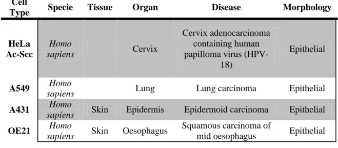
Table 1 - Growth media used in incubation growth of Basidiomycetes strain species. . 19 Table 2 - Cell lines characteristics (data obtained by ATCC and Sigma)
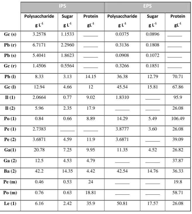
(http://www.lgcstandards-atcc.org/ and http://www.sigmaaldrich.com) ... 27 Table 3 - Polysaccharide, sugar and protein content (g L-1) of samples used during the
elaboration of the present work. ... 30 Table 4 - SO scavenging (%) of IPS (results are shown as average ± SD, and all
experiments were carried out in triplicate); AA (200 µg mL-1) was used as a positive control. ... 31 Table 5 - SO scavenging (%) of IPS undergone ethanol precipitation; (results are shown
as average ± SD, and all experiments were carried out in triplicate); AA (200 µg mL-1) was used as a positive control. ... 32 Table 6 – Assay of phenolic compound in fresh leaves and lyophilized samples; as
referred (a) and (b) represent younger and older leaves respectively; values are represented as µg/mL. ... 40
1
1 State of the art
1.1 What is Cancer?
Cancer is widely known nowadays as the disease of the century. Not only by the number of infected people worldwide but also by the amount of research dedicated to this field and its increasing success rate.
Neoplasms, which are abnormal masses or colonies of cells produced by a relatively autonomous new growth of tissue, arise normally by the clonal expansion of a single cell that has undergone mutation in the chromosomal DNA, caused by a chemical, physical or biological agent, (Zaidman et al., 2005) both external (tobacco, alcohol, chemicals, infectious agents and radiation) and internal (hormones, immune conditions, inherited mutations, and mutations occurring in metabolism) that directly and irreversibly alter the cell genome. (Zong et al., 2012)
Neoplastic cells are characterized by the loss of some specialized functions and acquisition of new biological proprieties, like self-sufficient growth signals, insensitivity to growth inhibitory signals, evasion of apoptosis, limitless replicative potential, sustained angiogenesis, invasion of surrounding tissues, metastasis to other tissues and organs and finally the passage of those biological proprieties to progeny cells (Zaidman et al., 2005; Zong et al., 2012).
This can further be clinically considered benign or malignant.
Cancer is a generic term used to describe malignant neoplasms which manifest a greater degree of autonomy, capability of metathesized expansion and capacity to resist treatment and cause death. Anaplasia is a representative propriety and denotes a lack of normal structural and functional cell characteristics (Zaidman et al., 2005).
The ability to invade and metastasize is the defining characteristics of a cancer. After the transformation from a normal cell into a malignant cell via genetic mutation, cancerous cells proliferate rapidly, break off from the parent lump, migrate around the body in the blood or the lymphatic system, and set up secondary foci of cancerous
2
growths at distant sites. Metastasis is responsible for 90% of the cancer death rate (Zong
et al., 2012).
A report released by the World Health Organization (WHO) has showed that an estimated 12.7 million people globally were diagnosed with cancer and about 7.6 million people died of it in 2008. As estimated in this report, more than 21 million new cancer cases and 13 million deaths are expected by 2030 (Zong et al., 2012).
Although the efficiency of chemotherapy for the majority of cancers has improved over the last three decades, the drugs used in this treatment, while formidable, contain high toxic effects that cause severe reduction in quality of life, causing problems in clinical medicine. Therefore, it is important to develop novel potent, but low toxic anti-cancer reagents, including natural products (Cao et al., 2010).
1.2 Polysaccharides
Polysaccharides are biopolymers comprised of monosaccharides linked together through glycosidic bonds. These structures can be linear or contain branched side chains. Polysaccharides have a general formula of Cx (H2O) y where x is usually a large
number between 200 and 2500. Considering that the repeating units in the polymer backbone are often six-carbon monosaccharides, the general formula can also be represented as (C6H10O5) n where 40 ≤ n ≤ 3000 (Zong et al., 2012).
Polysaccharides can be classified into two groups based on their source. Natural polysaccharides are obtained from several organisms such as algae, plants, microorganisms or animals and semi-synthetic polysaccharides, produced by chemical or enzymatic modification of the parent macromolecules. Polysaccharides obtained from different sources and by different chemical manipulations exist in a variety of chemical compositions, molecular weights and structures. They possess several physic - chemical properties including gelation, solubility, low osmotic effect and surface properties depending on their composition and architecture (Zong et al., 2012).
Since they are a structurally diverse class of macromolecules, polysaccharides play diverse and important roles in many biological processes. As well as serving as stores of energy (e.g. starch and glycogen) and structural components (e.g. cellulose in plants and
3
chitin in arthropods), polysaccharides and their derivatives can participate in signal recognition, cell – cell communication and also play key roles in the immune system, fertilization, pathogenesis prevention, blood clotting and system development (Zong et
al., 2012).
Recently, oligosaccharides have demonstrated a broad spectrum of biological effects, such as antibiotic, antioxidant, anti-mutant, anticoagulant and immuno-stimulation activities. The anti-cancer efficiency of polysaccharides was first recognized by Nauts et al., in 1946 when she discovered that certain polysaccharides could induce complete remission in patients with cancer (Zong et al., 2012).
1.2.1 Mushroom polysaccharides
Since the discovery that Lentinan, a polysaccharide from Lentinula edodes, inhibited mouse sarcoma 180 and displayed very low toxicity (when compared with chemical antitumor drugs) a number of polysaccharides with antitumor activity have been reported (Cao et al., 2010). This led to new scientific and medical studies, increasingly demonstrating the medicinal proprieties of mushroom-extracted compounds for the prevention and treatment of cancer (Zaidman et al., 2005).
It has been well established that many mushroom-extracted compounds, like polysaccharides, are commonly used as immunomodulators or biological response modifiers (BRM) (Zaidman et al., 2005; Wasser, 2010; Liang et al., 2011) ie, they cause no harm and place no additional stress on the body, but help it to adapt to environmental and biological stress, being mainly used as an adjuvant therapy approach in clinical trials (Sarangi et al., 2006; Liang et al., 2011)
In literature, a “mushroom” is: a macro-fungus with a distinctive fruiting body that is large enough to be seen by the naked eye and to be picked up by hand and commonly occurs in two classes: Basidiomycetes and ,sometimes, Ascomycetes (Zhang et al., 2007). Figure 1 represents there life cycle, including the structures where polysaccharides can be found.
Many higher Basidiomycetes mushrooms contain biologically active polysaccharides in its fruit bodies, cultured mycelium and cultured broth (Wasser,
4
2002). Data on mushroom polysaccharides is summarized for approximately 700 species of higher Hetero - and Homobasidiomycetes (Sarangi et al., 2006;Wasser, 2010; Wieter et al., 2011).
Figure 1 - Diagrammatic representation of mushroom life cycle (Lull et al., 2005)
Mushrooms have been part of a normal human diet for thousands of years and, in the last decade, the amounts consumed have risen greatly, involving a large number of species (Jayakumar et al., 2011).
In general, the total composition of mushrooms is water (90%), protein (2-40%), carbohydrates (1-55%), fiber (3-32%) and ash (8-10%). The ash is made up mainly of salts and metals like calcium and magnesium (Ishii et al., 2011). They are valuable health foods since they are low in calories, fats, and essential fatty acids, and high in vegetable proteins, vitamins and minerals. Mushrooms are the only non-animal-based food containing vitamin D (Jayakumar et al., 2011).
For millennia, medicinal mushrooms have established a history of use in traditional ancient therapies and contemporary research has validated and documented much of this ancient knowledge. The interdisciplinary field of science that studies medicinal mushrooms has been increasingly developing and demonstrating the potent and unique properties of compounds extracted from a range of mushroom species (Wasser, 2010).
5
Figure 2 - Pleurotus ostreatus and Pleurotus eryngii
(http://www.morelmushroomhunting.com/pleurotus_castreatus.htm)
Figure 3 –: Irpex lacteus (www.hoosiermushrooms.com/blog/) and Bjerkandera adusta (www.herbarium.iastate.edu/fungi/fungispecies.php?sp=Bjerkandera+adusta+%28Willd.%3AFr.%29+Karst.)
Numerous bioactive polysaccharides or polysaccharide – protein complexes from medicinal mushrooms are described as a manner to enhance innate and cell-mediated immune responses and exhibit antitumor activities in animals and humans (Wasser, 2010).
The stimulation of immune defense systems has significant effects on the maturation, differentiation and proliferation of many kinds of immune cells in the host. Many of these mushroom polymers were reported to have immunotherapeutic properties by facilitating growth inhibition and destruction of tumor cells (Wasser, 2010).
Recently, some of the medicinal effects associated to mushrooms included antitumor, immunomodulating, antioxidant, radical scavenging, cardiovascular, antihypercholesterolemia, antiviral, antibacterial, antiparasitic, antifungal,
6
detoxification, hepatoprotective and antidiabetic effects. Mushroom polysaccharides can prevent oncogenesis, show direct antitumor activity against several synergetic tumors and prevent tumor metastasis, being especially beneficial when used in conjunction with chemotherapy Although the mechanism of their antitumor actions is still not completely understood, stimulation and modulation of key host immune responses by these mushroom polymers appears to be central (Carbonero et al., 2008; Wasser, 2010).
Figure 4 - Ganoderma apllanatum (http://www.first-nature.com/fungi/ganoderma-applanatum.php)
Thus, the benefits of mushroom compounds on different clinical conditions have attracted the interest of the scientific community in the last decade in order to understand the molecular mechanisms responsible for their actions. Several classes of mushroom compounds such as proteins, polysaccharides, lipopolysaccharides and glucoproteins have been classified as molecules with potent effects on the immune system that may restore and enhance immunological responses of host immune effector cells (Wasser, 2010).
Polysaccharides or extracts mainly containing polysaccharides from more than 30 medicinal mushrooms species have shown anti-cancer activity in animal models, but only a few of them have been further taken to clinical assessment in humans (Wasser, 2010; Zong et al., 2012).
7
1.2.2 Structural and physical properties
A wide range of antitumor or immune-stimulating polysaccharides of different chemical structures have been investigated. Polysaccharides with strong anti-tumor action may differ greatly in their chemical structures. Antitumor activity is exhibited by a wide range of glycans extending from homopolymers to highly complex heteropolymers (Zhang et al., 2007; Yang & Zang, 2009; Synytsya et al., 2009).
A large group of bioactive polysaccharides is called heteroglycans which are classified as galactans, fucans, xylans and mannans depending on the individual sugar components in the backbone. Heteroglycan side chains may contain arabinose, mannose, fucose, galactose, xylose, glucuronic acid and a glucose moiety as a main component, or in various combinations (Zhang et al., 2007; Yang & Zhang, 2009).
The structure of naturally occurring glycans is so diversified that it is difficult to define a universal protocol for their analysis. The building blocks (derived from sugar residues) of complex polysaccharides exhibit very similar structures, but their differentiation (diversified linkage style) is widely varied (Zhang et al., 2007). The primary structure of a polysaccharide is defined by monosaccharide composition and sequence, configuration and position of glycosidic linkages as well as the nature, number and location of appended non-carbohydrate groups (Zhang et al., 2007).
Figure 5 - Repeating unit of immunomodulatory β-glucans (a) from Grifola frondosa and (b) from
8
Polysaccharides are usually composed of several monosaccharides linked with different glycosidic bonds. Some have hyper-branched structures, high molecular weights and tend to form aggregates in solution that may mask the behavior of individual micro molecules (Yang & Zhang, 2009).
In some mushroom species, polysaccharides are bound with proteins or peptides to form complexes which show higher potent antitumor activity (Zhang et al., 2007).
The major component in the biologically active polysaccharides was identified as β-D-glucans (Wong et al., 2011). The anti-tumor activities may be related to the triple helical conformation of the (1-3) – β-D-glucan backbone chain for some, such as lentinan from Lentinus edodes (Yang & Zhang, 2009). The few human tested polysaccharides are rich in β-D-glucans or β-D-glucans-proteins complex showing greater immunopotentiation activity than free glucans (Wasser, 2010; Zong et al., 2012).
In addition to the well-known antitumor (1-3) – β D glucan, a wide range of biologically active glucans with other structures are described. These glucans are linear or branched molecules with a backbone composed of α-or β-linked glucose units and some of them contain side chains that are attached at different positions (Zhang et al., 2007; Synytsya et al., 2009).
Compared with the intensive investigations of β-D-glucans, the biological activity of α-(1 - 3)-D-glucan, which is especially apparent after chemical and structural modification (carboxymethylation, sulfation, aminopropylation, or hydroxyethylation), has been reported only rarely in the literature. But carboxymethylated fungal α-(1 - 3)-D-glucans have a biological activity potential, i.e. they express cytotoxic or mitochondria metabolism-modulating in Lentinula edodes, Pleurotus ostreatus,
Piptoporus betulinus, and Laetiporus sulphureus has been recently denoted (Wasser,
2010).
For example, 43 terpenoids were separated from Ganoderma lucidum, including 6 new compounds. The chemical structures were elucidated and all of the compounds were assayed for their cytotoxicity against human HeLa cervical cancer cell lines. The C-3 carbonyl group, the double bonds (∆7, 8, ∆9, 11), the type of the side-chain and the number of hydroxyl group all played an important role in the structure–activity relationships (Cheng et al., 2010).
9
Besides a typical structure-function relationship that can be found between the anti-tumor activities and structural characteristics of β-D-glucans, a wide range of polysaccharides with different molecular masses ranging from 1x10⁴ to 1x10⁶ kDa can be isolated during extraction. This molecular weight difference can cause diverse immunomodulatory effects and it is, generally, found that those polysaccharides with higher molecular weight (1x10⁵ kDa or above) would exert stronger anti-tumor and immunomodulatory effects (Wong et al., 2011).
1.2.3 Anti-tumor activity and cellular mechanism
The importance of polysaccharides in tumor treatment was first recognized more than 100 years ago when it was found that certain polysaccharides could induce complete remission in patients with cancer. Since antitumor activity of macrofungal polysaccharides was first published by Chihara in the 1960’s, researchers have isolated structural diversified polysaccharides with strong antitumor activity. Unlike traditional antitumor drugs, they produce antitumor effect by activating various immune responses in the host causing no harm to the body (Zhang et al., 2007).
In time mushroom polysaccharides have already shown widely inhibitory effects towards many kinds of tumors suchs as sarcoma 180 solid cancers, Ehrlich solid cancer, sarcoma 37, Yoshida sarcoma and Lewis loung carcinoma (Zhang et al., 2007).
The proposed mechanism by which mushroom polysaccharides exert antitumor effect include: 1) prevention of the oncogenesis by oral administration of polysaccharides isolated from medicinal mushrooms (cancer-preventing activity); 2) enhancement of immunity against the bearing tumors (immuno-enhancing activity); and 3) direct antitumor activity to induce the apoptosis of tumor cells (direct tumor
inhibition activity) (Zhang et al., 2007).
Figure 6 shows the protein-bound polysaccharide K (PSK) isolated from Coriolus
versicolor, a major studied polysaccharide and the different ways it is known to act
(Maehara et al., 2012). First, PSK improves host immunocompetence by inhibiting of or neutralizing immunosupressive substances that are increaed in cancer. Second, it activates immune cells such as lymphocytes, either directly or by regulating the production of various cytokines, and third, acts directy in cancer cells.
10
Figure 6 - Tumor microenvironment and actions of PSK. (1) Suppressed production or neutralization of immunosuppressive factors. (2) Activation of immune cells and regulation of cytokine
production. (3) ´Direct action on tumor cells [induction of apoptosis, enhanced expression of major histocompatibility complex (MHC) class I antigen] (Maehara et al., 2012)
The cancer preventing activity of medicinal mushroom polysaccharides owes its mechanisms to the immunopotentiation of polysaccharides. This cancer preventing activity has been observed in farmers whose main occupation was producing medicinal mushrooms such as Agaricus blazei in Brazil. These farmers cancer death rate was 40% lower than in the general population. Consequently, an animal study was conducted by comparison of the administration of an ordinary diet (control) and a A. blazei diet (test). After the inoculation with tumors, the number of mice that developed cancer on the test diet was significantly lower than the ones in the control group, indicting the cancer preventing activity of polysaccharides (Zhang et al., 2007).
Mushroom polysaccharides exert their antitumor action mainly via activation of the immune response of the host organism (immuno-enhancing activity). They help the host adapt to several biological stresses and exert a nonspecific action supporting some or all of the major systems (Zhang et al., 2007). They enhance a host mediator immunomodulatory response activating the immune system to fight against cancer (Wong et al., 2011). Mushroom polysaccharides have been shown to produce over 50% reduction in tumor size and prolong the survival time of tumor bearing mice (Zhang et
11
Direct tumor inhibition activity, has also been documented in many mushroom
polysaccharides. Even though the anti-proliferative effect of polysaccharides towards tumor lines in vitro remains unclear, some studies indicate that incubation of polysaccharides with tumor cells could alter the inner cell signaling expression. Cell cycle arrest and induction of apoptosis could explain the in vitro anti-proliferative effect of polysaccharides. These results suggest that mushroom polysaccharides not only stimulate the proliferation of T lymphocytes and the immune function through the immunopotentiation but also have a direct action on the tumor cells even though little is known about its direct effect on cancer cells (Zhang et al., 2007).
Recent studies on direct cytotoxicity have indicated that polysaccharides can directly inhibit the cancer cell proliferation in a dose- and time-dependent manner mediated through processes like up-regulation of p21 and of a pro-apoptosis Bax protein and down-regulation of cyclin D1 (Zhang et al., 2007).
Cancer cells originate from normal cells and a novel agent that can select and destroy only the transformed cells without affecting the normal ones will be an ideal chemotherapeutic agent against cancer. In this respect, the mushroom polysaccharides and polysaccharide – protein complexes should be ideal because they are non-toxic and their anti-cancer effects are mainly host mediated (Sarangi et al., 2006).
1.2.4 Extraction and purification
Mushroom polysaccharides exist mainly as a structural component of the fungal cell wall, composed of two major types of polysaccharides: a rigid fibrillar of chitin (or cellulose) and a matrix-like β-glucan, α-glucan and glycoproteins. Schizophyllan (glucan chitin complex) a water soluble 1-6 branched β-(1-3) - glucan loosely attached to the outer layer of the cell wall, is secreted to the extracellular matrix where exo- or extracellular polysaccharides (EPS) can be found. These water-soluble glucans fill the outer layer resisting external pressure (Zhang et al., 2007).
In general, the extraction method involves elimination of low molecular weight substances from mushroom material. Selection of an extraction method depends on the cell wall structure, and hot water has been the most popular approach yielding water soluble polysaccharides. Methods can vary based on the structure and water solubility
12
of polysaccharides, but the basic rule is to break the cell wall from outer layer to inner layer with mild-to-strong extraction conditions (pH and temperature) (Zhang et al., 2007).
Extracted polysaccharides can be further purified using a combination of techniques such as ethanol precipitation, fractional and acidic precipitation with acetic acid, ion-exchange chromatography, gel filtration and affinity chromatography. The ethanol precipitation excludes the impurities from the polysaccharides (Zhang et al., 2007).
The polysaccharide with the broad polydispersity can be fractioned by the methods referred above, yielding polysaccharides with different molecular weights and low polydispersity. It should be noted that particular fractionation procedures scheme in each case depends on the composition of the polysaccharide of the original material involving molecular weight branching degree and pattern (Zhang et al., 2007).
1.2.5 Antioxidant activity
Free radicals play an important positive physiological role but may, at the same time, exert toxic effects (Kozarski et al., 2011). It is well known that reactive oxygen species (ROS) such as hydroxyl radical, superoxide anion and hydrogen peroxide are related to the pathogenesis of various diseases (Cheng et al., 2010).
ROS are mostly generated by normal metabolic processes or from exogenesis factors and agents (Hu et al., 2010), as a side product of oxidative phosphorylation. They participate in the regulation of signal transduction and gene expression, activation of receptors and as nuclear transcription factors, also playing a vital role in phagocytosis (Kozarski et al., 2011).
On the other hand, there is increasing evidence that free radicals may play a causative role in a variety of diseases including heart disease, liver damage, cancer, Parkinson’s and Alzheimer’s diseases, impairment of immune function, cataracts and muscular degeneration in elderly people (Hu et al., 2010; Kozarski et al., 2011).
It is pertinent to note that antioxidant defense systems may only partially prevent oxidative damage. Hence, there is currently a great interest in using dietary supplements
13
containing antioxidants with a new view to protect the components of the human body from oxidative damage (Jayakumar et al., 2011).
The antioxidant status of the human body reflects the dynamic balance between the antioxidant defense and prooxidant conditions (Kozarski et al., 2011). The oxidative damage to DNA, proteins and other cellular determinants seem inevitable when the concentration of ROS exceeds tolerability of cells (Sun et al., 2004) and impaired physiological functioning may occur, resulting in diseases and accelerated ageing (Kozarski et al., 2012). Anti-oxidants could attenuate this oxidative damage of a tissue indirectly by increasing the cells’ natural defenses and/or directly by savaging the free radical species (Sun et al., 2004).
Many synthetic chemicals such as phenolic compounds are found to be strong radical scavengers, but they usually have side effects. For this reason, natural antioxidants are generally preferred in food applications (Kozarski et al., 2012).
Natural antioxidants occur in fruits, vegetables, mushrooms and other natural edible resources. In general, several categories of antioxidants, including phenolic compounds, vitamins, proteins, peptides and amino acids, are widely used as ingredients in dietary supplements and have been investigated for the prevention of diseases such as cancer, coronary heart disease and even altitude sickness (Kozarski et al., 2012).
Among sources of various natural antioxidants, mushrooms have a prominent role. Extracts prepared from mycelium and fruiting bodies of non-toxic (i.e. edible) Agaricus
bisporus, Lentinula edodes, Ganoderma lucidum, Pleurotus sp. among other mushroom
species, show antioxidative activity in vitro and may have potential for application in
vivo (Kozarski et al., 2011; Kozarski et al., 2012).
Polysaccharides are potentially useful biologically active ingredients for pharmaceutical use, such as immune regulation, anti-radiation, anti-blood coagulation, anti-cancer, anti-HIV and hypoglycemic activities. Mushroom derived polysaccharides like lentinan, schizophyllan and krestine have been accepted as immunoceuticals in Japan, Korea and China (Kozarski et al., 2011).
Antioxidant supplements or foods containing high concentrations of antioxidants may help to reduce oxidative damage. Identification of new antioxidants remains a highly active research area (Kozarski et al., 2011).
14
Among the large resource of fungi, medicinal higher Basidiomycetes, are unlimited sources of biologically active polysaccharides as well as fruit bodies and cultured mycelia. Again, the most bioactive mushroom polysaccharides are β-(1-3) (1-6) glucans (Kozarski et al., 2011), although a wide range of biologically active glucans of other structures have been described (Kozarski et al., 2012).
The hydroxyl radical (OH•) is the most reactive of the ROS and as such can induce severe damage to adjacent biomolecules. The superoxide radical (O2•‾), which is
generated in numerous biological reactions, is also a highly toxic specie. Although O2•‾
radicals cannot directly initiate lipid oxidation, they serve as potential precursors of highly ROS, such as the OH• (Jayakumar et al., 2011).
In the present work, we decided to investigate the anti-oxidative activity of intrapolysaccharides (IPS) of our extracts by assaying the superoxide radical scavenging.
As we have seen, it is extremely advantageous and valuable to explore novel anti-tumor products from medicinal mushrooms (Liang et al., 2011).
1.3 Olive leaves
In recent years, traditional Arab-Islamic herbal medicine has been gaining interest in the scientific community, and more specifically, regarding cancer treatment (Zaid et
al., 2012)
The olive tree (Olea europaea) is an evergreen tree, from the Oleaceae family, (Zaid et al., 2012) with elongated leaves, present in the Mediterranean region for over 7000 years ago (Lalas et al., 2011).
Throughout the history, Olea europaea leaves have been heavily exploited for the prevention or the treatment of hypertension, carcinogenesis, diabetes, atherosclerosis and several other traditional therapeutic uses (Bouallagui et al., 2011; Zaid et al., 2012). Historically, olive leaf has also been used as a remedy for fever and other diseases such as malaria. (Lee et al., 2009)
15
The incidence of cancer overall in the Mediterranean basin has been thought to be the lowest compared to the other parts of the world. This is mostly accounted for the incidence of cancer of the breast, endometrium, prostate, colon and leukemia. (Bouallagui et al,. 2011; Zaid et al., 2012)
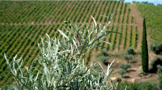
Figure 7 - Verdeal olive tree leaves
(http://www.quintadoromeu.com/index.php?p=paginas&op=azeite&idioma=eng)
Almost all of the 16 countries bordering the Mediterranean Sea are sharing a common Mediterranean dietary pattern characterized by a high consumption of fruits and vegetables, bread, wheat and other cereals, olive oil and fish. These food supplies are highly rich in bioactive com pounds such as vitamins and polyphenols. A large body of epidemiological evidence, together with data from animal and in vitro studies, has brought the proof of polyphenols cancer-preventive properties (Bouallagui et al., 2011).
Typical phenols of the O. europaea are the secoiridoids, complex molecules with a molecular weight up to 600 composed of one or two hydroxiaromatics linked to several other non aromatic compunds (Leonardis et al., 2008).
The major active components in olive leaf are known to be oleuropein and its derivatives such as hydroxytyrosol and tyrosol, as well as caffeic acid, p-coumaric acid, vanillic acid, vanillin, luteolin, diosmetin, rutin, luteolin-7-glucoside, apigenin-7-glucoside, and diosmetin-7-glucoside (Lee et al., 2009; Han et al., 2009) .Concentration
16
of phenols is several times higher in leaves when compared to several oil fractions, and its content varies with time and climate. (Han et al., 2009)
The lower incidence of certain cancers is most likely associated with the antioxidant activity of active ingredients of the olive oil. Oxidative stress has been shown to contribute to cancer development, and antioxidants are believed to reduce the risk of mutagenesis and carcinogenesis. Hydroxytyrosol (HT) or 3,4 – dihydrophenylethanol, is a very bioactive alchoolic ortho-diphenol, component of the secoroiroids. Several studies have demoonstrated its antioxidant and antimicrobial activities, which bennefict in general the cardio-vascular system and prevent several human diseases (Leonardis et al., 2008) throughout its capacity to protect cells from hydrogen peroxide damage and DNA peroxy nitrite-induced damage by blocking cell cycle progression at the G1 phase and inducing apoptosis (Zaid et al., 2012)
Native HT can only be found free in nature with the exception of ripened olive leaves, where the hidrolization of its main component, the oleuropein, occurs (Leonardis
et al., 2008). It is found in leaves and fruits as the most important constituent of
oleuropein (Han et al., 2009).
In vivo and in vitro studies on the activity of oleuropein have found that, in addition
to antioxidant properties, it has antiangiogenic action and inhibits cell growth, motility, and invasiveness, for example in lung cancer (Zaid et al., 2012).
Oleuropein was also found to cause cell rounding, which disrupts the cell actin cytoskeleton, providing direct antitumor effects due to cell disruption. In in vivo animal studies, rapid tumor regression was observed hen mice were given one percent oleuropein in drinking water. Saturated animal fats and polyunsaturated plant fats in the diet have been implicated in colon, breast, prostate, and several cancers. The vast usage of olive oil in the mediterranean diet may explain its aparent cancer-protective effect, rather than the amount of fat consumed. Furthermore, in recent study, evaluating the antioxidant and antiproliferative activity of water and methanol olive leaves extracts in cancer and endothelial cells, olive leaf crude extracts were found to inhibit proliferation of cell from a human breast adenocarcinoma, cells from human urinary bladder carcinoma (T-24), and cells from bovine brain capillary endothelial tumor (Lee et
17
2 Thesis aims
In the present study, we analyzed polysaccharide extracts from fruiting bodies and mycelia of several mushroom strings such as Ganoderma, Pleurotus, Irpex,
Bjerkandera, Piptoporus, and their biological activity, i.e. free radical scavenging
activity, and cytotoxic effect of their polysaccharide content against four tumor cell lines. Phenol compounds from different olive leafs were also analyzed and its effect on human carcinoma cells verified.
It was our intent to perform a systemic study globing some of the following procedures:
1) In vitro growth of human carcinoma cell lines, as well as obtaining parameters such as cell viability.
2) Extraction of intra and extra polysaccharides from several mushroom strings such as Ganoderma, Pleurotus, Irpex, Bjerkandera, Piptoporus, and dosage of protein and polysaccharide content, trough coomassie blue and phenol-sulfuric acid methods.
3) Addition of different concentrations of such polysaccharide extracts to the carcinoma cell lines to study cell viability in their presence through the tetrazolium dye (MTT) colorimetric assay.
4) Phenol extraction of lyophilized and fresh green olive leaves, from 5 different Portuguese varieties of Olea europaea – Madural, Cobrançosa, Arbequina,
Verdial and Galega.
5) Addition of different concentrations of such phenolic compounds to the carcinoma cell lines to study the cell viability in their presence through the tetrazolium dye (MTT) colorimetric assay.
18
3 Materials and Methods
3.1 Sampling
From this moment on and until the end of this dissertation, fungi samples will be represented as following: Ganoderma apllanatum – Ga; Ganoderma carnosum – Gc;
Pleurotus ostreatus – Po; Pleurotus eriinges – Pe; Irpex lacteus –Il; Bjerkandera adjusta – Ba; Piptoporus betulius – Pb.
The name of the fungus is often followed by (α) in which α symbolizes the biomass origin, (1) and (2) for mycelia growth in medium 1 and 2 respectively, (m) from when the biomass was removed from the fruiting body of full grown mushrooms, (s) fungus grown in straw and (r) grown in rice husk (both (s) and (r) were already grown in the straw and rice husks when delivered to analysis).
Figure 8 - Pleurotus ostreatus grown mushroom sample
As far as olive samples are concerned, the five varieties are represented by name,
Madural; Cobrançosa; Arbequina; Verdeal and Galega followed by (a) in younger
19
3.2 Mushroom cultivation for production of polysaccharides
Mushroom mycelia cultivation in laboratory was elaborated following some of the rules presented by Gern et al., 2008.
As far as laboratory mushroom mycelia growing strains, a piece of agar plated fungi (i.e Basidiomycetes strain) was transferred to an Erlenmeyer flask containing one of the two medium types described in Table 1.
The inoculation growth was accomplished by the transfer of agar plated fungi to a 500 mL Erlenmeyer containing 100 mL of culture medium. The culture was incubated and left to grow at 25ºC and 150 rpm for a week. The culture content was then transferred to a 2L Erlenmeyer containing 500 mL of the similar culture medium. The culture was left to grow another week under the same experimental conditions of encubation.
Glucose is used as a carbon source, and nitrogen source varies from medium 1 – yeast extract, to medium 2 – tomato pomace.
Table 1 - Growth media used in incubation growth of Basidiomycetes strain species.
Medium 1 Boiled wheat water with 40 gL
-1
of glucose and 5 gL-1
of yeast extract pH 6
Medium 2 Boiled wheat water with 40 gL
-1
of glucose and 5gL-1 of tomato pomace – pH 6
3.3 Isolation of extracellular (EPS) and intracellular (IPS)
polysaccharides
Isolation of polysaccharides was accomplished as described by Cui et al., 2007, with some modifications.
Mycelial biomass from the culture broth was separated by centrifugation at 5.000 rpm for an hour, in 250 mL centrifuge flasks.
20
The culture supernatant was treated with four volumes of 95% (v/v) ethanol to promote precipitation of the EPS. This mixture was left overnight at 4ºC. The EPS were recovered by centrifugation at 20.000 rpm for 45 min, and the pellet was re-suspended in a minimal volume of 50 mM phosphate buffer pH 7.0 (2ml for EPS and 1 ml for IPS).
The mycelial biomass pellet was re-suspended in one volume of distillated water (w/v) and the mixture was heated in a water bath (100ºC) for 6h to release the IPS. The IPS in the extract was recovered using the same procedure as the EPS recovery.
Figure 9 - Isolation of extracellular and intracellular polysaccharides (adapted from Cui et al., 2007)
EPS and IPS levels were quantified by the phenol/sulphuric acid method using glucose and poligalacturonic acid as standards. The key steps involved in EPS/IPS isolation and quantification are shown in Figure 9.
21
3.4 Polysaccharide determination
Among many colorimetric methods for carbohydrate determination, the phenol– sulfuric acid method is the easiest and most reliable method for measuring neutral sugars in oligosaccharides, proteoglycans, glycoproteins, and glycolipids since phenol in the presence of sulfuric acid can micro determinate sugars and their methyl derivatives, oligosaccharides, and polysaccharides. The phenol-sulfuric acid method is widely used because of its sensitivity and simplicity (Dubois et al., 1956; Masuko et al., 2005
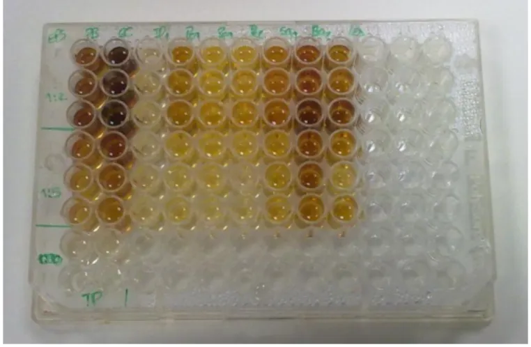
Figure 10 - Plate representing the Phenol - Sulphuric acid method
The quantification of polysaccharides was accomplished using the method described by Masuko et al., 2005. This method was developed in order to reduce the
amounts of chemicals used and is performed in a microtiter plate format, (Figure 10) which allowed to increase the number of samples to be assayed such as the column chromatographic fractions.
In a well of a 96-well flat-bottomed polysorp microtiter plates, 150 µl of concentrated sulfuric acid were rapidly added to 50 µl of sample to cause maximum mixing, followed immediately by the addition of 30 µl of 5% phenol in water. After incubating for 5 min at 90º C in a static water bath , the microtiter plate was cooled to room temperature for 5 min in another water bath and wiped dry to measure the absorbance at 490 nm by an microplate reader BIO-RAD model 680. All assays were carried out in triplicate.
22
Standard curves were carried out by using anhydrous D - glucose and polygalacturonic acid as standards.
3.5 Protein Assay
Quantification of protein was carried out by protein-dye binding method described for the first time by Bradford, 1976, and then by Sedmak and Grosseberg in 1977.
Figure 11 - Coomassie G250 brilliant blue assay
This method, represented in Figure 11, is based on the principle that, under certain conditions, acidic and alkali groups of proteins react with organic dyes forming colored precipitate, and so coomassie brilliant blue G 250 in dilute acid, which possesses a brownish-orange color reacts with protein amino groups, converting into an intense blue color.
In order to measure the protein content, polystyrene 96 well plates were used. The reaction occurred by mixing 100µl of coomassie brilliant blue G250 in 0.06% HCl 0.6 N to 100 µl of the appropriate sample. This mixture was then incubated at room temperature for about 5 minutes before reading its absorbance at 655 nm in a microplate reader BIO-RAD model 680.
The standard curve was carried out by using bovine serum albumin (BSA) solution as standard.
23
3.6 FTIR analysis
Fourier Transformed Infrared spectroscopy (FTIR) is used to investigate the vibrations of molecules and polar bounds between the different atoms. Structures of polysaccharides, such as monosaccharide types, glycosidic bonds and functional groups, can be analyzed using the FTIR spectroscopy (Yang & Zhang, 2009).
Infrared radiation (IR) refers broadly to that part of the electromagnetic spectrum between the visible and microwave regions. Of greatest practical use to the organic chemist is the limited portion between 4000 and 400 cm-1 (Silverstein et al., 2005).
Although the IR spectrum is characteristic of the entire molecule, it is true that certain groups of atoms give rise to bands at or near the same frequency regardless the structure of the rest of the molecule (Silverstein et al., 2005).
An FTIR analysis of intrapolysaccharides from Ganoderma carnosum (s) and
Pleurotus eryngii (m) as well as extrapolysaccharides from Ganoderma apllanatum (1)
and Pleurotus ostreatus (1) was carried out in a Bruker Vertex 70 in KBr pellets with a resolution of 2 cm-1 in a range of 500–4000 cm-1.
3.7 Purification of polysaccharides by gel filtration chromatography
Extrapolysaccharides from Ganoderma apllanatum (1) and Pleurotus ostreatus (1) were purified by gel filtration chromatography on a Sephacryl S-300 HR column (1 cm × 100 cm), previously equilibrated and eluted with 50 mmol L-1 phosphate buffer (pH 7.0) at a flow rate of 30 mL h-1.
Fractions (3ml) were collected and analyzed for polysaccharide and protein contents.
After collection each fraction was submitted to the phenol-sulphuric acid, the coomassie protein assay and a measure of absorbance at 200 and 280 nm to detect the presence of polysaccharides and proteins respectively.
24
3.8 Purification of Polysaccharides by affinity chromatography
Affinity chromatography was performed in 96 well microtiter plate as shown in Figure 12.
Figure 12 - Affinity chromatography columns in 96 well microtiter plates
250 µl of epoxy-activated Sepharose 6B-urea, embedded in 0.001% (m/v) sodium azide, were added to each well. About 15 min were given to the resin to compact as much as possible. A quick centrifuge for 2 minutes at 2500 rpm was performed to consolidate.
Supernatant was recovered and discarded. 150 µl of 50 mM phosphate buffer pH 7.0 was added to each well, centrifuged for 2 min at 2500 rpm and supernatant discarded four times as part as the equilibrium phase
Addition of 100 µl of polysaccharide extract (diluted in buffer solution) to each column (well) was performed and allowed to “sink in” for 15 minutes, with at least 3 refluxes performed.
In the washing process, the plate was then again centrifuged 2 min at 2500 rpm and now 100µl of the supernatant were removed, but collected in the next row of wells (supernatant from well A1 is deposited in well B1).
The washing process is repeated 3 times saving the supernatant in all following rows (as continuing the example, in C1, D1 and E1).
25
At the fourth time the washing 50 mM phosphate buffer was substituted by the elution buffer (25 mM phosphate buffer with 2M KCl).
This buffer allowed the molecules grouped with the resin to detach and release into the elution buffer, which we continued to remove from the resin column and transfer to new wells. This procedure was again repeated 3 times.
After that time, a phenol-sulphuric acid assay was performed, on the supernatant recovered in the wells, in order to dosage the release of the polysaccharide throughout the washing and elution phase.
3.9 Superoxide radical scavenging
The O2•‾ is considered to be a highly potent oxidant, and can react with all biomolecules operative in liver cells. The O2•‾ are produced in vivo and can result in the formation of H2O2 by the dismutation reaction.
In vitro O2•‾ were generated from dissolved oxygen through the phenazine
methosulfate (PMS) - b-nicotinamide adenine dinucleotide (NADH) – nitro blue tetrazolium (NBT) system.
PMS/NADH coupling reduces NBT, which increases absorption at 560 nm. (Liu et
al., 1997; Kodali & Sen, 2008)
In order to optimize this method 25 Mm Tris-HCL buffer, pH 8.0, was added to 780 mM NADH and to 500 µM NBT in a proportion of 4:1:1. 60 µl of this mixture was then added to 20 µl of polysaccharide extract sample in a 96 wells microplate with an automatic multichannel micropitete.
To start the reaction, 10 µl of PMS 100 µM, were added to the well and the reaction mixture was incubated at room temperature for 5 min. Absorbance was measured at 550 nm in a microplate reader BIO-RAD model 680, and Ascorbic Acid (AA) was used as positive control.
The scavenging activity (%) was calculated from the following equation: [1-(Asample / Ablank)]*100
26
were Ablank is the absorbance of the control reaction and Asample is the absorbance of
the sample reaction.
In order to investigate if the time affected the results, kinetic studies were performed between 5 and 30 minutes.
As described by Sun et al., 2004 and Kodali and Sen, 2008, ascorbic acid was used in a concentration of 200 µg/mL and revealed an O2•‾ scavenge of about 80%.
All assays were carried out in triplicate.
3.10 Extraction of phenolic compounds
The extraction of phenolic compounds was performed according to Han et al, 2009. Lyophilized and fresh green olive leaves, provided by Professor Eduardo Rosa, from the department of Agronomy, UTAD were collected from 5 different Portuguese varieties of Olea europaea – Madural, Cobrançosa, Arbequina, Verdeal and Galega – and two types of leaves were extracted – Older and young - in order to find out if ageing would affect their biological properties.
10 g of fresh or lyophilized leaves were extracted in 100 ml of 70% ethanol for 2 weeks in the dark. Extracted solution [10% (w/v)] was than filtered with a Millipore, (pore size 0.22 µm) filter unit.
3.11 Phenolic compounds Assay
Experiments were performed according to Cicco et al., 2009, with some modifications. 100 μl of properly diluted samples, calibration solutions or blank were pipetted into separate eppendorfs tubes and 100 μl of Folin - Ciocalteau reagent was added to each tube. The mixture was mixed well and allowed to equilibrate.
After exactly 2 min, 800 μl of a 5% (w/v) sodium carbonate solution was added. The mixture was swirled and put in a static water bath at 40 °C for 20 min. Then, eppendorfs tubes were rapidly cooled on the rocks and the colour generated was read at its maximum absorption, i.e.765 nm in the case of gallic acid (standard solution). The