1
Cohesion failure and Mitosis:
From Molecular Mechanisms to
Organismal Consequences
Mihailo Mirkovic
Dissertation presented to obtain the PhD degree in Molecular Biology
Instituto de Tecnologia Química e Biológica António Xavier | Universidade Nova de Lisboa
Research work coordinated by: Instituto Gulbenkian de Ciencia
Oeiras, May 2018
2 Table of Contents Declaration………...…...6 Declaração...6 Summary...7 Resumo...11 Acknowledgments...15 List of Publications...17 Introduction: 1.0 General Introduction………...…...19
1.1 Cohesin-Molecular Glue and much more………...22
1.1.1 The importance of gluing DNA molecules……….…..…..23
1.1.2 The Cohesin Cycle………..…...34
A) The cohesin cycle: Chromatin Loading……..……...34
B) The cohesin cycle: Cohesion establishment………...36
C) The cohesin cycle: Prophase and Cohesion retention at the centromere………...38
D) The cohesin cycle: The final cut……….…..……….…….41
1.1.3 Multiple-step cohesin removal……….……...42
1.1.4 Sister Chromatid Resolution………..…...43
1.1.5 Inner-centromere defining platfor……….…....51
1.1.6 Force Balance………...53
1.1.7 Anaphase sharpness……….….56
3
1.2 The Guardians of Mitotic Fidelity………70
1.3 Aneuploidy and its Consequences……….86
Results:
Chapter I:
Premature loss of cohesion and Mitosis……….……100
I.1 Premature Loss of Sister Chromatid Cohesion Does Not Elicit a Robust SAC Response………...…104 I.2 Loss of Sister Chromatid Cohesion Activates EC Mechanisms during Early Mitosis………...…..107 I.3 Attachments of Single Chromatids to the Mitotic Spindle Are Progressively Stabilized……….…….113 I.4 Cyclin B Is Gradually Degraded during Cohesin Cleavage….119 I.5 Mathematical Modeling of Multiple Feedback across the Mitotic Network……….…….…...…120 I.6 Cells with Premature Loss of Sister Chromatid Cohesion Are Ultrasensitive to Cdk1 Inhibition………....…127 I.7 Discussion……….…....…..131 I.8 Materials and Methods……….……..……...132
4 Chapter II:
Spindle Assembly Checkpoint aggravates cohesin defects in mitosis……….………..….139
II.1 Drosophila wing modifier screen reveals that depletion of Mad2 and Mps1 suppresses the developmental defects associated with loss of cohesion………..…...…..141 II.2 SAC inactivation rescues chromosome segregation defects associated with loss of cohesion……….…...146 II.3 SAC inactivation suppresses chromosome shuffling after loss of cohesion………...…155 II.4 SAC inactivation restores cell survival after loss of
cohesion………...…...…..163 II.5 Discussion………....…165 II.6 Materials and Methods……….…..…168
Chapter III:
Cohesin loss and Aneuploidy in the developing fly………...180
III.1 A genetic system for acute and time-controlled generation of aneuploidy in a developing organism…………..…………...…183
III.2 Reversible removal of cohesin results in a single round of mitotic abnormalities and consequent aneuploidy…………...186 III.3 Larvae challenged with aneuploidy during development hatch into impaired adults……….…...196
5
III.4 Aneuploidy results in chromosomal instability and chromosome
accumulation in the Neuroblasts……….…...….199
III.5 Karyotype restrictions in the proliferating aneuploid Neuroblast population……….…….208
III.6 Aneuploidy elicits a stress response in the brain tissue.….210 III.7 Neural stemness delays aneuploidy stress response……..214
III.8 Developmental aneuploidy does not alter significantly adult brain size and shape………..…...217
III.9 Protecting only the developing brain from induced aneuploidy rescues the lifespan of the ecloded flies………...…219
III.10 Discussion………..…….…………222
III.11 Materials and Methods………..……229
6 Declaration
I declare that this dissertation and the data presented are the result of my own work, developed between 2014 and 2018 in the laboratory of Dr. Raquel Oliveira at the Instituto Gulbenkian de Ciência in Oeiras, Portugal. Specific author contributions are indicated in each chapter, in the Acknowledgements section.
Financial support was granted by Fundação para a Ciência e a Tecnologia, doctoral fellowship PD/BD/52438/2013 and ERC Starting Grant (StG), LS3, ERC-2014-STG-638917, Marie Curie Career Integration Grant (MCCIG321883/CCC) and an EMBO Installation Grant (IG2778).
Declaração
Declaro que esta dissertação de doutoramento e os dados nela apresentados são o resultado do meu trabalho, desenvolvido entre 2014 e 2018 no laboratório do Dr.Raquel Oliveria no Instituto Gulbenkian de Ciência em Oeiras, Portugal. As contribuições de cada autor são indicadas em cada capítulo na secção dos Agradecimentos/Acknowledgements.O apoio financeiro foi concedido pela Fundação para a Ciência e Tecnologia, através da bolsa de doutorame nto PD/BD/52438/2013e porfundos do Conselho Europeu de Investigação ERC Starting Grant (StG), LS3, ERC-2014-STG-638917, Marie Curie Career Integration Grant (MCCIG321883/CCC), EMBO Installation Grant (IG2778).
7 Summary
Mitosis is a dynamic culmination of the cell cycle, resulting in generation of two daughter cells from one mother. In order for this to happen, the cell must package its DNA into chromosomes and divide it equally amongst progeny. To ensure this process happens accurately, the cell glues identical chromosomes together so it can segregate them in symmetrical fashion during anaphase. The glue holding chromosomes together is a molecule called cohesin, which encompasses replicated DNA fibers via topological entrapment. The aim of this thesis was to study the immediate mitotic response to premature cohesion loss, as well as the long term consequences of such perturbed mitosis for the cell and the whole organism. In order to study cohesion loss in mitosis, we utilized an established acute system for cohesin depletion, via the use of TEV protease, which cleaves TEV sites inserted into cohesin within hours after heat shock induction, or minutes after injection. To study cohesion loss in the entire organism, we modified the existing TEV tool in D.melanogaster to include a cohesin rescue step, generating a transient cohesin loss, which would impair mitosis in a window time, while minimizing chronic damage to the organism.
The Chapter I of the thesis focuses on the interplay between cohesin loss and mechanisms protecting mitotic fidelity. It has previously been demonstrated that premature cohesion loss triggers the activation of the Spindle Assembly Checkpoint (SAC), a system for mitotic delay generated by unattached chromosomes, whose main role is to provide more time for Aurora B, the main error correction protagonist, do destabilize erroneously attached chromosomes and allow for biorientation. However, cells escape
8
SAC surveillance upon cohesion loss relatively fast, resulting in aneuploidy. Our work provided additional insight on why cohesin loss is not robustly detected by the SAC. We have demonstrated that upon premature cohesion loss, chromosomes undergo cycles of attachment and detachment to the mitotic spindle, which are Aurora B dependent. However these cycles of detachment and consecutive SAC generation decline during the arrest and result in aberrant mitotic exit. The likely reason behind this is the fact that Aurora B, as well as SAC are dependent on the activity of Cdk1-Cyclin B complex. On the other hand, the stability of Cdk1-Cyclin B is SAC dependent, as SAC abolishment leads to Cyclin B degradation. To add an additional layer of complexity, Aurora B activity, which leads to SAC generation, is also Cyclin B dependent.
This places the entire system in a positive feedback state, where the activity decline in any of the three main modules (SAC, Aurora B and Cyclin B) results in a mitotic exit, despite the dire consequences for the cell.
In the Chapter II of the thesis, we examined situations that alleviate mitotic defects caused by premature loss of cohesin. An interesting modulator screen of our collaborators, in the Drosophila wing disc, revealed that cohesion defects can be suppressed if the SAC is downregulated. This is a very counterintuitive result, as the SAC is one of the main guardians of mitotic fidelity. However, we demonstrate that the prolonged mitosis due to SAC activation in the absence of cohesin is actually detrimental to the symmetry of genome segregation, when compared to mitosis without cohesion where SAC is not active. The culprit behind this aggravated
9
asymmetry when mitosis is prolonged is the error correction and Aurora B activation, which results in continuous cycles of chromosome shuffling, attachment and detachment. Live imaging and the quantification of centromere segregation at the anaphase in the embryo, as well as the wing disc, demonstrated that mitotic fidelity can be enhanced in the absence of cohesin in two major ways. The first is the SAC inhibition, which shortens mitosis and allows for mitotic exit without excessive chromosome shuffling and motion. The second is the inhibition of Aurora B, which results in a similar rescue of symmetry, as it prevents chromosome spindle disengagement and inhibits SAC in the process. Surprisingly, even in the complete absence of cohesin, the initial chromosome-microtubule capture is quite accurate, and additional rounds of trying to correct an unfixable error only make the situation worse.
The Chapter III of the thesis examines the consequences of mitosis without cohesin at the level of a developing organism. Since cohesin has numerous interphase roles, we developed a system in which the initial cleavage of cohesin is followed by a rescue with a TEV-resistant, wild type variant. This tool was adapted to use in the entire developing Drosophila melanogaster, and when used to generate cohesin loss and subsequent genome imbalance, it resulted in eclosion of adult flies with severe motion defects and an extremely short lifespan. We traced the fate of aneuploid cells in two tissues, the epithelial wing disc, and the stem cell of the nervous system, the Neuroblast. We demonstrate, as previously published, that aneuploidy in the wing results in cell death and compensatory proliferation. However, the brain stem cells, Neuroblasts, display a high tolerance for aneuploidy,
10
undergoing multiple aneuploid cell cycles, accumulating chromosomes, and showing a delayed appearance of aneuploid stress response. These cells displayed chromosomal instability just hours after becoming aneuploid, further contributing to their karyotype diversity. We then utilized drosophila genetics to examine organ sufficiency when faced with developmental aneuploidy. We did so by protecting only the brain from developmental aneuploidy with the use of Neuroblast-specific drivers to express the non-cleavable version of cohesin constitutively. Protecting only the brain, but not the rest of the developing organism from aneuploidy induction completely rescues the motion defects and the lifespan of adults. This result points to the brain as a limiting tissue in metazoan aneuploid development.
11 Resumo
A mitose é o culminar dinâmico do ciclo celular, que resulta na geração de duas células filhas a partir de uma mãe. Para que isso aconteça, a célula deve empacotar o DNA em cromossomas e dividi-lo igualmente entre as células descendentes. De forma a garantir que este processo aconteça com precisão, a célula “cola” os cromossomas idênticos um ao outro para assim os segregar simetricamente durante a anafase. A cola que mantém os cromossomas juntos é uma molécula chamada coesina, que engloba as fibras de DNA vizinhas, prendendo-as topologicamente. O principal objetivo desta tese foi estudar a resposta mitótica imediata à perda prematura dessa coesão, bem como as consequências a longo prazo de uma mitose perturbada por tal, para a célula e para o organismo como um todo. Para estudar a perda da coesão na mitose, este trabalho baseou-se num sistema de perturbação agudo, estabelecido para depleção da coesina, através do uso da protease TEV. Esta cliva sequências TEV inseridas na coesina, horas após a indução de choque térmico ou minutos após a injeção. Para estudar a perda de coesão em todo o organismo, modificamos a ferramenta TEV existente em D. melanogaster para incluir uma etapa de resgate da coesina, gerando uma perda transitória desta, que prejudicaria a mitose numa janela de tempo limitada, minimizando os danos crónicos no organismo.
O Capítulo I deste trabalho foca na interação entre a perda de coesina e o mecanismo que protege a fidelidade mitótica. Foi demonstrado anteriormente que a perda prematura de coesão
12
desencadeia a ativação do ponto de controlo mitótico (SAC), um sistema de atraso mitótico gerado por cromossomas não ligados, cujo principal papel é o de fornecer mais tempo para a Aurora B, principal protetora de correção de erros, fixar cromossomas erroneamente ligados. No entanto, após perda de coesão, as células escapam à vigilância do SAC relativamente rápido, levando a células aneuploides. O nosso trabalho forneceu informações adicionais sobre como a perda de coesina não é detetada de forma robusta pelo SAC. Nós demonstramos que, após a perda prematura da coesão, os cromossomas sofrem ciclos de fixação e desprendimento do fuso mitótico, que são dependentes da Aurora B. No entanto, esses ciclos de desprendimento e ativação consecutiva do SAC diminuem durante o bloqueio e resultam numa saída mitótica aberrante. A razão provável por trás disso é o fato de que a Aurora B, bem como SAC, são dependentes da atividade do complexo Cdk1-Ciclina B. Por outro lado, a estabilidade da Ciclina B é dependente do SAC, uma vez que a supressão do SAC leva à degradação da Ciclina B. Para adicionar uma camada adicional de complexidade, a atividade da Aurora B, que leva à ativação do SAC, também é dependente da Ciclina B. Tal coloca o sistema num estado de feedback positivo, onde o declínio em qualquer um dos três módulos principais (SAC, Aurora B e Ciclina B) resulta numa saída mitótica, apesar das terríveis consequências para a célula.
No Capítulo II desta tese, examinamos situações que aliviam defeitos mitóticos causados pela perda prematura da coesina. Um screen fenotípico interessante a partir do disco imaginal da asa de
13
Drosophila revelou que os defeitos de coesão podem ser suprimidos se o SAC for regulado negativamente. Este é um resultado muito contra-intuitivo, uma vez que o SAC é um dos principais guardiões da fidelidade mitótica. No entanto, demonstramos que a mitose prolongada devido à ativação do SAC na ausência de coesina é realmente prejudicial à simetria da segregação do genoma, quando comparado à mitose sem coesão onde o SAC não está ativo. O culpado por trás dessa assimetria agravada quando a mitose é prolongada é a correção de erros e a ativação da Aurora B, que resulta em ciclos contínuos de reordenação, fixação e desprendimento dos cromossomas. Através de live imaging e da quantificação da segregação do centrómero na anafase no embrião, bem como no disco imaginal da asa, demonstraram que a fidelidade mitótica pode ser aumentada na ausência de coesina de duas formas principais. A primeira é através da inibição do SAC, que encurta a mitose e permite a saída mitótica sem o excessivo embaralhamento e movimento dos cromossomas. A segunda é através da inibição da Aurora B, que resulta num resgate semelhante de simetria, já que impede o desprender dos cromossomas do fuso mitótico, inibindo o SAC no processo. Surpreendentemente, mesmo na ausência completa de coesina, parece que a captura inicial dos cromossomas e microtúbulos é bastante precisa, e tentativas adicionais de corrigir um erro não-remediável só pioram a situação. O Capítulo III desta tese examina as consequências da mitose sem coesina ao nível do organismo em desenvolvimento. Uma vez que coesina tem numerosos papéis interfásicos, desenvolvemos um sistema no qual a clivagem inicial da coesina é seguida por um resgate com uma variante de tipo wild-type resistente a TEV. Esta
14
ferramenta foi adaptada de forma a poder ser usada durante todo o desenvolvimento da Drosophila melanogaster. Quando usada para gerar perda da coesina e subsequente desequilíbrio do genoma, resultou na eclosão de moscas adultas com defeitos de movimento severos e um tempo de vida extremamente curto. Traçamos o destino das células aneuploides em dois tecidos, o disco epitelial da asa e a célula estaminal do sistema nervoso, o neuroblasto. Demonstramos, como publicado anteriormente, que a aneuploidia na asa resulta em morte celular. No entanto, as células estaminais cerebrais, os neuroblastos, apresentaram uma alta tolerância à aneuploidia, sofrendo múltiplos ciclos celulares aneuplóides, acumulando cromossomas e apresentando um atraso no aparecimento das respostas dos sinalizadores de stress. Essas células apresentaram instabilidade cromossômica apenas algumas horas após se tornarem aneuploides, contribuindo ainda mais para a diversidade cariotípica. Utilizamos então a genética da Drosophila para examinar a suficiência de cada órgão quando confrontados com a aneuploidia ao nível do desenvolvimento. Fizemos isso protegendo apenas o cérebro da aneuploidia no desenvolvimento, fazendo com que a versão não-clivável da coesina fosse expressa constitutivamente e especificamente em neuroblastos. Proteger apenas o cérebro, mas não o resto do organismo em desenvolvimento da indução de aneuploidia, resgata completamente os defeitos de movimento e o tempo de vida dos adultos. Este resultado aponta para que o cérebro seja um tecido limitante no desenvolvimento aneuplóide metazoário.
15
Acknowledgments
I was told that this is the most difficult part of Thesis writing, and I was told correctly. The section is way too short to properly thank everyone who influenced me and my work during this five year period.
To Raquel, I joined your lab after being unceremoniously kicked out from another, and I cannot thank you enough for everything. As a founding member of the lab, your (infinite) patience, insane work ethic and scientific rigor, made me feel I really have someone to look up to as a model scientist. Trough endless microscope sessions, discussions, cigarette and coffee breaks, quarrels about what is the “right” experiment, I have never felt my opinion, however naïve it may have been, was not listened to, or my curiosity unanswered. This truly motivated me to grow, and as a supervisor, there is no greater gift you can give to a novice scientist; Apart from teaching them not to do a western blot on a piece of cardboard. Thank you, I could not have asked for a better supervisor.
To Ana, these five PhD years were full of doubt for me. Without your support, nothing would be possible, nor worth it, as your endless kindness and love kept me afloat and sane trough many difficult times. I can only hope that one day I can give back for all the good things you gave to me through these intense years. Hvala ti duso!
To the members of the CHR lab, past and present, Pedro, Ewa, Xana, Sara, Mariana, Lina, Cintia, Catarina, Margarida, and the endless stream of interns and summer students, thank you for
16
putting up with me for all these years and beer hours, I know it was quite a task. Among such a large group of people, there was not a single bad apple, and every day the lab felt like a comfortable place to work. I know that wherever I go, I will likely work in a place with an atmosphere far more somber than the one we have created as a group.
To Gaston, thank you for teaching me to view science as an art form, and to always embrace crazy experiments and unexpected outcomes.
To Lars and the entire Epilab, thank you for “adopting” me when I was a lone student in CHR group, thank you for all the beautiful moments in and outside the IGC. Joao, thank you for all the fishing trips!
To Monica, Elio, Florence, Alekos, thank you for the scientific and non-scientific discussions and guidance. And to Alekos, thanks for all the tzatziki!
To the staff of the IGC Microscopy Facility (especially Nuno Martins) and the Fly room (Lilliana Vieira), thank you for your fine work which made mine possible.
Finally, to my Mother and Father, Varja and Zarko, to my brothers and sister, Jakov, Luka, Olga, and the entire family, thank you for the continuous support in my somewhat peculiar interests and endeavors.
17
List of Publications (6), in chronological Order:
1) Centromere-Independent Accumulation of Cohesin at Ectopic Heterochromatin Sites Induces Chromosome Stretching during Anaphase
Raquel A. Oliveira , Shaila Kotadia , Alexandra Tavares, Mihailo Mirkovic, Katherine Bowlin, Christian S. Eichinger, Kim Nasmyth, William Sullivan .
Published: October7, 2014, PLOS Biology
2) Premature Sister Chromatid Separation Is Poorly Detected by the Spindle Assembly Checkpoint as a Result of System-Level Feedback
Mihailo Mirkovic, Lukas H. Hutter, Béla Novák, Raquel A. Cell Reports, Volume 13, Issue 3, p469–478, 20 October 2015
3) Centromeric Cohesin: Molecular Glue and Much More.
Mirkovic M, Oliveira RA.
Progress in Molecular and Subcellular Biology. 2017;56:485-513.
4) Absence of the Spindle Assembly Checkpoint restores mitotic fidelity upon loss of sister chromatid cohesion
Rui D. Silva*, Mihailo Mirkovic,*, Leonardo G. Guilgur, Om S. Rathore, Rui Gonçalo Martinho, Raquel A. Oliveira
*Equal Contribution
18
5) Neuronal development restricts organism recovery upon reversible loss of cohesin and consequent aneuploidy
Mihailo Mirkovic*, Leonardo G. Guilgur*, Diogo Santos , Raquel A. Oliveira (Submitted)
*Equal Contribution
6) A quantitative view on cohesin decay in mitotic fidelity
Sara Carvalhal, Alexandra J. Tavares, Mariana B. Santos, Mihailo Mirkovic, and Raquel A. Oliveira
JCB 2018
19 1.0 General Introduction
Mitosis as the culmination of the cell cycle
Cell division is a fundamental process of life. It allows the transmission of information, encoded in the genome or the cytosol, from one generation to another, while simultaneously providing the capacity for rejuvenation. As such, this process is repeated numerous times during the lifetime of unicellular organisms, and billions of times during the development and growth of metazoans. Just the sheer numerical scale of this process requires extreme accuracy as errors in cell division can lead to cell death, decline of cell fitness or the rise of a disease.
Cell division is the last stage of the process known as the cell cycle. Cell cycle consists of the G1 stage, mainly characterized by rapid growth and cell volume increase, S phase, where DNA is replicated and the cell is already committed to division. After replication takes place, G2 stage represents a time when the cell undergoes large scale biosynthesis in order to prepare for mitosis. Each of these stages and their transitions are highly regulated events, with multiple checkpoints. The G1 to S transition is mainly dependent on cell growth, allowing the cell to measure its own size before committing to DNA replication and consequent division. S phase is marked by replication of genomic loci, and has a checkpoint of its own, as errors during DNA replication can recruit the DNA damage and repair machinery. G2 to Mitosis transition is regulated by the tug of war between Cyclin B-Cdk1 accumulation and activation, and its inhibitors, such as the Wee phosphatase. Once the cell commits to mitotic entry, there is no going back.
20
Mitosis in its self is a fascinating process. To put on a bit of a personal twist on this introduction, after the first time of observing mitosis at the microscope, I had no qualms about studying this process during my PhD. I knew nothing about it (and still know very little), but I knew I wanted to study it. The sheer speed and magnificent orchestration of this process is the reason why every microscopist remembers his first live encounter with a dividing cell. Cell division happens on the timescale from minutes to up to an hour in metazoans, and is by far the shortest stage of the cell cycle. However, in that time, the changes in the cell architecture are both rapid and profound. In metazoans, the nuclear envelope, the great barrier between the cytosol and the DNA is destroyed by the activity of Cdk1-Cyclin B mitotic complex. At the same time, the migration of duplicated centrosomes, coupled with the polymerization of tubulin give rise to the mitotic spindle, which fills the space vacated by the nuclear envelope. This mitotic spindle aims to make contact with the DNA, which itself undergoes rapid and profound changes.
The DNA undergoes a poorly understood process known as condensation, in which the entire genome is compacted into distinct units, called chromosomes. This takes place by extensive and dynamic DNA looping and reorganizing, chaperoned by the activity of Mitotic kinases and numerous molecules regulating chromatin structure. Among the key structural molecules involved in chromosome organization and architecture are cohesin and condensin. One ensures that the chromosomes which are identical are linked, while another ensures their discrete existence as compact units, structurally independent from each other.
21
All these rapid changes result in a classical mitotic image which is present in any elementary school textbook; Metaphase, a stage where condensed chromosomes are bioriented on the mitotic spindle, in the middle of the cytosol.
The end of mitosis is carefully orchestrated and rapid. Simultaneously, Cohesin is destroyed, allowing the spindle to pull the chromosomes to the poles, and at the same time, Cyclin B-Cdk1 activity is downregulated, unleashing the activity of phosphatases and allowing for cytokinesis and mitotic exit to take place. This is followed by the reformation of the nuclear envelope, which results in two daughter cells arising from a common mother.
22
Introduction 1.1- Cohesin and its role in the cell cycle
This chapter is adapted from:
Centromeric Cohesin: Molecular Glue and Much More. Mirkovic M, Oliveira RA.
Progress in Molecular and Subcellular Biology. 2017;56:485-513. ABSTRACT
Sister chromatid cohesion, mediated by the cohesin complex, is a pre-requisite for faithful chromosome segregation during mitosis. Premature release of sister chromatid cohesion leads to random segregation of the genetic material and consequent aneuploidy. Multiple regulatory mechanisms ensure proper timing for cohesion establishment, concomitant with DNA replication, and cohesion release during the subsequent mitosis. Here we summarize the most important phases of the cohesin cycle and the coordination of cohesion release with the progression through mitosis. We further discuss recent evidence that has revealed additional functions for centromeric localization of cohesin in the fidelity of mitosis in metazoans. Beyond its well-established role as “molecular glue”, centromeric cohesin complexes are now emerging as a scaffold for multiple fundamental processes during mitosis, including the formation of correct chromosome and kinetochore architecture, force balance with the mitotic spindle, and the association with key molecules that regulate mitotic fidelity, particularly at the chromosomal inner-centromere. Centromeric chromatin may be thus seen as a dynamic place where cohesin ensures mitotic fidelity by multiple means.
23
1.1.1 The importance of gluing DNA molecules
Mitosis is the most dynamic period in the life of the cell. In a short period of time, the cell condenses its DNA into discrete chromosomes, aligns them on the metaphase plane, and finally, destroys the forces that hold equal-DNA molecules together, creating two identical daughter nuclei in the process. The fidelity of this process relies on cells’ ability to keep the two identical sister chromatids together from the moment of DNA replication until the later stages of mitosis, once (and only when) the conditions for their separation are met.
Sister chromatid cohesion provides cells with the ability to determine chromosome identity, as cohesed sister chromatids are identical and therefore need to be pulled to opposite poles. Moreover, sister chromatid cohesion provides the counterforce that resist the pulling force of the spindle, thus preventing premature sister chromatid separation (Oliveira et al., 2010; Tanaka et al., 2000), and random chromosome segregation. Cohesin is also essential for the correct geometry of the kinetochore region which promotes effective, stable capture of the kinetochores by the mitotic spindle, leading to the biorientation of chromosomes during metaphase (Ng et al., 2009; Sakuno et al., 2009; Stephens et al., 2013).
Therefore, to align chromosomes at the metaphase plane and segregate them symmetrically, chromosomal cohesive state must be maintained until anaphase at all cost. Premature separation of chromosomes renders the cell unable to align chromosomes correctly, causing random segregation of the genetic material and
24
consequent aneuploidy (Figure 1), which is usually lethal and a common cause of human pathological conditions (Box 1).
Fig. 1 Sister chromatid cohesion during mitosis. Cohesin is essential
for biorientation of chromosomes on the metaphase plane and the symmetry of subsequent anaphase. Defects in sister chromatid cohesion result in premature separation of sister chromatids, resulting in random chromosome segregation and aneuploidy
25
Box 1 – Sister Chromatid cohesion defects and human disease
Proteins involved in keeping the two sister DNAs together have been linked to several human-health and reproduction conditions. Defects in cohesion and mechanisms regulating cohesin are common amongst cancer cells (De Koninck and Losada, 2016; Losada, 2014). Cancer cells display Chromosomal Instability (CIN) characterized by frequent gain or loss of chromosomes (Holland and Cleveland, 2009). CIN enhances the speed at which the cancer cells can evolve, by gaining or losing whole chromosomes, making them highly adaptable to any possible treatment. Interestingly, recent studies have been able to reverse the chromosomal instability of multiple cancer-derived cells lines by reinstating the network associated with protection of cohesin (Tanno et al., 2015).
Age-related female infertility has also been proposed to relate with cohesion decay, giving rise to genetic abnormalities such as Down’s syndrome (Reviewed in (Webster and Schuh, 2016). “Cohesion fatigue”, evidenced by decreased levels of cohesion is followed by segregation defects and decreased fertility in oocytes (Patel et al., 2015; Zielinska et al., 2015). It is currently thought that the meiotic cohesin variant is loaded into an oocyte only during the germ-line development (pre-meiotic S-phase) without significant turn-over (Burkhardt et al., 2016; Tachibana-Konwalski et al., 2013). This would mean that oocytes solely rely on cohesion established during their creation, and maintain it throughout the entire reproductive life cycle of the female, which lasts for decades in humans. Studies in human oocytes have shown that cohesin deficiency, present in older females, contributes to the increased
26
distance between bivalents in meiosis and leads to aberrant kinetochore attachments and segregation errors, resulting in the increased frequency of aneuploidy (Patel et al., 2015; Zielinska et al., 2015).
Other rare developmental disorders have also been linked to the cohesion process and are now known as “Cohesinopathies” (reviewed in references (Dorsett, 2007; Liu and Krantz, 2008; Remeseiro et al., 2013)). Most of these diseases are linked to the non-mitotic roles of the cohesion apparatus (e.g. regulation of transcription and genome architecture). However, a certain number of Cohesinopathies, such as the Roberts or Warsaw breakage syndromes exhibit cohesion defects between replicated chromatids during mitosis, resulting in aneuploidy and mitotic defects (Tomkins et al., 1979; van der Lelij et al., 2010).
_____________________________________________________
In order to understand how defects in chromosome cohesion take place, it is fundamental to understand the molecular structure of the cohesin complex, as well as the principle mechanisms underlying its loading, establishment and release during the cell cycle. Here we summarize our current knowledge on the regulation of sister chromatid cohesion. We further highlight the importance of such dynamic regulation for the efficiency of mitosis, in mechanisms that go far beyond cohesin’s primary role in sister chromatid cohesion.
Cohesin: the molecular glue that holds chromosomes together
The molecule responsible for the pairing of replicated chromosomes is called cohesin (Guacci et al., 1997; Michaelis et
27
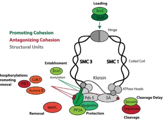
al., 1997) (Figure 2). Cohesin is a tripartite ring complex, which topologically entraps replicated DNA molecules keeping them together until the onset of anaphase (Haering et al., 2008; Ivanov and Nasmyth, 2005). The core of this ring complex is composed out of three molecules: SMC 1 and SMC 3 (belonging to the Structural Maintenance of Chromosomes protein family) and the kleisin subunit Scc1, which connects them (Nasmyth and Haering, 2009; Peters et al., 2008). (Figure2). Additional proteins directly associate with the cohesin complex (Scc3/SA, Pds5, WAPL, Sororin) and are thought to have critical roles in cohesin dynamics, and consequently mitotic fidelity (summarized in Box 2).
Figure. 2 The cohesin complex. Cohesin complex forms a ring-shaped
28 Cohesin and its regulators
Figure 3: Cohesin and associated molecules. The cohesin complex and
the different associated molecules that modulate cohesin’s function. Molecules are color-coded according to their influence on the stability of cohesin’s association with chromatin (molecules that promote cohesion are in green; cohesion antagonists in red and proteins with dual effect in orange)
30
The most popular, and soundly tested cohesin ring model postulates that cohesin keeps sister chromatids together by entrapping sister DNA fibers within the same cohesin ring (Haering et al., 2008). EM-studies support that cohesin rings are about 40 nm in diameter (Haering et al., 2002) thus providing sufficient space for enclosing two 11 nm fibers. Other models have been proposed, such as the “handcuff” model, in which cohesion is mediated by two interlinked cohesin complexes, each entrapping its own DNA fiber (Diaz-Martinez et al., 2008; Guacci, 2007). In either case, solid evidence supports that cohesin’s interaction with DNA is of a topological nature (Haering et al., 2008; Ivanov and Nasmyth, 2005), emphasizing that regulation of cohesin binding and function relies on the opening and closing the interphases between the core components (discussed below).
Besides its role in sister chromatid cohesion, cohesin also regulates transcription, contributes to the DNA repair mechanisms, and participates it the organization of the genome in mitotic and post-mitotic tissues (Nasmyth and Haering, 2009; Peters et al., 2008)
The distribution and presence of cohesin on chromatin during the cell cycle coincides with its multiple roles. Cohesin is loaded onto chromatin during G1 phase in budding yeast (Guacci et al., 1997) , and already in telophase in vertebrates (Losada et al., 1998). During G1 phase, Fluorescence Recovery After Photo-bleaching (FRAP) studies have shown that cohesin is dynamically interacting with the DNA (Gerlich et al., 2006). Similar dynamics was observed in cells that are not undergoing mitotic divisions, for example, endocycling Drosophila Salivary glands (Eichinger et al., 2013). This highly dynamic nature of cohesin-DNA interaction in non-dividing or non-replicated cells is believed to relate to
31
cohesin’s role in transcription regulation and interphase genome architecture.
Following the onset of S phase, a fraction of cohesin molecules establishes cohesion between newly replicated sister chromatids. Specific changes on the cohesin complex (discussed below) ensure the post-replicative stabilization of cohesin-DNA interaction concomitantly or right after replication fork passage. This cohesive state is then maintained until the subsequent mitosis.
In early mitosis, the majority of the cohesin complexes are released from chromosome arms. By the time cells reach metaphase, cohesion is solely maintained by a small pool of cohesin molecules retained at the centromeric and pericentromeric regions (Losada et al., 1998; Waizenegger et al., 2000; Warren et al., 2000).
At the onset of anaphase, remaining centromeric cohesin is destroyed in a rapid and acute manner by a cysteine protease named Separase, allowing the segregation of sister chromatids by the spindle (Uhlmann et al., 1999). This enzyme cleaves the kleisin subunit Rad21/Scc1 releasing sister chromatids from topological entrapment. The destruction of cohesin during anaphase marks the point of no return for the mitotic cell: once cohesin is cleaved, separation of the chromatids is rapid and irreversible. Consequently, release of cohesin from mitotic chromosomes is a highly regulated affair. The key surveillance mechanism governing cohesin release is the Spindle Assembly Checkpoint(SAC) (Reviewed in (Musacchio and Salmon, 2007). The SAC regulates cohesin cleavage by delaying the onset of anaphase until all the chromosomes are bioriented on the metaphase plane. SAC mediates this delay by directly inhibiting the Anaphase Promoting
32
Complex/Cyclosome (APC/C), whose activity is needed for anaphase events. APC/C mediates cohesin cleavage trough indirect activation of Separase, the protease responsible for proteolytic opening of the cohesin ring.
Loss of cohesin or cohesin-regulators in virtually all organisms results in premature separation of sister chromatids (Guacci et al., 1997; Losada et al., 1998; Michaelis et al., 1997; Mirkovic et al., 2015; Sumara et al., 2000; Vagnarelli et al., 2004), arguing that cohesin is the most significant force that counteracts spindle forces. Nevertheless, it is conceivable that other forces may additionally play a role in chromosome cohesion. In particular, DNA-DNA intertwines (catenation) have long been argued to contribute to cohesion during mitosis (Reviewed in (Diaz-Martinez et al., 2008; Guacci, 2007; Liu et al., 2009b). Due to the helical nature of the DNA molecule, the replication fork passage creates tangles between replicated DNA molecules. These catenations need to be resolved before the onset of anaphase; otherwise, the entanglements will cause chromosome bridges and breakages in the DNA molecule. Topoisomerase II is the molecule responsible for de-catenation of these linkages and inhibition of this enzyme leads to accumulation of catenations, which are sufficient to confer cohesion even in the absence of cohesin proteins (Toyoda and Yanagida, 2006; Vagnarelli et al., 2004).
How much residual catenation contributes to cohesion during normal mitosis is a matter of debate. Although residual catenation has been observed even in anaphase segregating chromatids (Baumann et al., 2007), inhibition of topoisomerase specifically during metaphase has only a small effect on the efficiency of chromosome segregation (Oliveira et al., 2010). This suggests that
33
residual catenation may contribute to chromosome cohesion; yet, it is insufficient to resist the drastic spindle forces affecting chromosomes during mitosis. More importantly, unlike cohesin’s destruction, which requires SAC silencing and APC/C activation, there is little to no evidence that removal of residual catenation is delayed by cell cycle progression checkpoints which control mitosis. SUMOylation of topoisomerase II has been proposed to restrict centromeric de-catenation during mitosis (Bachant et al., 2002; Dawlaty et al., 2008; Ryu et al., 2010), but there is no evidence that this reaction is under surveillance of the SAC. Thus, regulation of the cohesive state of chromosomes is mechanistically linked to the control of cohesin’s association with chromatin throughout the cell cycle, which will be discussed below.
Fig. 4 Overview of the cohesin cycle. Cohesin is loaded in telophase or
G1, and is dynamically associated with chromatin. Upon replication, cohesion is established, connecting two replicated strands. Non-centromeric cohesin is removed from chromosome arms during prophase in metazoans, resulting in X-shaped chromosomes in metaphase. Finally, cohesin is cleaved during anaphase, allowing for the separation of sister chromatids.
34 1.1.2 The Cohesin Cycle
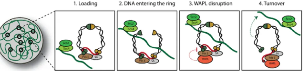
A) The cohesin cycle : Chromatin Loading
Fig. 5 Cohesin loading and turnover. Cohesin loading onto DNA
depends on the Scc2/4complex. DNA loading involves opening of the SMC1/3 interface, the hinge. Before replication, this interaction is dynamic, as loaded cohesin can be destabilized by WAPL, which opens the SMC3/Kleisin interface and releases cohesin from the chromatin.
Cohesin loading onto chromatin is dependent on a two-protein complex known as Scc2/4 , also known as NIPB (Nipped-B) in D.melanogaster, or NIPBL (NIPB-Like) complex in humans (Ocampo-Hafalla and Uhlmann, 2011). The Scc2/Scc4 loading complex is essential for sister chromatid cohesion during G1/S phase, but not during G2 (Ciosk et al., 2000; Uhlmann and Nasmyth, 1998). This would entail that the Scc2/Scc4 has a primary function of loading cohesin onto the chromatin, but not in its stabilization or maintenance.
Given the ring-like architecture of cohesin, its loading onto chromatin requires opening of the ring. Elegant experiments with fusion of interfaces between different cohesin components support that the entry gate for cohesin loading resides at the interface of the SMC1 and SMC3 hinge domains, in an ATP-dependent process (Arumugam et al., 2003; Gruber et al., 2006; Weitzer et al.,
35
2003). Nevertheless, the molecular mechanism by which Scc2/4 promote cohesin’s loading remains unknown.
Sites of cohesin loading do not necessarily coincide with cohesin’s accumulation. This is mostly due to the fact that once loaded, cohesin complexes can slide on the DNA molecule (Hu et al., 2011; Lengronne et al., 2004; Ocampo-Hafalla et al., 2016; Stigler et al., 2016). Additionally, before DNA replication, the cohesin molecules display a highly dynamic association with DNA (Gerlich et al., 2006). Dissociation of cohesin from un-replicated DNA molecules is mediated by Wings-apart like protein (WAPL) (Gandhi et al., 2006; Kueng et al., 2006; Verni et al., 2000). Upon binding to the cohesin complex, WAPL removes cohesin from chromatin by disrupting the interface between SMC3 and Rad21/Scc1 subunits (Buheitel and Stemmann, 2013; Eichinger et al., 2013).
Cohesin loading is not a uniform event across the chromatin landscape and is found to be enriched at the centromeric/pericentromeric regions in most species studied so far (Blat and Kleckner, 1999; Glynn et al., 2004; Oliveira et al., 2014). Studies in budding yeast support that cohesin enrichment at the centromere is dependent on centromeric DNA sequences as well as proteins involved in kinetochore assembly (Megee and Koshland, 1999; Tanaka et al., 1999; Weber et al., 2004). However, species with longer centromeric sequences, such as fission yeast, rely on heterochromatin rather than centromeric sequences for cohesin enrichment (Bernard et al., 2001; Nonaka et al., 2002). In accordance, recent studies in D. melanogaster showed that cohesin enrichment at ectopic regions of pericentromeric heterochromatin occurs in the absence of a proximal centromere, most likely due to preferential binding of the
36
cohesin loading factor Scc2/Scc4 (Nipped B) (Oliveira et al., 2014). The preferential activity of Nipped B at the centromeric region is thought to be due to the specific state of pericentromeric heterochromatin, mainly H4K20 and H3K9 methylations and the presence of HP1 protein, tough clear links have been controversial (Hahn et al., 2013; Koch et al., 2008).
B) The cohesin cycle II: Cohesion establishment
Fig. 6 Cohesion establishment during S phase. Upon DNA replication,
a fraction of cohesin becomes stable on the chromatin. This happens due to SMC3 acetylation by Eco1 and recruitment of Sororin, protecting the cohesin complex from WAPL removal. This stable fraction of cohesin is considered “cohesive” cohesin, stably binding sister chromatids until the end of mitosis
Cohesin establishment occurs during replication, at the time the newly replicated DNA molecule is being formed. Disruption of cohesin loading during G1 results in sister chromatid defects, while disruption during G2 does not. This means that the “effective” cohesion is established during S phase, during DNA replication (Uhlmann and Nasmyth, 1998). At the onset of replication, the dynamic properties of cohesin turnover change and a new pool of
37
stable, “cohesive” cohesin can be identified by FRAP (Gerlich et al., 2006).
Stabilization of cohesin complexes upon replication depends on the Eco1 acetyl transferase (Skibbens et al., 1999; Tanaka et al., 2000; Toth et al., 1999). This enzyme acetylates cohesin associated with replicated DNA at specific lysine residues on SMC3 and failure to acetylate leads to cohesion defects and cell death. The mechanism by which SMC3 lysine acetylation prevents cohesin de-association once it is bound to chromatin is contentious (reviewed in (Rudra and Skibbens, 2013)). Some studies propose models in which the acetylation locks the SMC3/kleisin interface, effectively closing the ring; however, these findings are inconsistent with the fact that SMC3 can be acetylated before replication (Rudra and Skibbens, 2013). SMC3 acetylation during the S phase has also been shown to confer cohesin protection by aiding the recruitment of Sororin, which favors cohesion establishment by protecting acetylated cohesin complexes from WAPL-mediated removal (Nishiyama et al., 2010).
These stably associated cohesin molecules (~30% of total chromatin bound cohesin (Gerlich et al., 2006)) are responsible for sustaining cohesion from the time of DNA replication until the subsequent mitosis.
38
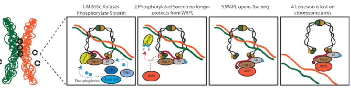
C) The cohesin cycle: Cohesin’s Prophase Release and retention at the centromere
Fig. 7 Cohesin release during early mitosis and centromeric protection. In metazoans, cohesin is removed from the arms by the
prophase pathway. Mitotic kinases phosphorylate Sororin and SA. Phosphorylation induces Sororin displacement, which allows WAPL to destabilize cohesin. Centromeric cohesin complex are protected from this removal process as Shugoshin/PP2A complex protects centromeric cohesion from WAPL-mediated removal
Once the cell enters mitosis, profound changes in the distribution of cohesin begin to take place. Cohesin at the chromosome arms is removed while centromeric cohesion is retained (Losada et al., 1998; Waizenegger et al., 2000; Warren et al., 2000). The loss of arm cohesion, coupled with centromeric retention gives the characteristic “X” shape to the metaphase chromosomes. The removal of cohesin from the arms in early mitosis is a consequence
39
of the “prophase pathway” which mainly relies on action of WAPL protein (Gandhi et al., 2006; Kueng et al., 2006).
WAPL imposes opening of the cohesin ring by disrupting the interface between SMC3 and Rad21/Scc1 subunits (Buheitel and Stemmann, 2013; Eichinger et al., 2013). Consequently, WAPL mutations or knockdown leads to the loss of the characteristic X shape of chromosomes, with cohesin remaining all over chromosome arms (Gandhi et al., 2006; Haarhuis et al., 2013; Kueng et al., 2006).
Several mitotic kinases contribute to the process of cohesin removal, by phosphorylating key proteins involved in the cohesin cycle. Aurora B and Cyclin Dependent Kinase 1 (Cdk1) were shown to antagonize Sororin by phosphorylation, resulting in its dissociation from chromosome arms during prophase (Dreier et al., 2011; Nishiyama et al., 2013). WAPL and Sororin directly compete for the binding to the cohesin-associated protein Pds5 (Nishiyama et al., 2010). The removal of Sororin from chromosome arms during prophase favors WAPL binding, and consequently the removal of cohesin complexes from chromosome arms. In addition to antagonizing Sororin, Aurora B seems to participate in WAPL activation, thus directly promoting cohesin removal (Nishiyama et al., 2013)
Polo Like kinase (Plk) is another key mitotic kinase participating in the cohesin cycle. The phosphorylation activity of Plk1 is crucial for the release of cohesin during the prophase pathway by phosphorylation of SA (Hauf et al., 2005; Lenart et al., 2007; Sumara et al., 2002). The net result of these changes in the
40
cohesin complex results in the removal of most of cohesin from chromosome arms but not from the centromeric region.
How are centromeric complexes protected from prophase pathway removal?
A key molecule in the protection of centromeric cohesion is called Shugoshin, meaning “Guardian Spirit” in Japanese (also known as MEI-S332 in D. melanogaster). Shugoshin confers protection of cohesin specifically at the centromere of both mitotic and meiotic cells (Kerrebrock et al., 1992; Kitajima et al., 2004; McGuinness et al., 2005).
Shugoshin is moved to the centromeric chromatin in complex with the PP2A phosphatase at the onset of mitosis (Kitajima et al., 2006; Liu et al., 2013b). Sugoshin-PP2A complex protects centromeric cohesin from WAPL-mediated removal by several means:
It antagonizes the Aurora B/Cdk1 mediated phosphorylation of Sororin and thereby favors Sororin interaction with Pds5, shifting the WAPL/Sororin competition for cohesin binding towards Sororin, preventing WAPL-mediated removal (Dreier et al., 2011; Liu et al., 2013b; Nishiyama et al., 2013). Aurora B and Cdk1 also phosphorylate and aid in the centromeric localization and activation of Shugoshin (Kitajima et al., 2006; Liu et al., 2013b; Tanno et al., 2010). This means that Cdk1 and Aurora B have conflicting roles in cohesin maintenance. They destabilize Sororin and thereby promote cohesin dissociation along chromosome arms, while at the same time localize and activate Shugoshin at the centromere, allowing for cohesin protection. Shugoshin-PP2A also protects cohesion by counteracting Plk1-mediated phosphorylation of SA
41
(Hauf et al., 2005; Kitajima et al., 2006; McGuinness et al., 2005) and by directly competing with WAPL for the binding to cohesin (Hara et al., 2014).
This protection mechanism is of outmost importance as centromeric cohesin complexes are the only ones that suffice cohesion maintenance during prometaphase and metaphase, while chromosomes are under drastic pulling and pushing forces exerted by the mitotic spindle to accomplish chromosome alignment.
D) The cohesin cycle: The final cut
Mitosis is a process of trial and error, with a few decisive breakpoints. Mitotic events of chromosome attachment, substrate phosphorylation, and biorientation are mostly redundantly regulated, and reversible. This allows for ample error correction in an otherwise error prone process. However, once the metaphase is formed, and chromosomes are bioriented, the cell reaches the point of no return: cohesin cleavage.
The cleavage of cohesin at the metaphase-to-anaphase transition is conducted by a large cysteine protease called Separase, which cleaves the kleisin subunit, distancing the heads of SMC1 and SMC3 subunits (Lin et al., 2016; Uhlmann et al., 2000). This opens the cohesin ring, releasing sister DNA molecules from the proteinaceous cage.
Once the forces that hold chromosomes together are released, there is no going back: therefore, centromeric cohesin cleavage must occur only after multiple safeguard mechanisms have been satisfied. Separase activity is tightly regulated and inhibited through multiple mechanisms until the onset of anaphase.
42
Firstly, Separase is inhibited by the binding of Securin, whose degradation is a prerequisite for sister chromatid separation (Ciosk et al., 1998; Hirano et al., 1986; Zou et al., 1999). Securin inhibits Separase by binding to its active site and abolishing its interaction with other substrates (Hornig et al., 2002; Lin et al., 2016). However, mutants for Securin in several organisms do not suffer from premature loss of cohesion, evidencing that other mechanisms of Separase inhibition must be in place (Alexandru et al., 2001; Hellmuth et al., 2015) (see below). Furthermore, Securin has been proposed to work as a Separase chaperone by binding to bind to the nascent Separase and aiding in its proper folding and activity (Jallepalli et al., 2001). Consequently, Securin was shown to be required for sister chromatid separation in fission yeast and D. melanogaster (Funabiki et al., 1996; Stratmann and Lehner, 1996).
The second layer of Separase inhibition is mediated by the Cdk1-Cyclin B complex. Cdk1-Cyclin B-Cdk1 phosphorylates Separase and this phosphorylation promotes Cdk1-CycB-separase binding, preventing Separase activation until the onset of anaphase (Gorr et al., 2005; Stemmann et al., 2001). The dual inhibition of Separase by CycB-Cdk1/Securin is lifted by the APC/C, an E3 ubiquitin ligase, which is the main effector of anaphase (reviewed in (Primorac and Musacchio, 2013; Sullivan and Morgan, 2007)). The APC/C ubiquitinates both Securin and Cyclin B, targeting them for the degradation by the proteasome, releasing the Separase from its double leash. This, in turn, leads to cohesin cleavage and the onset of anaphase (Oliveira and Nasmyth, 2010).
Given the importance of this transition, the APC/C itself is tightly regulated during mitosis by a surveillance mechanism known as
43
the Spindle Assembly Checkpoint (SAC) (Musacchio and Salmon, 2007; Sullivan and Morgan, 2007). The key effector of this mechanism is the Mitotic Checkpoint Complex (MCC). Unattached kinetochores catalyze the formation of this inhibitory complex, which sequesters Cdc20, a key activator required for APC/C activity (Musacchio and Salmon, 2007; Sullivan and Morgan, 2007). The MCC complex is composed of Mad2, BubR1, Bub3 and Cdc20, and that form a complex that actively binds and inactivates the APC/C (Primorac and Musacchio, 2013). As long as the SAC is active and the MCC is being produced at unattached kinetochores, the APC/C will not be activated by cdc20, Cyclin B and Securin will remain intact, Separase inactive, and cohesin will not be cleaved. This equilibrium changes once metaphase is achieved and chromosomes are bioriented. Stable chromosome attachments result in SAC satisfaction and the release of Cdc20 from the inhibitory MCC complex (Primorac and Musacchio, 2013; Sullivan and Morgan, 2007). Once this happens, APC/C binds Cdc20 becoming active to ubiquitinate Cyclin B and Securin. Ubiquitination promotes the proteasome-mediated degradation of these targets and consequently the release of Separase from its inhibition. Anaphase is imminent.
Since chromosome biorientation and microtubule attachment are highly dynamic processes, once all the chromosomes are bioriented, the decision to commit to anaphase must be rapid and the execution swift. Indeed, live imaging analysis revealed that separase-mediated cohesin cleavage happens within a few minutes during the metaphase-to-anaphase transition (Gerlich et al., 2006; Oliveira et al., 2014; Yaakov et al., 2012).
44
In order to achieve this sharp metaphase to anaphase transition and rapid cohesin cleavage, multiple positive feedback mechanisms are needed to create a molecular switch. Firstly, Separase has autocatalytic activity, and once released from its Cyclin B-Cdk/Securin inhibition, it is able to cleave itself, and convert to an even more enzymatically potent form (Waizenegger et al., 2002). Furthermore, APC/C is constantly ubiquitinating the MCC and trying to pry away the Cdc20 subunit away from it, weakening the SAC signal in the process (He et al., 2011; Uzunova et al., 2012). In this way APC, accelerates its own release from SAC inhibition during anaphase.
In addition (or in parallel) to separase-mediated cleavage, the cohesin protection machinery is also released from centromeres at the metaphase to anaphase transition, which may accelerate cohesin release. Release of Shugoshin/PP2A from the centromeres may additionally promote the Plk1-mediated phosphorylation of Rad21/Scc1 (Plk1-mediated), which enhances its cleavage by the Separase (Alexandru et al., 2001; Hornig and Uhlmann, 2004).
Moreover, both Shugoshin and Sororin, two key molecules involved in cohesin protection, are directly targeted for degradation by the APC/C (Karamysheva et al., 2009; Rankin et al., 2005). Whether or not removal of the mechanisms involved in cohesin protection actively contribute to the sharp cohesion release process remains to be determined.
As discussed above, cohesin cleavage is only initiated once chromosome biorientation is achieved. Thus, given that chromosomes at this stage are being pulled by mitotic spindle,
45
release of cohesin is on its own sufficient to trigger pole-ward chromosome movement (Oliveira et al., 2010; Uhlmann et al., 2000). This, however, is insufficient for efficient anaphase chromosome movement. Sister chromatid separation, when triggered alone, results in ~1/3 slower movements, and concomitant re-activation of the SAC and error-correction mechanisms (Mirchenko and Uhlmann, 2010; Oliveira et al., 2010) . Uncoupling cohesin cleavage from Cyclin B destruction leads to similar failures in chromosome segregation (Parry et al., 2003; Vazquez-Novelle and Petronczki, 2010; Vazquez-Novelle et al., 2014). Successful anaphase onset thus relies not only on a sharp anaphase transition but also on a synchrony between sister chromatid cohesion release and cell cycle progression. The fact that cohesin cleavage is regulated by the APC/C, which cleaves both securin (cohesin release) and Cyclin B (cohesin release + cell cycle transition) should in principle provide this synchrony. Additional feedbacks, however, further ensure that sister chromatid separation occurs in synchrony with inactivation of Cdk1 (reviewed in (Kamenz and Hauf, 2016)).
46
Fig. 8 Cohesin cleavage at the metaphase-to-anaphase transition
In the presence of unattached kinetochores, the spindle assembly checkpoint is activated and generates the formation of the mitotic checkpoint complex (MCC) that prevents anaphase promoting complex/cyclosome activation. Separase is kept inactive by securin and Cdk1/CyclinB binding. b Upon bipolar attachment, the SAC signal is extinguished and the APC/C is activated. Active APC/C ubiquitinates securin and Cyclin B and targets them for degradation. c Active separase cleaves the Rad21/Scc1 subunit and causes ring opening. This opening allows the spindle to drag sister chromatids to opposite poles
47
1.1.3 Functional implications for a multiple-step cohesin removal
Cohesin binding and release is a dynamic and multi-step process whose mechanisms are mostly conserved across species. Exception goes for the dual-step removal for cohesin during mitosis. In budding yeast, unlike in metazoans, arm cohesion is not removed at the onset of mitosis and the entire cohesin pool is removed at the metaphase to anaphase transition by Separase. The question does arise as to why do metazoans have a two-step removal of cohesin? Does accumulation and retention of cohesin specifically at the centromeric region play any specific function in metazoans? When considering the biological significance of multiple steps for cohesion removal present during mitosis, one must have interphase functions of cohesin in mind. During prophase removal of cohesin, the Scc1 subunit is not cleaved, but disengaged from SMC3 (see above), leaving intact cohesin complexes in the cytoplasm. This cohesin is not reloaded during mitosis, possibly due to the dissociation of the Scc2/4 loading complex from chromosomes (Watrin et al., 2006; Woodman et al., 2014). However, this cohesin can load freely during the impending telophase/G1 and preform roles in transcription regulation and interphase genome architecture early in the subsequent cell cycle. Thus, the prophase pathway may be seen as a recycling mechanism, protecting the majority of cohesin from cleavage during anaphase. It is nevertheless becoming more and more evident, however, that the concentration of cohesin specifically around the centromere fulfills important functions for the efficiency of mitosis, as outlined below.
48 1.1.4 Sister Chromatid Resolution
Fig. 9 Cohesin and sister chromatid resolution. Cohesin entrapment
prevents efficient decatenation by topoisomerase II. Cohesin removal from chromosome arms ensures proper sister chromatid resolution. Abnormal retention of cohesin on the arms results in residual entanglements and consequently mitotic defects
49
During replication, sister DNA molecules become heavily intertwined as a consequence of the unwinding of parental DNA strands and/or colliding replication forks. In order to segregate these tangled sister molecules into two daughter cells, their catenations must be resolved. Failure to resolve such DNA intertwines by topoisomerase II leads to breaks in the DNA molecules during anaphase, when chromosomes are pulled to the poles by the spindle. Cohesin was shown to block the action of Topoisomerase II (Farcas et al., 2011; Sen et al., 2016), possibly by keeping the two sisters in such close proximity that disfavors their efficient decatenation. Thus, cohesin removal from chromosome arms during prophase is believed to aid sister chromatid resolution along chromosome arms, providing Topoisomerase II with enough space to resolve catenations.
The degree to which sister chromatid resolution can occur in the presence of chromosome-bound cohesin has been hard to estimate. A recent study has elegantly shown that in the absence of WAPL, when cohesin is retained all over chromosome arms, most of sister chromatid resolution can be observed, at least at the limit of the cytological method applied to differentially label individual sister chromatids (Nagasaka et al., 2016). Thus, although cohesin may impair efficient decatenation, the degree of chromosome intertwines even in the presence of cohesin must be residual.
These residual levels of chromosome intertwines are nevertheless sufficient to impair efficient chromosome segregation. When cohesin is not removed from chromosome arms in a timely manner, which happens if WAPL is down-regulated and the prophase pathway inhibited, chromosomes lose their characteristic “X-shape”
50
and cells undergo an erroneous anaphase, marked by detectable chromosome bridges during anaphase (Haarhuis et al., 2013; Tedeschi et al., 2013). Similar results were observed in cells expressing a modified version of Sororin that lacks its Cdk1-phosphorylation site. This version is not removed from chromosomes arms at the onset of mitosis leading to over-cohesion of metaphase chromosome arms and lagging chromosomes during anaphase (Nishiyama et al., 2013). Moreover, chromosome rearrangements that misplace pericentromeric heterochromatin away from the centromere were shown to abnormally accumulate non-centromeric cohesin (Oliveira et al., 2014). These chromosomes also exhibit chromatin stretching during anaphase, specifically at ectopic cohesin-retention sites. Thus, the spatial and temporal positioning of cohesin on the mitotic chromosome is crucial for timely chromosome resolution. Any disturbance, such as prolonged retention or enrichment of cohesin along chromosome arms leads to incomplete sister chromatid separation, followed by mitotic errors.
51
1.1.5 Inner-centromere defining platform:
Fig. 10 The inner centromere network. Cohesin sets the blueprint for
the inner centromere network, regulating chromosome architecture and microtubule attachment. Cohesin is needed for the recruitment of Haspin kinase, which triggers the cascade resulting in recruitment of CPC and Shugoshin to the pericentromeric region
Centromeric cohesin has recently emerged as a core component of the inner centromeric network and thereby influences the localization of important machinery that regulates mitotic fidelity. Kinetochore microtubule attachments are regulated by the actions of Aurora B, a key mitotic kinase that destabilizes erroneous kinetochore-microtubule attachments. It is well established that Aurora B destabilizes attachments that are not under tension through the phosphorylation of key kinetochore substrates (Biggins and Murray, 2001). This phosphorylation results in microtubule
52
detachment and the creation of unattached kinetochores that can trigger SAC signaling. Aurora B, together with its regulatory partners INCENP, Borealin and Survivin, forms the Chromosome Passenger Complex (CPC). This complex decorates the entire chromosome length during early mitotic stages but dynamically shifts its localization towards prometaphase/metaphase, becoming highly enriched at the inner centromeric region (Reviewed in (Carmena et al., 2012).
Cohesin’s importance for CPC localization has been documented in several studies (Carretero et al., 2013; Haarhuis et al., 2013; Kenney and Heald, 2006; Mirkovic et al., 2015; Sonoda et al., 2001; Vass et al., 2003) but only recently the mechanistic details for this interaction are being elucidated. CPC localization to the inner centromere was shown to depend on two histone marks: Histone H3 phosphorylation on Threonine 3 (H3pT3) and histone 2A-serine 121 (H2A-S121) phosphorylation (Yamagishi et al., 2010). The cohesin subunit PDS5A interacts with the Haspin Kinase, which is the kinase responsible for H3T3 phosphorylation (Yamagishi et al., 2010). Depletion of Pds5 or Cohesin subunits result in delocalized Aurora B and possibly impaired error correction (Carretero et al., 2013; Mirkovic et al., 2015; Yamagishi et al., 2010). Interestingly enough, “too much” cohesin produces a similar phenotype, as WAPL depleted cells also exhibit delocalized Aurora B signals and defective error-correction capacity (Haarhuis et al., 2013).
In addition to CPC localization, cohesin also plays a role in the localization of another key inner centromere component: Shugoshin. Shugoshin interacts directly with cohesin and requires this interaction for its activity (Liu et al., 2013a; Liu et al., 2013b). In
53
this way, cohesin enhances its own centromeric protection but also contributes to other events that are governed by Sgo1 at the centromeres, namely biorientation of sister chromatids, localization of the CPC and SAC silencing (reviewed in (Marston, 2015)). Thus, while enhancing its own protection, cohesin plays a pivotal role in the establishment of the inner centromere network.
1.1.6 Force Balance
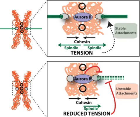
Fig. 11 Force balance. Cohesin is the major force resisting the mitotic
spindle during metaphase. The antagonism between cohesin and the spindle results in sufficient tension that is required to stabilize the attachments of microtubules to the kinetochore. Erroneous attachments (e.g. mono-oriented chromosomes or chromosome with the two kinetochores bound to the same pole)are not under sufficient tension. This reduced tension leads to destabilization of these interactions by Aurora B kinase
54
Centromeric retention of cohesin has profound roles in mitotic fidelity, as it is the condition for biorientation of chromosomes and symmetrical segregation of the genome. The binding and stability of microtubule attachments to the kinetochore is enhanced by the tension between the spindle and the kinetochore, both in vivo and in vitro (reviewed in (Biggins, 2015)). Tension-dependent stabilization of kinetochore-microtubule interactions depends on an intrinsic stabilization ability of the mechanical force exerted by the microtubule pulling forces (Akiyoshi et al., 2010), as well as on biochemical changes that promote the stabilization of kinetochore-microtubule interactions. The latter are regulated by Aurora B kinase, responsible for the correction of erroneous microtubule-kinetochore interactions through the phosphorylation of key kinetochore substrates. Upon bipolar attachment, i.e. maximal tension, the increase in the distance between the inner-centromeric Aurora B and the kinetochore is believed to displace Aurora-B away from its targets thus reverting Aurora-B mediated destabilization of microtubule attachments (Liu et al., 2009a).
How chromosome tension is established, sensed and ultimately regulates kinetochore- microtubule interactions has been widely investigated. Bipolar attachment increases tension across the entire pericentromeric domain (inter-kinetochore tension), but also within each individual kinetochore, marked by the increase in the distance between the proteins of inner and outer kinetochore (reviewed in (Maresca and Salmon, 2010). Both intra- and inter-kinetochore stretch require a counterforce to the spindle to generate stable microtubule attachment and tension. The cohesin ring presents the only force at the centromere that is able to resist the pulling forces of the spindle. Thus, centromeric cohesion